Download presentation
Presentation is loading. Please wait.

1
Anatomy and Physiology I The Skeletal System Physiology of the Skeletal System

2
Skeletal System Bones are made of several tissues Primarily made of collagen and calcium salts About 206 bones in the human body

3
Functions of Skeletal System SUPPORT: Hard framework that supports and anchors the soft organs of the body. PROTECTION: Surrounds organs such as the brain and spinal cord. MOVEMENT: Allows for muscle attachment therefore the bones are used as levers. STORAGE: Minerals and lipids are stored within bone material. BLOOD CELL FORMATION: The bone marrow is responsible for blood cell production.

4
Parts of the Skeletal System Axial skeleton –Skull and bones that support it –Includes vertebra and ribs –80 bones Appendicular skeleton –Limbs –126 bones

5
Features of a Long Bone: Epiphysis: Ends of the bone. Diaphysis: The shaft of the bone which surrounds the medullary cavity. Articular Cartilage: Cushions the ends of the bones and allows for smooth movement. Epiphyseal Plate: Areas made of cartilage allowing for the growth of the bone.

6
Joints Where bone meets bone Ligament – holds bone to bone Types of joints: –Immovable - skull –Ball-and-socket - shoulder –Hinge - knee –Pivot – forearm –Gliding - vertebrae

7
Joints Cartilage covers ends of movable bones –Reduces friction Lubricated by fluid from capillaries

8
Cartilage

9
Bone Structure Periosteum – hard outer covering –Cells for growth and repair Compact bone – hard strong layer –Bone cells, blood vessels, protein with Ca and P Spongy bone – at ends of long bones –Has small open spaces to lighten weight Marrow cavity – hollow in middle of long bones

10
Bone Structure

11
Bone Marrow Red marrow – produces blood cells and clotting factors –Found in humerus, femur, sternum, ribs, vertebrae, pelvis –Produces RBC 2 million per second Yellow marrow – stores fat –Found in many bones

12
Haversian System Structure of compact bone Rings of bone tissue with blood vessels and nerves in the center

13
Haversian System

14
Shows lacunae and caniculi

15
Bone Development Initial skeleton of cartilage in infants Replaced with bone by osteoblasts More than 300 bones at birth – fuse to 206 Always growing and breaking down –Osteoblasts – form new bone cells –Osteoclasts – break bone cells down –Osteocytes – mature bone cells

16
Broken Bones Fracture is a break of the bone Simple or Complex fracture Regrowth of bone: –Spongy bone forms in first few days –Blood vessels regrow and spongy bone hardens –Full healing takes 1-2 months

17
Ankle Fracture Surgery Fibular Fracture

18
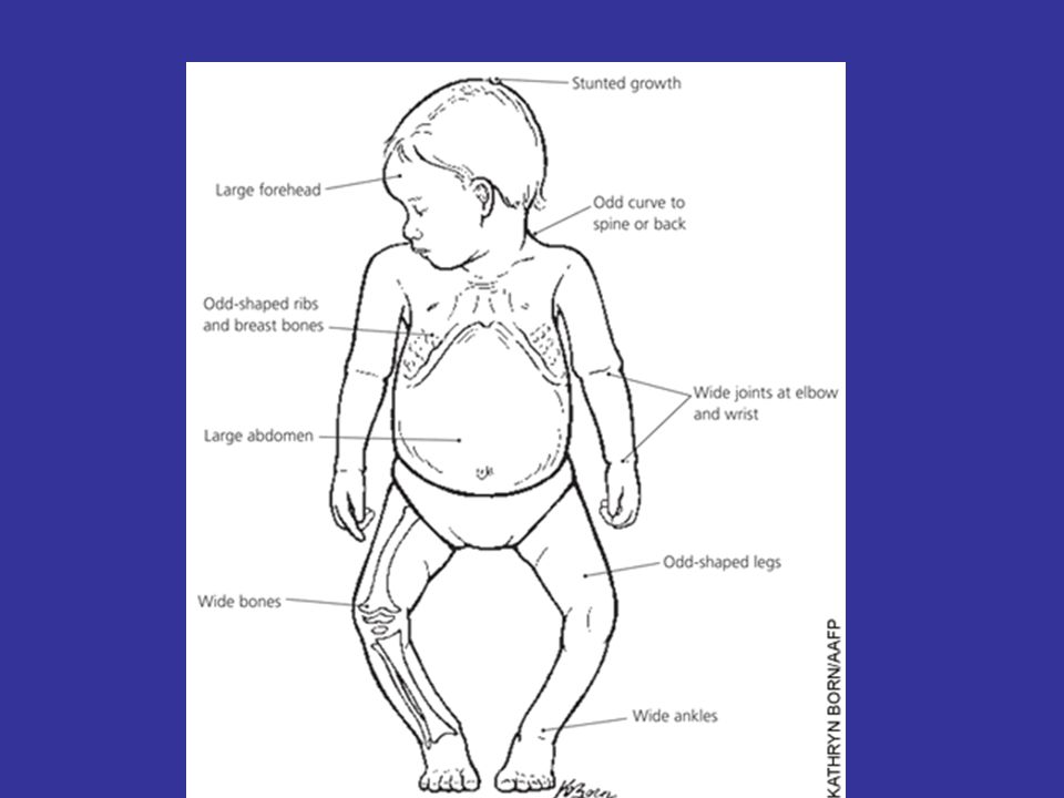
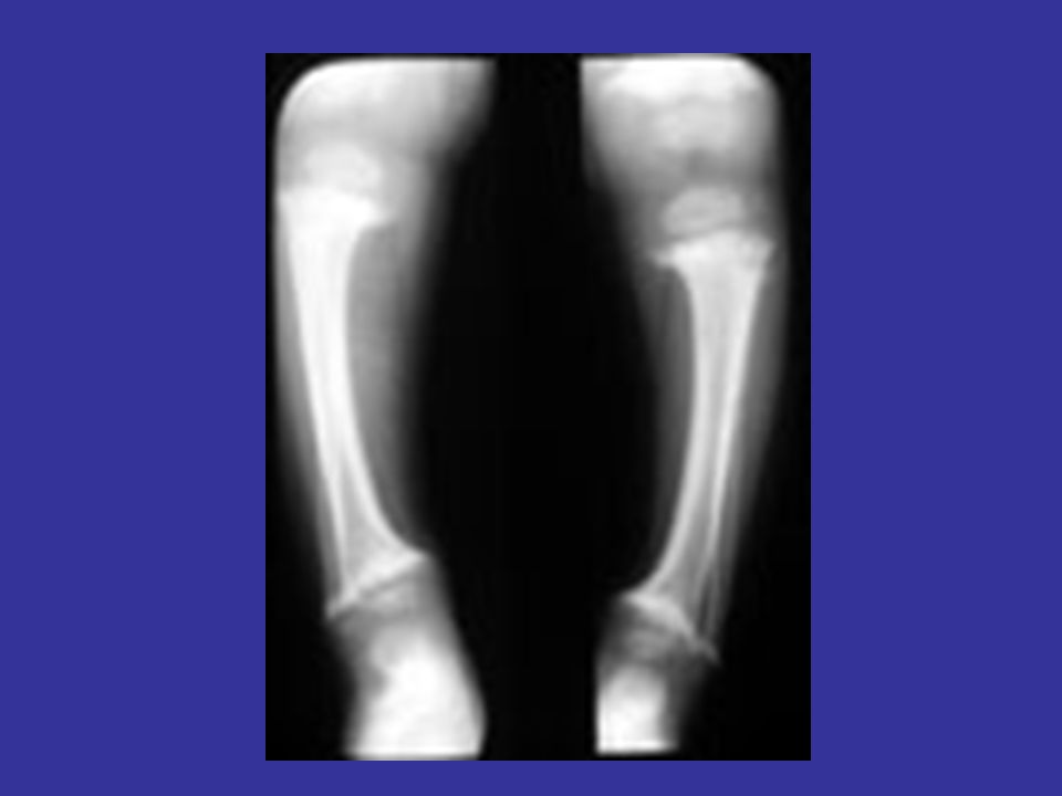
Homeostatic Imbalances Rickets Disease of children due to a lack of vitamin D. Calcium is not deposited in bones. Bones become soft. Bowing of the bones, and other deformities occur.

21
Homeostatic Imbalances Osteoporosis Bone reabsorption is greater than bone deposition. Due to any of the following: Lack of estrogen in women. Lack of exercise to stress the bones. Inadequate intake of calcium and phosphorus. Abnormalities of vitamin D metabolism. Loss of muscle mass.

22
Osteoporosis Decline in Bone Density Bone Resorption > Bone Deposition Increase Risk for Fracture compression fractures of vertebrae hip fractures Role of calcium, vitamin D, estrogen, exercise Calcitonin vs. Parathyroid Hormone

23
Osteoporosis

25
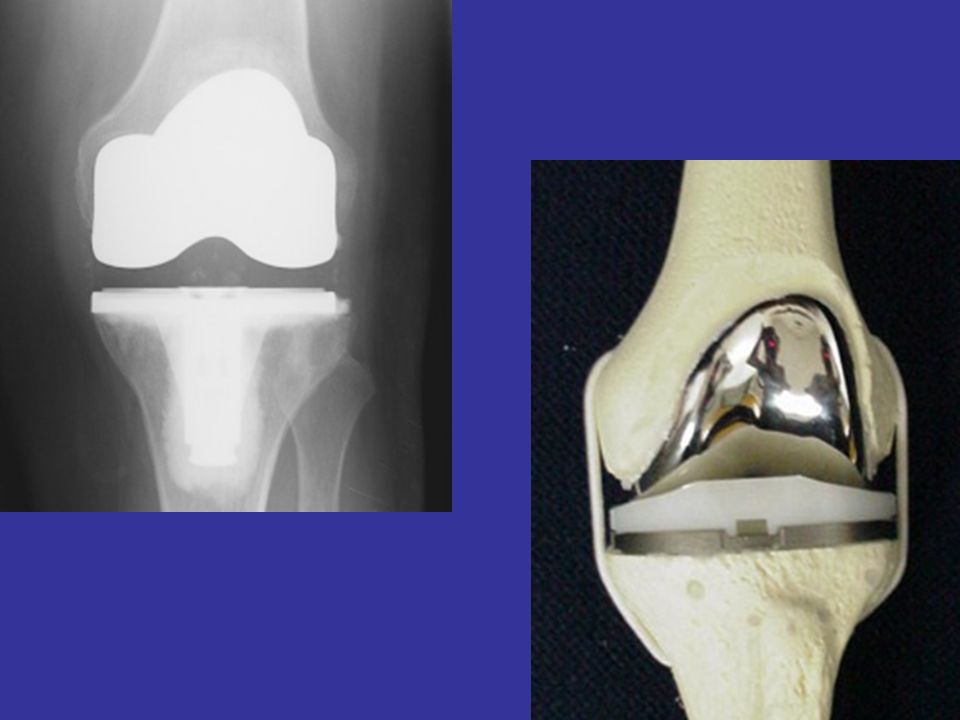
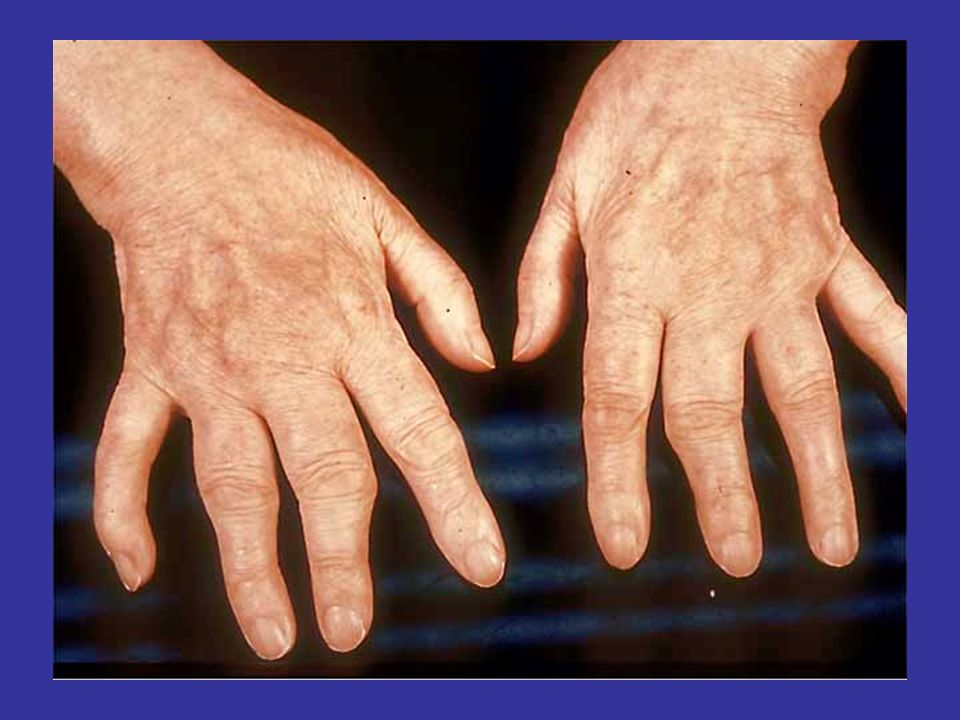
Age Related Dysfunctions Arthritis: Osteoarthritis- 90% of pop. By age 40 chronic inflammation of articular cartilage can be normal age-dependent change can also be pathology due to: age-related changes decrease blood supply trauma

26
Osteoarthritis

31
Haversian System

33
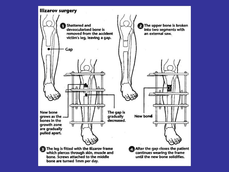
BONE LENGTHENING Recommended for children whose bones are still growing. This series of treatments involves several surgical procedures, a lengthy convalescence period, and considerable risks -- but it can add up to 6 inches of length to a leg.

34
Under general anesthesia, the bone to be lengthened is cut. Metal pins or screws are inserted through the skin and into the bone. Pins are placed above and below the cut in the bone, and the skin incision is stitched closed.

35
A metal device (usually some sort of external frame) is attached to the pins in the bone and will be used later to gradually pull the cut bone apart, creating a space between the ends of the cut bone that will fill in with new bone. The lengthening device is used very gradually to ensure adequate filling of the bone and stretching of the soft tissues.

36
Later, when the leg has reached the desired length and has healed (usually after several months), another surgical procedure will be done to remove the pins.
, another surgical procedure will be done to remove the pins.")
Similar presentations


 or an organ –Bone referred to as a connective tissue consists of: cells extracellular.>")


 2.Protection: skull, vertebrae,>")

 Joints ► Cartilages Ligaments ► Divided.>")


 Joints Cartilages Ligaments Divided into two divisions Axial skeleton –>")