Download presentation
Presentation is loading. Please wait.

2
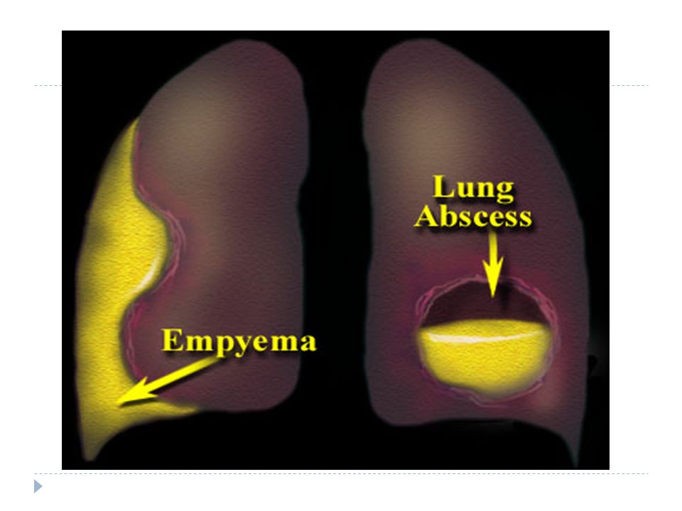
EMPYEMA

3
Empyema The presence of pus in the pleural space. Which may be thin as serous fluid or thick and impossible to aspirate by ordinary needle. Microscopically, neutrophil leucocytes are present in large number. It may involve the whole pleural space or loculated (encysted) Empyema is almost invariably unilateral.
 Empyema is almost invariably unilateral.")
4
Empyema Aetiology Empyema is always secondary to infection in a neighboring structure, usually the lung; 1-Bacterial pneumonia &tuberculosis. 2-Over 40% of patients with community-acquired pneumonia develop an associated pleural effusion And 15% of them develop secondary bacterial infection and empyema. 3-Infection of haemothorax,& rapture of subphrenic abscess. 4-Delay in the diagnosis and instigation of appropriate therapy.

5
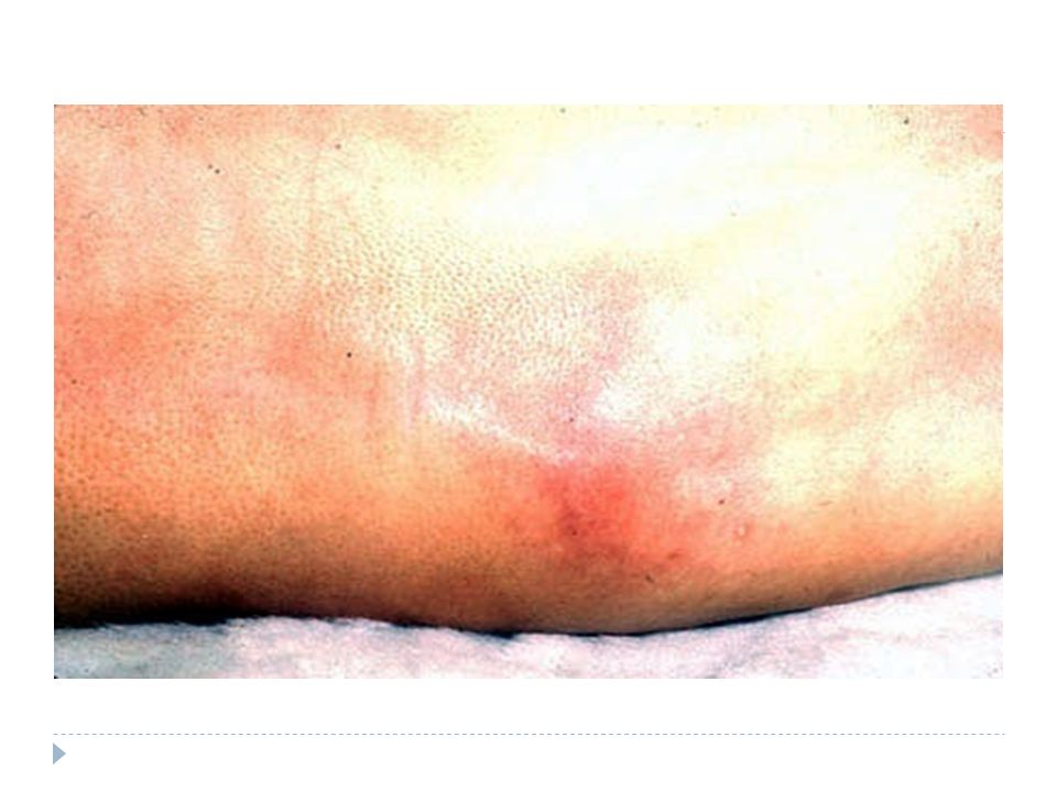
Pathology Both layers of pleura are covered with a thick, shaggy inflammatory exudate. The pus in the pleural space is often under considerable pressure and if the condition is not adequately treated pus may rupture into a bronchus causing a bronchopleural-fistula and pyopneumothorax, Or track through the chest wall with the formation of a subcutaneous abscess or sinus. Empyema can heal by eradication of the infection. Obliteration of the empyema space and apposition of the visceral and parietal pleural layers. Re-expansion of the compressed lung is secured at an early stage by removal of all the pus from the pleural space.

6
Factors interfere with the re-expansion of the lungs
1-The visceral pleural becomes grossly thickened and rigid due to delayed treatment or inadequate drainage of the infected pleural fluid. 2-The pleural layers are kept apart by air entering the pleura through a bronchopleural fistula. 3-There is underlying disease in the lung, such as bronchiectasis, bronchial carcinoma or pulmonary tuberculosis preventing re-expansion.

7
The Stages of Empyema Stage I - “Exudative”
sterile pleural fluid develops secondary to inflammation without fusion of the pleura Stage II - “Fibrinopurulent” a fibrinous peel develops on both pleural surfaces limiting lung expansion Stage III - “Organizing” in-growth of capillaries & fibroblasts into the fibrinous peel

8
Clinical features 1-Pyrexia,usually high and remittent
Systemic features ; 1-Pyrexia,usually high and remittent 2-Rigors,sweating,malaise and weight loss 3-Polymorphonuclear leucocytosis, high (C-Reactive Protein) CRP. Local feature ; 1-Pleural pain; breathlessness; cough and sputum usually because of underlying lung disease; copious purulent sputum if empyema rupture into a bronchus (bronchopleural fistula) 2-Clinical signs of fluid in the pleural space
 CRP. Local feature ; 1-Pleural pain; breathlessness; cough and sputum usually because of underlying lung disease; copious purulent sputum if empyema rupture into a bronchus (bronchopleural fistula) 2-Clinical signs of fluid in the pleural space.")
9
Differential diagnosis
Pleural involvement occurs in up to 5% of patients with rheumatoid arthritis. Pleural malignancy Chylothorax and pseudochylous effusion Pulmonary embolism Esophageal rupture

10
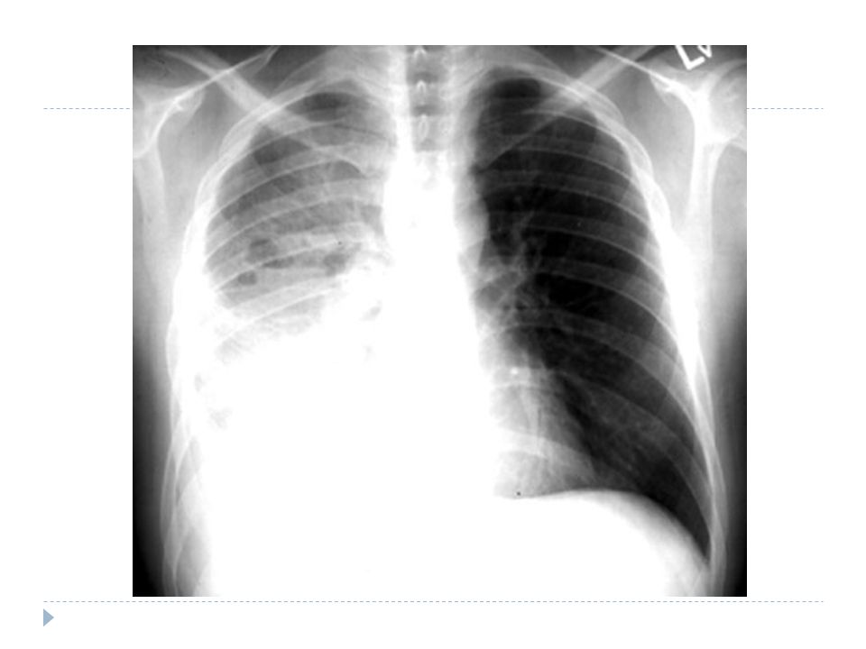
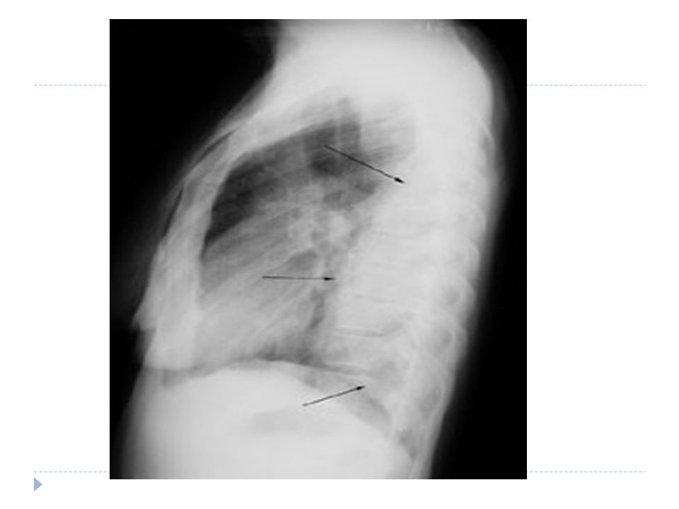
Investigation Radiological examination;
The appearance is often indistinguishable from those of pleural effusion. When air is present in addition to pus (pyopneumothorax). A horizontal 'fluid level' marks the interface between the liquid and air . Ultrasound and CT are extremely valuable in defining the extent of pleural thickening and location of the fluid . And in the case of CT assessing the underlying lung parenchyma and patency of the major bronchi.
. A horizontal fluid level marks the interface between the liquid and air . Ultrasound and CT are extremely valuable in defining the extent of pleural thickening and location of the fluid . And in the case of CT assessing the underlying lung parenchyma and patency of the major bronchi.")
13
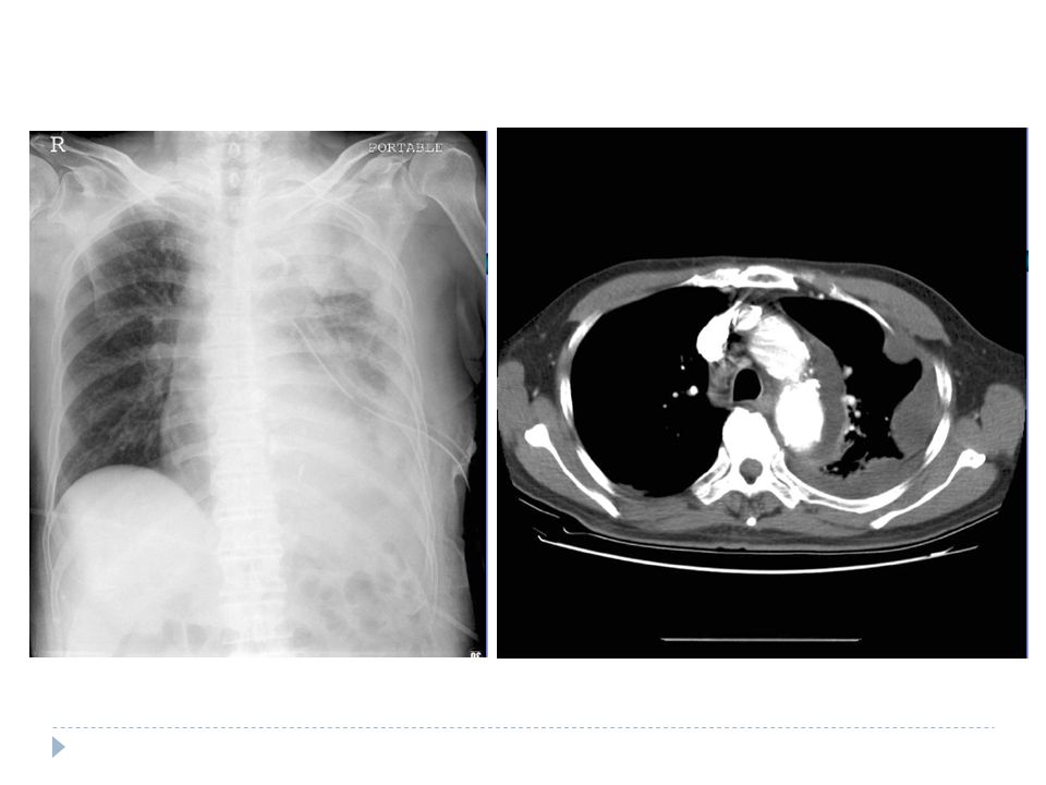
Radiology Examination Cont..
The presence of fever, pulmonary infiltrates and fluid should always alert clinician to the possibility of a parapneumonic collection. Ultrasound is good visualizing septations within loculations that are not usually seen on CT images, but may not identify some separate fluid loculations in inaccessible areas of the thorax.

18
Bacteriology Aerobic organisms are the most frequent organisms identified from infected pleural fluid. These are most commonly Gram-positive organisms from Streptococcal species, followed by Staphylococcus aureus. Gram-negative empyema is more frequent in patients with underlying diseases, especially those with diabetes and alcoholism. Staphylococcus aureus and Gram-negative enteric bacteria such as Klebsiella pneumonia have a particular propensity to cause pleural infection.

19
Bacteriological data. Streptococcus pneumoniae: 15-20%
Increased resistance Staphylococcus:15-30% Streptococcus spp Gram Negative: 20-50% Klebsiella, Enterobacter, Pseudomonas, Hemophilus, E.Coli Anaerobes: Fusobacterium, Bacteroides fragilis

20
Aspiration of pus This confirms the presence of an empyema. Ultrasound or CT is recommended to identify the optimal place to undertake pleuracentesis, which is best performed using a wide-bore needle. The pus is frequently sterile when antibiotics have already been given. The distinction between tuberculous and non-tuberculous disease can be difficult and often requires pleural histology and culture.

21
Management Treatment of non-tuberculous empyema; If the pus is thin an intercostal tube should be inserted under ultrasound or CT guidance into the most dependent part of the empyema space and connected to a water-seal drain system. If the initial aspirate reveals turbid fluid or frank pus, or if loculations are seen on ultrasound, the tube should be put on suction (5-10 cm water) and flushed regularly with 20 ml normal saline.
 and flushed regularly with 20 ml normal saline.")
22
Chest catheter drainage
Optimal size of catheter? Excellent outcomes may be achieved with such small catheter especially when combined with fibrinolytic therapy. Drainage may fail if the fluid is of high viscosity and direct blocks the tube. The balance of forces drawing it down the tube is inadequate. If the fluid is partitioned by fibrinous septae. The rapidity of chest tube drainage might be improved by increasing the drain size, but the successful drainage is unchanged. Here again, provide that the catheter is patent, its bore is irrelevant.

23
Management… cont.

24
Intrapleural fibrinolytics
1949 Tillet and Sherry: partial purified streptococcal fibrinolysin Highly purified streptokinase: IU Urokinase: IU It form a complex with plasminogen that converts additional circulating plasminogen to plasmin. Plasmin lyses fresh fibrin clot and digests prothrobin and fibrinogen. Improvement in the chest radiograph and greater volume pleural drainage, not outcome of mortality, surgical frequency, or hospital stay. Tube drainage with streptokinase and early surgical intervention showed reduced length of hospitalization Potential side effect: hemorrhage, pleuritic pain and fever

25
Management… cont. Thoracocentesis and pleural fluid analysis help in deciding therapy . Streptokinase and Urokinase can be used in multiloculated empyema. Video assisted thoracoscopic surgery or thoracoscopy with debridement are equally effective for multi-loculated empyema. Cases not responding to above measures need decortication

26
Management cont…. Antibiotic directed against the organism causing the empyema should be given for 2-4 weeks. An empyema can often be aborted if the previously mentioned measures are started early. If the intercostal tube is not providing adequate drainage (when the pus is thick or loculated) surgical intervention is required . Surgical 'decortications' of the lung If gross thickening of the visceral pleura has developed and is preventing re- expansion of the lung.
 surgical intervention is required . Surgical decortications of the lung If gross thickening of the visceral pleura has developed and is preventing re- expansion of the lung.")
27
Antibiotic treatment As soon as the bacteriologic sample are recovered
Pneumonia Amoxicillin, 3GC or 3GC +/- Metronidazole Amox-clavulanic acid Dosage of the molecule Nosocomial Tazobactam or Imipenem +/- Aminoglycoside or Quinolone Not Pneumococcus directed molecules Adapted to the laboratory results

28
Treatment of tuberculous empyema
Antituberculosis chemotherapy must be started immediately The pus in the pleural space aspirated through a wide-bore needle until it ceases to re-accumulate . Intercostal tube drainage is often required. In many patients required to ablate a residual empyema space.

29
Surgery for pleural infecetion
No definite data that define the point at which a patient with empyema should proceed to surgical intervention. Open thoracotomy with decortication Mini-thoracotomy Video-assisted thoracoscopic surgery (VATS) Rib resection with open drainage VATS: reduced hospital inpatient time, postoperative complications and length of operating time VATS: failures are with empyema in the organizing stage of the disease
 Rib resection with open drainage. VATS: reduced hospital inpatient time, postoperative complications and length of operating time. VATS: failures are with empyema in the organizing stage of the disease.")
Similar presentations


 Dr. Essam H. Jiffri.>")


. -Cytological tests (>")


