Download presentation
Presentation is loading. Please wait.

1
POSTSURGICAL EMPYEMA Second most frequent cause of empyema.
Incidence– 20%. Pneumonectomy %. Lobectomy-- 1-3%.

5
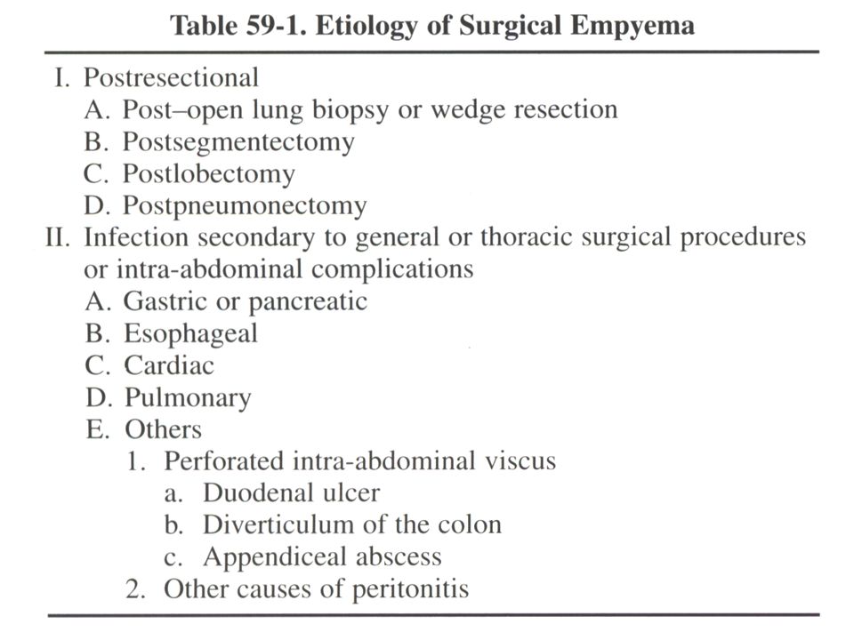
Nonresectional postsurgical empyema
Esophageal surgery leak into pleural space. Subdiaphragm surgery of stomach, pancrease, spleen. Rupture of infected pleural bleb. Lung abscess.

6
Empyema after lung resection
In early postoperative period. Pleural space contaminated at time of pulmonary resection. Develop of bronchopleural or esophagopleural fistula. Blood-borne source. More often when the pleural space is incompletely filled by expansion of the remaining lung, mediastinum shift, elevation of diaphragm.

7
Symptoms and Signs Expectoration of serosanguineous liquid.
Purulent discharge from wound or drain. Radiology— Pleural opacity, air-fluid level. After pneumonectomy– Decrease fluid level early postoperatively, appearance of a new fluid level.

8
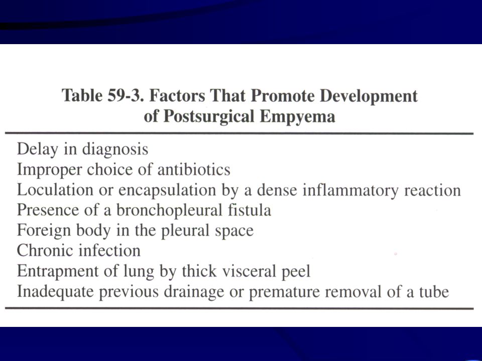
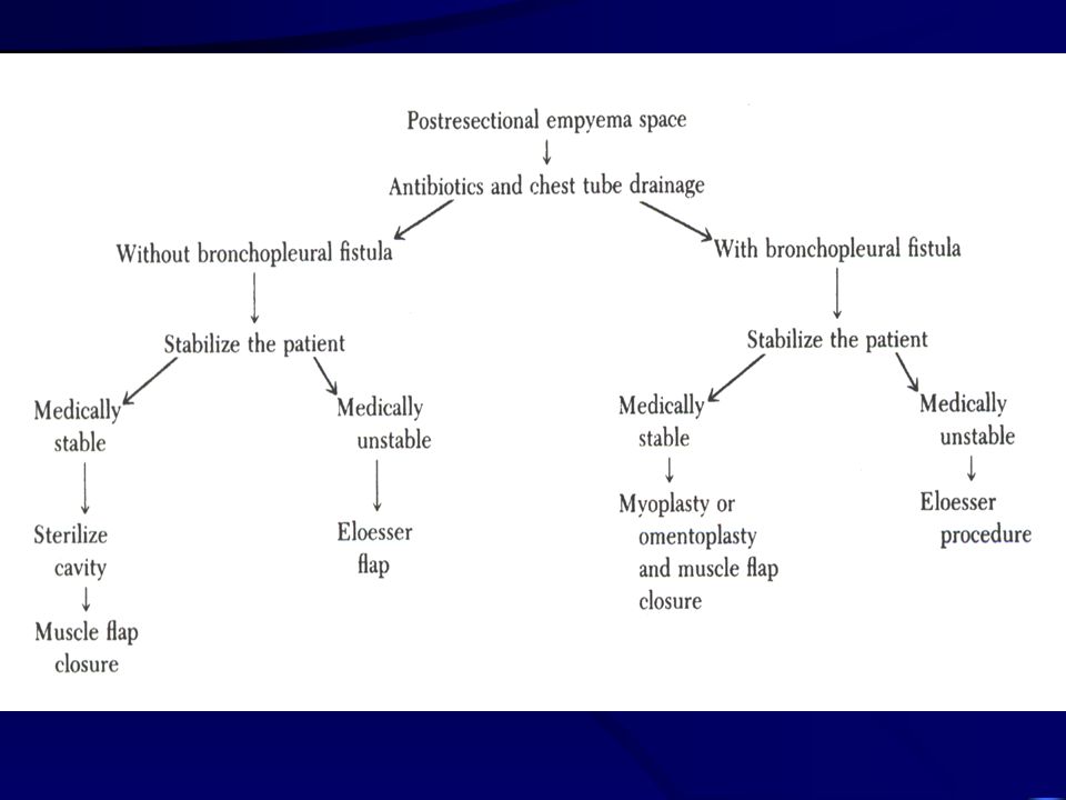
General principles of treatment
Patient with bronchopleural fistula--- Close chest tube thoracostomy. Antibiotics. Patient stabilized– days– Close the bronchopleural fistula– By myoplasty or omentoplasty, single stage muscle flap closure the remaining space. Patient unstable– Close tube drainage change to open drainage by Eloesser’s procedure.

9
General principles of treatment
Only empyema space without bronchopleural fistula--- .Patient stable– Irrigation with antibiotics, single stage muscle flap closure the remaining space. . Patient Unstable– Open Eloesser’s flap.

11
Postpneumonectomy empyema
40% with bronchopleural fistula. 20% fistula close spontaneously. Treatment– see fig 59-2

13
Postpneumonectomy empyema
Clagett’s procedure— 1). Second small chest tube inserted into the second intercostals space. 2). Continue inflow-outflow irrigation. 3). 2g cephalosporin in 500ml D5W, rate 50ml/hour. 4). Gram-negative organism % neomycin. 5). Success rate achieves 50%.
. Second small chest tube inserted into the. second intercostals space. 2). Continue inflow-outflow irrigation. 3). 2g cephalosporin in 500ml D5W, rate 50ml/hour. 4). Gram-negative organism % neomycin. 5). Success rate achieves 50%.")
14
Closure of the postpneumonectomy empyema space
Transposition skeletal muscle flap--- Best way. Single-stage. Execellent blood supply. Pedicle flap to reach almost any location in the pleural space. Rib resected for entry.

15
Muscle flap Latissimus dorsi 30-40%. Serratus anterior 10-15%.
Pectoralis major 20-30%, minor 0-2%. Omentum 5-15%. Rectus abdominis 5-15%.

17
Omentum flap As flap or free graft.
Neovascularization within 48 hours. Excellent vascular supply. Brought up through anterior opening of diaphragm.

18
Pectoralis major flap Blood supply from thoracoacromial artery and internal mammary artery. For sternal infection.

19
Latissimus dorsi flap Most commonly used.
Blood from thoracodorsal artery.

20
Serratus anterior flap
Second choice for muscle transfer flap.

21
Rectus abdominis flap For lower third sternal defect.

22
Single-stage muscle flap closure
For persistent postpneumonectomy empyema space. Indication: 3 months for benign disease and 6 months to 1 years for malignant disease. Two predominant point: 1. No residual space. 2. Sufficient number of flap.

23
Single-stage muscle flap closure
Six basic step: 1. Appropriate antibiotics. 2. Original incision open. 3. Cavity debride widely. 4. Close the bronchopleural fistula, with omentum flap. 5. Appropriate muscle flap fill the pleural space. 6. Begun with latissimus dorsi flap.

24
Two-stage muscle flap closure
First stage– 1. Reopening the thoracotomy wound. 2. Leaving open, 5-7 days. 3. Bronchopleural fistula close primarily and covered by serratur anterior muscle. 4. Wound left open packed daily for 6 weeks to 3 months. Second stage– 1. Filling the pleural space by antibiotics solution 2. Closing the wound in layers.

25
Postresectional lobectomy empyema
Lower lobe lobectomy--- Bronchopleural fistula close by myoplasty– By intercostals muscle, obliteration the space by LD and SA. Upper lobe lobectomy--- Reverse pectoralis major through second intercostals space.

Similar presentations


, FCCP>")


