Download presentation
Presentation is loading. Please wait.

1
Bone Development & Growth

2
Bone Development Bones form by replacing connective tissue in the fetus Some form with sheet-like layers of connective tissue (intramembranous bones) and other replace masses of cartilage (endochondral bone)
 and other replace masses of cartilage (endochondral bone)")
3
Intramembranous Bones The flat bones of the skull form as intramembranous bones that develop from layers of connective tissue Osteoblasts deposit bony tissue around themselves Once osteoblasts deposit bone in the lacunae, they are called osteocytes Cells of the membranous connective tissue that lie outside of the developing bone give rise to the periosteum.

4
Intramembranous Bones

5
Endochondral Bones Most of the bones in the skeleton fall into this category They first develop as hyaline cartilage models and are then replaced with bone Cartilage is broken down in the diaphysis and progressively replaced with bone while the periosteum develops on the outside

6
Endochondral Bones- cont. Cartilage tissue is invaded by blood vessels and osteoblasts that first form spongy bone at the primary ossificatin center Osteoblasts beneath the periosteum lay down compact bone outside the spongy bone Secondary ossification centers appear later in the epiphysis A band of hyaline cartilage, the epiphyseal disk, forms between the two ossification centers

7
Endochondral Bones- cont. Layers of cartilage cells undergoing mitosis make up the epiphyseal disk Osteoclasts break down the calcified matrix and are replaced with bone-building osteoblasts the deposit bone in the place of the cacified cartilage Epiphyseal disks are responsible for lengthening bones while increasing the thickness are due to intramembranous ossification under the periosteum A medullary cavity forms in the region of the diaphysis due to the activity of the osteoclasts.

8
Formation of bone collar around hyaline cartilage model. 1 2 3 4 Cavitation of the hyaline cartilage within the cartilage model. Invasion of internal cavities by the periosteal bud and spongy bone formation. 5 Ossification of the epiphyses; when completed, hyaline cartilage remains only in the epiphyseal plates and articular cartilages Formation of the medullary cavity as ossification continues; appearance of secondary ossification centers in the epiphyses in preparation for stage 5. Hyaline cartilage Primary ossification center Bone collar Deteriorating cartilage matrix Spongy bone formation Blood vessel of periostea l bud Secondary ossification center Epiphyseal blood vessel Medullary cavity Epiphyseal plate cartilage Spongy bone Articular cartilage Stages of Endochondral Ossification Figure 6.8

9
Homeostasis of Bone Tissue Osteoclasts tear down and osteoblasts build bone throughout the lifespan, with an average of 3-5% of bone calcium exchanged annually

10
Blood Cell Formation Two kinds of marrow occupy the medullary cavity of bones: –Red marrow functions in the formation of red blood cells, white blood cells an platelets and is found in the spongy bone of the skull, ribs, sternum, clavicles, vertebrae and pelvis –Yellow marrow occupies the cavities of most bones and stores fat

11
Storage of Inorganic Salts The inorganic matrix of bone stores inorganic mineral salts in the form of calcium phosphates Calcium in bone is a reservoir for body calcium; when blood levels are low, osteoclasts release calcium from bone

12
Storage of Inorganic Salts- cont. Calcium is stored in bone under the influence of calcitonin when blood levels of calcium are high Bone also stores magnesium, sodium, potassium, and carbonate ions Bones can also accumulate harmful elements, such as lead, radium and strontium

13
Osteoporosis Causes bones to become brittle As bones grow longer, they also become thicker and denser. In young adults, the density of bone usually remains constant as bone tissue is constantly being broken down and replaced During middle age, bone replacement gradually become less efficient and bones may become less dense. The loss of bone density is called osteoporosis and can cause bone to become light and brittle and therefore easily broken

14
Osteoporosis- cont. Although both men & women lose bone as they age, women are at a greater risk for osteoporosis –1. Women’s bones are usually smaller and lighter than men’s bones –2. The production of female sex hormones declines rapidly after menopause and sex hormones help to maintain bone density; this decline leads to an increase in bone loss

15
Osteoporosis- cont. Bone density can only be increased during your teens and twenties. Regular exercise and healthy diet will make you healthier now and in the future The stronger your bones are now, the less likely you are to be affected by osteoporosis later. More than 600,000 bone fractures a year result from osteoporosis in the US.

16
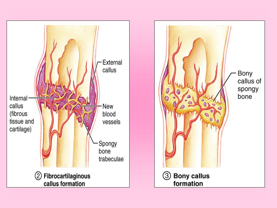
Bone Repair Even simple fracture involve significant bone damage that must be repaired if the bone is to resume normal function Fragments of dead bone or damaged bone must be removed. Osteoclasts (reabsorb bone matrix as part of growth or repair) dissolve and reabsorb the calcium salts of bone matrix
 dissolve and reabsorb the calcium salts of bone matrix.")
17
Bone Repair Osteoclasts remove the damaged and dead bone fragments Then new bone must be produced to replace the part taken away The inner layer of the periosteum contains osteoblasts that are activated when bone is damaged. The osteoblasts produce bone matrix to knit the broken ends back together. Holding these bones still speeds healing, that is why a cast is used.

19
Bone Repair Since most bone has a good blood supply, the repair process is usually relatively rapid and simple fracture often heal within 6 weeks. Other factors that influence the repair include the age of the person, general state of health and nutrition (sufficient calcium, phosphorus, vitamin D and protein, if any of these is lacking, bone repair will be a slower process.

Similar presentations


 - hematopoeisis.>")