Download presentation
Presentation is loading. Please wait.

1
Common Pediatric Hip Problem
Dr.Abdulmonem Al-Siddiky Dr.Kholoud Al-Zain Dr.Khalid Bakarman Assistant Professors Consultant Pediatric Orthopedic Surgeons

2
Common Pediatric Hip problems
DDH SCFE Perth's

3
DDH

4
Nomenclature CDH : Congenital Dislocation of the Hip
DDH : Developmental Dysplasia of the Hip

5
Pediatric Hips Dislocation
Types: Idiopathic isolated pathology Teratologic: Neurologic as: patient with C.P or MMC Muscular as: Arthrogryposis Syndromatic as: Larsen syndrome Miscellaneous: Complication to hip septic arthritis Traumatic

6
Pediatric Hips Dislocation
Note delivery in its self (OBGY Dr.) does not dislocate a hip DDH occurs in the 3ed trimester Teratologic usually in the 1st trimester
 does not dislocate a hip. DDH occurs in the 3ed trimester. Teratologic usually in the 1st trimester.")
7
Normal pelvis Adult Child

8
Normal pelvis adult child

9
Normal pelvis adult child

10
Normal pelvis adult child

11
DDH Normal hip Dislocated hip

12
DDH The pathology is of 2 components: Femoral head position
Acetabular development

13
1) Femoral Head Position
Normal hip Dislocated hip

14
2) Acetabular Development
Normal hip Dislocated hip

15
DDH Normal hip Dislocated hip

16
Patterns of disease Dislocated Dislocatable Sublaxated
Acetabular dysplasia

18
Causes (multi factorial)
Unknown Hormonal Relaxin, oxytocin Familial Lig.laxity diseases Genetics F 4-6x > M Twins 40% Mechanical Pre natal Post natal

19
Mechanical Causes Pre-natal: Post-natal swaddling , strapping Breach
Oligohydrominus Primigravida Twins Post-natal swaddling , strapping

21
Infants at Risk Parents who are relatives (consanguinity)
Positive family history: 10X 1st child Breach presentation: 5-10 X Oligohydrominus Twins: 40% A baby girl: 4-6 X Torticollis: CDH in 10-20% of cases Foot deformities: Calcaneo-valgus Metatarsus adductus Knee deformities: hyperextension and dislocation

22
DDH When risk factors are present the infant should be reviewed:
Clinically Radiologically

23
Examination The infant should be: Quiet Comfortable

24
DDH Look: External rotation Lateralized contour Shortening
Asymmetrical skin folds Anterior Posterior

26
DDH Move Limited abduction

27
DDH Special test (depending on the age): Galiazzi sign
Ortolani, Barlow test only till 4-6 m of age Hamstring Stretch test Trendelenburg sign older comprehending child Limping: Unilateral one sided limping Bilateral waddling gait (Trendelenburg gait)
")
28
DDH- Giliazi test

29
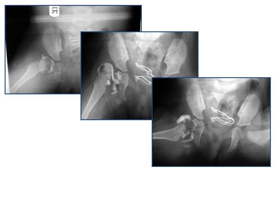
DDH- Ortolani test

30
DDH- Barlow test

31
DDH- Barlow &Ortolani tests

32
DDH- Hamstring Stretch Test

33
DDH- Trendelenburg Test

34
DDH- Trendelenburg Test

35
DDH- Investigations 3w -3m U/S
> 3months XR pelvis (AP + abduction) > 5-6m: More reliable Is when ossification centers normally appears If delayed or did not appear it’s one of the signs of DDH
 > 5-6m: More reliable. Is when ossification centers normally appears. If delayed or did not appear it’s one of the signs of DDH.")
36
DDH- Radiology

37
Radiology After 6 months: reliable

38
Radiology After 6 months: reliable

39
A concentrically, reduced, stable, painless, mobile hip joint.
Treatment - Aims A concentrically, reduced, stable, painless, mobile hip joint. Obtain concentric reduction Maintain concentric reduction In a non-traumatic fashion Without disrupting the blood supply to femoral head That is why: Refer to pediatric orthopedic surgeon

40
DDH- Treatment Method depends on age The earlier started:
Its easier Better the results (higher remodeling potential) Treatment is mainly non-operative Should be detected EARLY Either surgical or non-surgical
 Treatment is mainly non-operative. Should be detected EARLY. Either surgical or non-surgical.")
41
Treatment Birth – 6m 6-12 m: 12 - 18 m: 18 – 24 m: 2-8 years:
In OPD: reduce + maintain with Pavlik harness or hip spica (H.S) 6-12 m: GA + closed (? Open) reduction + maintain with H.S m: GA + open reduction + maintain with H.S 6w, then B.S cast for months 18 – 24 m: GA + open reduction + acetabuloplasty + H.S 6w, then B.S cast 6w 2-8 years: GA + open reduction + acetabuloplasty + femoral shortening + H.S 6w, B.S 4-6w Above 8 years: GA +open reduction + acetabuloplasty (advanced) + femoral shortening + H.S
 6-12 m: GA + closed ( Open) reduction + maintain with H.S m: GA + open reduction + maintain with H.S 6w, then B.S cast for months. 18 – 24 m: GA + open reduction + acetabuloplasty + H.S 6w, then B.S cast 6w. 2-8 years: GA + open reduction + acetabuloplasty + femoral shortening + H.S 6w, B.S 4-6w. Above 8 years: GA +open reduction + acetabuloplasty (advanced) + femoral shortening + H.S.")
42
Pavlik Harness Maximum to start it is 6m of age, if older use other method This is to achieve stable reduction It’s a dynamic splint Is kept on for 6w continuous, then use a rigid abduction splint

43
Abduction splint It’s a rigid splint This is to:
Maintain the reduction, And wait for improvement of the acetabular cover to be: A.I < 30° & with concavity

44
Normal Hip Arthrogram

45
Hip Arthrogram Guided Reduction
Dislocate view Reduced view

47
Hip Spica

48
Broom-Stick Cast

50
Example: Open reduction & Acetabuloplasty

51
Example: Open reduction & Acetabuloplasty & Femoral Shortening

52
DDH Late complications if not treated: Severe pain (hip area, back)
LLD (leg length discrepancy) Pelvic inequality (tilt) Early hip arthritis Early Lumbar spine degeneration
 Pelvic inequality (tilt) Early hip arthritis. Early Lumbar spine degeneration.")
53
SCFE

54
SCFE

55
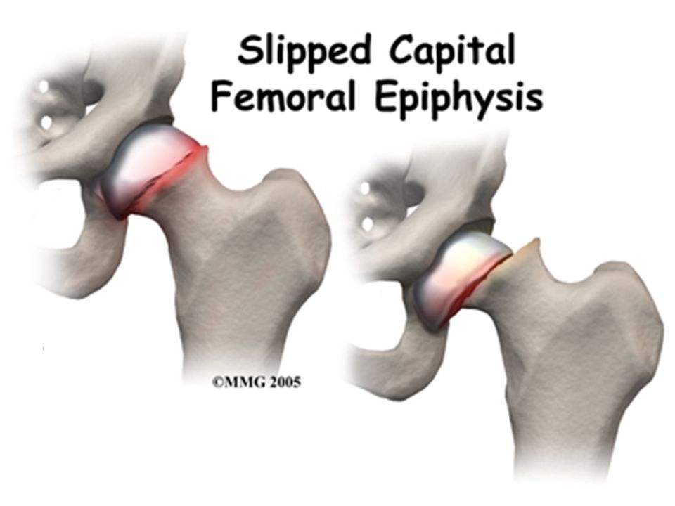
SCFE Slipped Capital Femoral Epiphysis At the level of physis
As if it is a Salter-Harris fracture, type-1 So it is an emergency

57
SCFE- Top View Anterior slippage

58
SCFE Types: When it’s acute or unstable urgent surgery Radiological:
Acute < 3w Chronic > 3w, can see start of callus formation Acute on chronic Clinical: Unstable can not weight bear on that limb Stable can put some weight (walk) When it’s acute or unstable urgent surgery
 When it’s acute or unstable urgent surgery.")
59
SCFE Causes (multifactorial): Unknown Hormonal:
Hypothyroid Abnormal G.H Hypogonadisum Metabolic Chronic renal failure Mechanical (obesity) Trauma
 Trauma.")
60
SCFE: Slipped Capital Femoral Epiphysis Where at level of growth plate Why: ? Hormonal ? Metabolic ? Mechanical, obesity ? Trauma ? Unknown

61
SCFE Typically: (8 – 12y) old Male Obese Dark skinned % chance that the other hip will be affected, within 18m post the 1st hip affection

62
SCFE: Typical : > 8-12y > in males > in obese > in black > if other side affected

63
SCFE History: Pain hip, anterior thigh, knee
Duration of C/O (more or less than 3w) Gait painful or painless Trauma minor or none Any known hormonal or metabolic issues
 Gait painful or painless. Trauma minor or none. Any known hormonal or metabolic issues.")
64
SCFE: History: > Hip pain/knee pain > Minor trauma > no trauma > Limping (painful)
")
65
SCFE Examination: The limb is in ext. rotation
With hip flexion the limb goes in spontaneous ext. rotation Limited int. rotation & abduction Painful hip R.O.M Gait can or can not (antalgic) weight bear on affected limb Thigh muscle wasting (disuse), esp. in chronic cases
 weight bear on affected limb. Thigh muscle wasting (disuse), esp. in chronic cases.")
66
SCFE

67
Hip in ER (external rotation) IR (internal rotation)
On Examination: Hip in ER (external rotation) IR (internal rotation) Abduction Usually painful ROM Limping (painful)
 IR (internal rotation) Abduction. Usually painful ROM. Limping (painful)")
68
SCFE Investigation: XR pelvis: XR knee is normal
AP standing & frog lateral See the actual slip Positive “Klein Line” Or just wide physis pre slip phase XR knee is normal MRI in unusual or unclear presentations

69
Investigations X-ray: If not clear but still doubtful MRI can help
Pelvis: Slippage positive or growth plate space (pre slip phase) Knee normal If not clear but still doubtful MRI can help
 Knee normal. If not clear but still doubtful MRI can help.")
70
SCFE- XR AP

71
SCFE- XR Frog Lateral

72
SCFE- Chronic

73
SCFE- Kline’s Line

74
SCFE- Kline’s Line

75
SCFE

77
SCFE- Example 1

78
SCFE- Example 2

79
SCFE Severity: Depends on degree of slip
The metaphysis is divided to 3 (1/3) The more the slip the worsted the severity
 The more the slip the worsted the severity.")
80
SCFE- Severity

81
SCFE Treatment: Acute or chronic its an emergency refer to Orthopedic urgently Aim prevent further slippage & fuse the physis

82
SCFE Treatment: Acute: Chronic salvage corrective osteotomies
Emergency in-situ fixation (no reduction done) Using 1 or 2 (6mm) screws Screw threads pass the physis to fuse it Screw stops 5mm before the articular surface to prevent “Chondrolysis” Do hormonal essay if any abnormality refer to endocrine Chronic salvage corrective osteotomies
 Using 1 or 2 (6mm) screws. Screw threads pass the physis to fuse it. Screw stops 5mm before the articular surface to prevent Chondrolysis Do hormonal essay if any abnormality refer to endocrine. Chronic salvage corrective osteotomies.")
83
SCFE

84
SCFE

85
Treatment: Refer to orthopedic as emergency case What they will do? In situ pinning – to prevent further damage to the vascularity Protected weight bearing for 3-4 weeks then full weight bearing No sport for 6 months

86
SCFE

87
SCFE Complications: Chondrolysis that causes early hip OA
Femoral AVN FAI ( Femoral Acetabular Impingement) Stiff hip joint Premature (early) hip O.A If not treated coxa vara (or valga) LLI (leg length inequality) Pelvic obliquity Early Lumbar spine degeneration
 Stiff hip joint. Premature (early) hip O.A. If not treated coxa vara (or valga) LLI (leg length inequality) Pelvic obliquity. Early Lumbar spine degeneration.")
88
SCFE- Chondrolysis

89
SCFE- Chondrolysis

90
SCFE- AVN

91
Late complications : FAI ( femoral Acetabular Impingement)
Early arthritis LLD (leg length discrepancy) Pelvic inequality Early Lumbar spine degeneration
 Pelvic inequality. Early Lumbar spine degeneration.")
93
Legg-Calve-Perth’s Disease (LCP)
")
94
Perthe’s Disease:

95
Perth’s Disease It is vascularity of head of femur (AVN) of an unknown cause. So a patient with SCA & femoral AVN does not have Perth’s disease.

96
Perth’s Disease

97
Legg-Calve-Perth’s Disease

98
Perth’s Disease Typically: 4-8 years old males obese
Bil in 10 – 12% of patients

99
Perth’s Disease Theories of its cause: Most agree its multifactorial
Minor trauma (hyperactive child) A.V malformation Virus infection Most agree its multifactorial
 A.V malformation. Virus infection. Most agree its multifactorial.")
100
Perth’s Disease Severity depends on how much of the head is involved

101
Of the disease depends on the amount of femoral head involvement
Severity Of the disease depends on the amount of femoral head involvement

102
Perth’s Disease Stages (weeks-years per stage): Vasculitis
Fragmentation Reossification / Healing Reossified / Healed

103
Perth’s Disease Prognosis: ( < 6y) of age: (6-9y) of age:
Good prognosis (heals well) Usually conservative treatment (6-9y) of age: Various outcomes Majority of patients present in this age gp ( > 9y) of age: Usually bad prognosis Needs surgical treatment (may be >1 operation)
 Usually conservative treatment. (6-9y) of age: Various outcomes. Majority of patients present in this age gp. ( > 9y) of age: Usually bad prognosis. Needs surgical treatment (may be >1 operation)")
104
Perth’s Disease- example
At 3y of age 5y 7y 9y

105
Perth’s Disease History:
Pain hip, anterior thigh, knee Antalgic gait C/O since weeks to months Trauma minor or none URTI few weeks earlier The usual a minor trauma few months ago with initial antalgic gait & now pain is better but still limping

106
History: Hip pain or knee pain Minor or no trauma Painful limping

107
Perth’s Disease Examination: Antalgic or limping gait
Restricted hip ROM in all directions, esp. with more sever head involvement Worse restriction for internal rotation & abduction Knee normal Thigh muscle wasting (disuse)
")
108
On Examination: Abduction IR (internal rotation) Usually painful range of motion Limping (painful)
 Usually painful range of motion Limping (painful) .")
110
Perth’s Disease Investigation: XR pelvis AP standing & frog lateral
XR knee is normal MRI: In unusual presentations Vary early in the disease even before classical XR changes

111
Perth’s Disease XR changes
AP standing Frog lateral

112
Perth’s Disease XR changes
Subchondral fracture, one of the 1st signs of LCP, best seen on frog lat XR Metaphyseal cysts

113
Perth’s Disease XR changes

114
Perth’s Disease

115
Investigations: X-ray: - knee normal - pelvis head size irregular shape If early – MRI can help

116
Perth’s Disease Treatment: Refer to Orthopedic Dr. as an urgent case.
Vary controversial, depending on age, stage & classification. Aim have a painless, contained, mobile hip joint

117
Perth’s Disease Treatment: But basic guidelines:
Pain relief (may) admit, skin traction few days, analgesia Increase hip ROM P.T, mobilize PWB or NWB Keep hips abducted: So head will mold better in the acetabulum, and less body weight on the femoral heads. By abduction splint or casting (Broom-Stick cast or Spica cast) While keeping the head contained: Do containment osteotomy in the fragmentation stage. If came in late reossification stage wait till heals then do salvage surgery
 admit, skin traction few days, analgesia. Increase hip ROM P.T, mobilize PWB or NWB. Keep hips abducted: So head will mold better in the acetabulum, and less body weight on the femoral heads. By abduction splint or casting (Broom-Stick cast or Spica cast) While keeping the head contained: Do containment osteotomy in the fragmentation stage. If came in late reossification stage wait till heals then do salvage surgery.")
118
Perth’s Disease

119
Perth’s Disease

120
Perth’s Disease

121
Treatment: Very controversial Refer to Orthopedics as an urgent case
Guidelines of treatment: > Control pain > Maintain ROM > Hip containment options

122
Perth’s Disease Complications:
FAI ( Femoral Acetabular Impingement) may need Chelectomy Heals in coxa magna (big), brevia (short), plana (wide) Stiff hip joint LLI (leg length inequality) Pelvic obliquity Premature (early) hip O.A Early Lumbar spine degeneration
 may need Chelectomy. Heals in coxa magna (big), brevia (short), plana (wide) Stiff hip joint. LLI (leg length inequality) Pelvic obliquity. Premature (early) hip O.A. Early Lumbar spine degeneration.")
123
Perth’s Disease Abduction Hinge

124
Late complications : Early arthritis LLD (leg length discrepancy)
Pelvic inequality Early Lumbar spine degeneration

125
Remember

126
Common Pediatric Hip problems
DDH SCFE Perthe’s

127
thanks

Similar presentations


 MB BS BSc MSc (SEM) MRCS (Eng) Diploma in MM (UIAA)>")
>")


 Dysplasia of the Hip. Natural History and Prevention Levels. Nicolas Padilla Professor of Pediatrics School of Nursing and Obstetrics.>")