Download presentation
Presentation is loading. Please wait.

1
Chapter 15: Phylum Nematoda

2
General Characteristics
Bilateral symmetry Unsegmented worms: not metemerised Triploblastic: 3 germ layers Complete mouth -> anus digestive tract Located everywhere ( fresh water, open seas, and soil from the polar to the tropics) No circulatory or respiratory tract
 No circulatory or respiratory tract.")
3
Where do nematodes live?
Image: Redrawn by Becky Westerdahl from Ayoub, S.M Plant nematology an agricultural training aid. Sacramento California Department of Food and Agriculture, Division of Plant Industry. On the world wide web at

4
General Characteristics
Mode of nutrition Can be free living- feeding on bacteria, yeast, fungus, and algae (can be harmful to plants) Can be predatory- eating rotifers, small annelids and other nematodes Can parasitize almost any animal and plant species Pseudocoelomates: contains a body cavity called a pseudocoel but it lacks the peritoneal lining (mesodermal epithelium)
 Can be predatory- eating rotifers, small annelids and other nematodes. Can parasitize almost any animal and plant species. Pseudocoelomates: contains a body cavity called a pseudocoel but it lacks the peritoneal lining (mesodermal epithelium)")
6
Benefits of a Pseudocoel
Even though it is not a true “coelomated” body cavity, having a pseudocoel is still an advancement over Platyhelminthes because: It increases movement Higher development and differentiation of digestive, excretory, and reproductive systems Simple means of circulation (distribute O2 and nutrients) Storage place for waste products Acts as a hydrostatic organ
 Storage place for waste products. Acts as a hydrostatic organ.")
7
General Structure Tapered at both ends Cylindrical in shape
No cilia or flagella (except one species) Has longitudinal muscles (no circular) No protonephridia: excretory system can be: 1 or more gland cells Canal system Combination of gland cells and canal systems
 Has longitudinal muscles (no circular) No protonephridia: excretory system can be: 1 or more gland cells. Canal system. Combination of gland cells and canal systems.")
8
General Structure Outer Body is made of many layers of non-cellular cuticle (made mainly of collagen) that is secreted by the underlying layer called the hypodermis Functions of the Cuticle Serves to maintain the hydrostatic pressure Made of crisscrossing fibers that give longitudinal elasticity but limits its ability to expand laterally
 that is secreted by the underlying layer called the hypodermis. Functions of the Cuticle. Serves to maintain the hydrostatic pressure. Made of crisscrossing fibers that give longitudinal elasticity but limits its ability to expand laterally.")
9
General Structure Hypodermis: under the cuticle and secretes the cuticle First true cellular layer Longitudinal muscles located under the hypodermis No circular muscles Muscles are divided into 4 quadrants

10
General Structure Hydrostatic Skeleton
Accomplished by the fluid-filled pseudocoel located between the digestive tract and the body wall Function is to transmit the force of the muscle contraction to the enclosed fluid Nematodes do not have circular muscles to antagonize the longitudinal -> therefore, the cuticle serves that function

12
Hydrostatic Skeleton As muscles on one side of the body contract they compress the cuticle on that side and the force of the contraction is transmitted by the fluid in the pseudocoel to the other side of the worm-> stretching the cuticle on that side (opposite side) Stretching and compressing of the cuticle serve to antagonize the muscles and are forces that return the body to the resting position Produces a thrashing motion for movement
 Stretching and compressing of the cuticle serve to antagonize the muscles and are forces that return the body to the resting position. Produces a thrashing motion for movement.")
13
Review of the Body Layers
Cuticle Hypodermis Longitudinal Muscles Hydrostatic Skeleton (fluid in pseudocoel)
")
14
Digestive System Mouth (encircled by sense organs) Muscular Pharynx
Non-muscular intestine Short Rectum Anus (single opening)
")
15
Digestive System Steps of Ingestion
Food is sucked into the pharynx when muscles in the anterior portion contract and opens the lumen of the pharynx Muscles anterior to the food relax and closes the lumen of the pharynx forcing food posterior toward the intestine Food moves posterior by adding more food and body movements (hydrostatic pressure) Feces formed and excreted by muscles that pull the anus open and push out feces by the force of the hydrostatic pressure that surrounds the gut
 Feces formed and excreted by muscles that pull the anus open and push out feces by the force of the hydrostatic pressure that surrounds the gut.")
16
Nervous System Composed of a nerve ring around the pharynx and 2 longitudinal nerve cords (1 dorsal and 1 ventral) No nerve branches to the muscles , but processes from the muscles go to the nerve cords

17
Reproductive System Most are dioecious: separate male and female sexes
Structure of the male: Males are usually smaller and has a curved posterior end with copulatory spicules (used for insemination of sperm against high hydrostatic pressure in the females body) Structure of the female: Females are longer and thicker than the male; no curved posterior end
 Structure of the female: Females are longer and thicker than the male; no curved posterior end.")
19
Female Reproductive Tract
Females contain: Ovaries-> Oviduct->Uterus->Vagina-> Genital Pore Eggs of the female are produced by the ovaries and travel through tubes called oviducts where they are fertilized by the male sperm The fertilized eggs develop in the uterus of the female until it is expelled out through the genital pore

20
Male Reproductive Tract
Males contain: Testes-> Vas Deferens-> Seminal Vesicles-> Ejaculatory Duct Sperm are produced in the testes and travel through a system of tubules to get expelled into the female Sperm lack flagella (once inside the female they move by pseudopodia)
")
21
Reproduction Fertilization is internal
Eggs are stored in the uterus until deposition Following Deposition is embryonation-> Juvenile worm (4 stages) each stage is separated by a shedding of the cuticle
 each stage is separated by a shedding of the cuticle.")
22
Nematoda Examples We will be looking at the following Nematoda parasitic examples: Ascaris lumbricoides Necator americanus (Hookworm) Trichina worm-Trichinosis Pinworms Filarial Worms Wuchereria bancrofti- Elephantiasis Onchocerciasus Dirofilaria immitis- Dog Heartworm
 Trichina worm-Trichinosis. Pinworms. Filarial Worms. Wuchereria bancrofti- Elephantiasis. Onchocerciasus. Dirofilaria immitis- Dog Heartworm.")
23
Ascaris lumbricoides Large round worm of humans (live in small intestine and lung) Infection occurs when parasite eggs are eaten with uncooked or poorly washed food (fruits or veggies) or when soiled fingers (feces) are put into mouth (Passive Transmission) Most damage is done when they migrate around bodily tissues then return to gut to reach maturity Females lay up to 200,000 eggs/day Males grow to 30 cm and female 40 cm Can also infect dogs, cats, pigs, and horses
 or when soiled fingers (feces) are put into mouth (Passive Transmission) Most damage is done when they migrate around bodily tissues then return to gut to reach maturity. Females lay up to 200,000 eggs/day. Males grow to 30 cm and female 40 cm. Can also infect dogs, cats, pigs, and horses.")
24
Distribution of Ascaris
Highly adapted for high temperatures and sunlight 64% of the south-eastern U.S. is infected and 1.2 billion world-wide High prevalence in tropical and subtropical areas with inadequate sanitation and where human feces are used as fertilizer

25
Ascaris lumbricoides Causes: Prevention:
Allergic reaction (release of waste or molting) Intestinal blockage/perforation Abdominal pain Peritonitis Pulmonary symptoms (coughing) Can emerge from anus or throat Can travel to trachea, Eustachian tube or middle ear Prevention: Sanitary habits Washing agricultural products
 Intestinal blockage/perforation. Abdominal pain. Peritonitis. Pulmonary symptoms (coughing) Can emerge from anus or throat. Can travel to trachea, Eustachian tube or middle ear. Prevention: Sanitary habits. Washing agricultural products.")
26
9.8 Male (top) and female Ascaris lumbricoides
 and female Ascaris lumbricoides")
27
Ascaris lumbricoides Female Male
Ascaris unfertilized egg. Ascaris egg containing a larva, which will be infective if ingested. Ascaris fertilized egg.

29
Ascaris lumbricoides

30
Class Nematoda - The Roundworms
Ascaris lumbricoides - Large Intestinal Roundworm Life cycle: (complex, involves a heart-lung cycle) Humans ingest embryonated eggs containing infective larvae. Larvae hatch from the eggs in the small intestine, penetrate the intestine wall, enter the bloodstream, migrate to the liver, travel to the lung via the blood stream. Larvae break out of lung capillaries into alveoli, travel to the bronchioles, and are coughed up to the pharynx. They are swallowed and return to the intestine. Two molts to 4th stage larvae take place in alveoli.
 Humans ingest embryonated eggs containing infective larvae. Larvae hatch from the eggs in the small intestine, penetrate the intestine wall, enter the bloodstream, migrate to the liver, travel to the lung via the blood stream. Larvae break out of lung capillaries into alveoli, travel to the bronchioles, and are coughed up to the pharynx. They are swallowed and return to the intestine. Two molts to 4th stage larvae take place in alveoli.")
31
Class Nematoda - The Roundworms
Life cycle: (continued) Larvae mature to adults in the small intestine. Worms do not attach to the intestinal wall, but maintain their position by constant movement. Worms have a life span of approximately 1 year. Undeveloped eggs are passed in the feces. These eggs develop in soil and are infective after two weeks to one month. The egg shell is very thick and resistant to environmental changes. Eggs can remain infective for up to 5 years if protected from direct sunlight and desiccation.
 Larvae mature to adults in the small intestine. Worms do not attach to the intestinal wall, but maintain their position by constant movement. Worms have a life span of approximately 1 year. Undeveloped eggs are passed in the feces. These eggs develop in soil and are infective after two weeks to one month. The egg shell is very thick and resistant to environmental changes. Eggs can remain infective for up to 5 years if protected from direct sunlight and desiccation.")
32
Ascaris Lifecycle Highlights
Mode of Infection Egg location Larvae location Adult location Symptoms Treatment Prevention Passive: unsanitary habits; eating eggs off fingers or contamin-ated food Soil->small intesti-ne Hatch in small intestine->migrate to liver->travel to lung via blood stream-> get coughed up and swallowed Larvae-> adults in small intestine Site of sexual reprodu-ction Eggs are excreted out in feces -blockage -coughing -cramping -allergic RXN Medication -wash hands -wash fruits and veggies

33
Intestinal roundworms
Ascaris lumbricoides Image: Courtesy of Richard Cyr. On the world wide web at

34
Another “NASTY NEMATODE” – THE HOOKWORM

35
Hookworm Named for the dorsal curve in their anterior end
Mouth has teeth/plates for grasping and cutting the intestinal lining of the host Smaller in size than Ascaris The parasite pumps blood through its gut, partially digesting it before excreting it Do not permanently attach to one spot-> they move around intestine and reattach when they are ready to feed Have anti-clotting factors that keep blood from clumping while feeding

37
Hookworms: Symptoms Hookworms suck more blood than they use-> causing anemia in host Stunt growth and development (physical and mental) in children Lack of energy Respiratory problems

38
Hookworm: Life Cycle Eggs pass out of feces and juveniles hatch in soil If human skin comes in contact with soil, infective juveniles burrow (ACTIVE TRANSMISSION) through skin to blood Juveniles travel in blood to lungs, are coughed up to be swallowed, and mature in the intestine
 through skin to blood. Juveniles travel in blood to lungs, are coughed up to be swallowed, and mature in the intestine.")
40
Hookworm life cycle

41
Hookworm Lifecycle Highlights
Mode of Infection Egg location Larvae location Adult location Symptoms Treatment Prevention Active: walking in contaminated areas - Parasite burrows into skin Feces -> soil Hatch in soil and burrow into skin ->travel to lung via blood stream-> get coughed up and swallowed Larvae-> adults in small intestine Site of sexual reprodu-ction Eggs are excreted out in feces -Anemia -coughing -delayed growth Medication -Protect exposed skin in contaminated areas

42
Trichinella spiralis Encysted larvae of Trichinella in pressed muscle tissue. Larvae of Trichinella, freed from their cysts, typically coiled.

43
Trichina Worm Responsible for the disease Trichinosis
Humans become infected eating undercooked or raw pork Located almost anywhere (China, Spain, France, Italy); not prevalent in U.S. due to meat inspection programs (only 100 cases/year) It is a parasite of carnivorous and omnivorous animals(foxes, wolves, bears, raccoons, rats, skunks)-> common in rats and swine fed uncooked garbage and scraps in slaughterhouse
; not prevalent in U.S. due to meat inspection programs (only 100 cases/year) It is a parasite of carnivorous and omnivorous animals(foxes, wolves, bears, raccoons, rats, skunks)-> common in rats and swine fed uncooked garbage and scraps in slaughterhouse.")
44
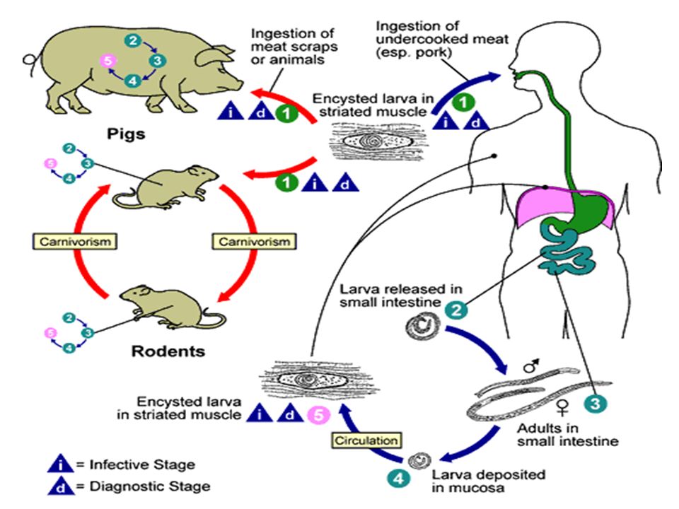
Trichina Worm: Life Cycle
Infected larvae cysts are ingested (passive transmission) in meat products Hatches from cyst in host’s gut and travel to small intestine where they mature and undergo sexual reproduction (adult expelled by immune system) Newborn larvae travel in blood and enters skeletal muscle where the larvae matures in the muscle and manipulates the hosts muscle DNA (lose striations) -> it recruits a blood supply to provide food to the cell and produce collagen to form a cyst and turns into a nurse cell complex This juvenile awaits ingestion by another host
 in meat products. Hatches from cyst in host’s gut and travel to small intestine where they mature and undergo sexual reproduction (adult expelled by immune system) Newborn larvae travel in blood and enters skeletal muscle where the larvae matures in the muscle and manipulates the hosts muscle DNA (lose striations) -> it recruits a blood supply to provide food to the cell and produce collagen to form a cyst and turns into a nurse cell complex. This juvenile awaits ingestion by another host.")
48
Trichina Worm Symptoms based on phases:
Intestinal: small intestine swelling, nausea, vomiting, abdominal pain, diarrhea, fever Migrational: High fever, swelling, cough, lung pain Muscle: acute, localized swelling and pain

49
Trichina Worm Lifecycle Highlights
Mode of Infection Egg location Larvae location Adult location Symptoms Treatment Prevention Passive- eating raw / undercooked pork and ingesting the encysted juveniles None- Larval stage is directly produced Encysted in pork-> ingested -> small intestine -> new larvae travel to skeletal muscle of host Larvae-> adults in small intestine See previous slide -fever -pain Medication -Cook pork completely

50
Enterobius vermicularis: Pinworms
Most common worm parasite in the U.S. Causes little disease Infects 30% of children and 16% of adults in the U.S. Adults (females about 12mm long)live in the large intestine and cecum Females migrate to anal region at night and lay eggs-> causes itching Scratching anal region contaminates hands and bedclothes Eggs develop rapidly and become infective within 6 hours at body temperature When swallowed, hatch in small intestine and mature in large intestine
live in the large intestine and cecum. Females migrate to anal region at night and lay eggs-> causes itching. Scratching anal region contaminates hands and bedclothes. Eggs develop rapidly and become infective within 6 hours at body temperature. When swallowed, hatch in small intestine and mature in large intestine.")
51
Enterobius vermicularis

52
Female pinworm from intestine Group of Pinworm eggs released from anus

53
GOT PINWORMS? Pinworm Symptoms
Perianal itching Children can have behavioral changes; restlessness, irritability, and insomnia Women-> pinworm can enter vagina and cause additional irritation

54
Pinworm Detection Look for eggs or worms on the perianal skin
Will not find eggs or worms in feces Detection can be done by examining the perianal region with a flash light to locate worms (during night/early morning hours) Eggs on perianal skin can also be detected by using “the Scotch tape method”
 Eggs on perianal skin can also be detected by using the Scotch tape method")
56
Enterobius vermicularis Enterobius vermicularis eggs
Female Male Enterobius vermicularis egg Enterobius vermicularis eggs

57
Pinworm Lifecycle Highlights
Mode of Infection Egg location Larvae location Adult location Symptoms Treatment Prevention Passive- eating eggs off fingers -unsanitary habits -auto-infection Perianal region->fingers-> ingestion Hatch in small intestine Larvae travel to large intestine where they mature in the large intestine and cecum -Perianal itching -behavior changes Medication -Sanitary habits

58
Filarial Worms Thread-like nematodes
Located in tropical regions (primarily) Humans are usually the definitive host 250 million people worldwide are infected Spread by an insect vector Microfilariae: modified eggs (juvenile) live in definitive host but cannot develop further until ingested by and intermediate host (mosquito in this case) 3 Type of Filarial Worms (actually there are 8) Wuchereria bancrofti-> Elephantiasis Onchocerciasus-> African River Blindness Dirofilaria immitis-> Dog Heartworm
 Humans are usually the definitive host. 250 million people worldwide are infected. Spread by an insect vector. Microfilariae: modified eggs (juvenile) live in definitive host but cannot develop further until ingested by and intermediate host (mosquito in this case) 3 Type of Filarial Worms (actually there are 8) Wuchereria bancrofti-> Elephantiasis. Onchocerciasus-> African River Blindness. Dirofilaria immitis-> Dog Heartworm.")
59
Wuchereria bancrofti Transmitted by mosquito bite
Microfilariae: sucked up by mosquito Very tiny worms but reproduce to block lymphatic vessels and cause edema (swelling) Repeated exposure can cause Elephantiasis: excessive growth of connective tissue leading to swelling of body parts (usually seen in extremities and scrotum)
 Repeated exposure can cause Elephantiasis: excessive growth of connective tissue leading to swelling of body parts (usually seen in extremities and scrotum)")
60
Wuchereria bancrofti: Symptoms
Swelling-> allergic reaction and reproduction of worms Obstruction of lymphatic vessels Elephantiasis Connective tissue swelling

63
Elephantiasis of leg caused by filarial worms 9.12

64
Surgery in elephantiasis
Diagnosis: Demonstration of microfilaria in blood molecular diagnosis using PCR Wuchereria bancrofti :iethylcarbamazine, Surgery in elephantiasis

65
Wuchereria bancrofti Lifecycle Highlights
Mode of Infection Egg location Larvae location Adult location Symptoms Treatment Prevention Mosquito bite Produced in humans (micro-filariae) sucked up by mosquito Micro-filariae develop further in mosquito and advanced form gets injected into humans Lymph vessels- make more micro-filariae -Swelling of C.T. in arms and legs -elephantiasis Some medication -not reversible -protection from bites
 sucked up by mosquito. Micro-filariae develop further in mosquito and advanced form gets injected into humans. Lymph vessels- make more micro-filariae. -Swelling of C.T. in arms and legs. -elephantiasis. Some medication. -not reversible. -protection from bites.")
66
Onchocerca volvulus Vector is a small black fly
Affects 30 million people in Africa, Central America, and South America Also called the “blinding filaria” Causes: African River Blindness: due to presence of microfilariae in ocular structures (ocular lesions can lead to blindness) When parasite dies the immune system produces a severe reaction resulting in inflammation->blindness Progressive allergic skin reaction-dermatitis and lizard skin
 When parasite dies the immune system produces a severe reaction resulting in inflammation->blindness. Progressive allergic skin reaction-dermatitis and lizard skin.")
68
Black flies of the genus Simulium.
Onchocerca volvulus Black flies of the genus Simulium. Nodules containing Onchocerca volvulus on the head of a man in Guatemala

69
Onchocerca volvulus Lifecycle Highlights
Mode of Infection Egg location Larvae location Adult location Symptoms Treatment Prevention Black fly bite Produced in humans (micro-filariae) sucked up by black fly Micro-filariae develop further in black fly and advanced form gets injected into humans (under skin) Sub-cutaneous tissue and blood -> reproduce and the larvae produced can travel all over including the eye -Skin rash -blindness (ARB) Some medication -blindness not reversible -protection from bites
 sucked up by black fly. Micro-filariae develop further in black fly and advanced form gets injected into humans (under skin) Sub-cutaneous tissue and blood -> reproduce and the larvae produced can travel all over including the eye. -Skin rash. -blindness (ARB) Some medication. -blindness not reversible. -protection from bites.")
70
Dog Heartworm Most common filarial worm in the U.S.
Causes heartworm in dogs Live in dogs heart and lungs Can do damage to the dogs heart-> often leads to death for the dog Killing the worm is difficult and dangerous for the dog Prevention of infection by regular dosing of a dog with drugs that kill circulating larvae is a better strategy

71
Diriofilaria immitis Dog heartworm 9.13

Similar presentations


>")


>")

