Download presentation
Presentation is loading. Please wait.

1
Spinal cord compression in spine tumours and injuries Chaloupka, R., Grosman, R., Repko, M., Tichý, V. Ortopedická klinika, FN Brno, Jihlavská 20, 625 00 Ortopedická klinika FN Brno-Bohunice

2
Pain is caused predominantly by growing of tumour tissue nerve structures compression neural symptoms Spine instability in extensive involvement of one or more vertebrae small force results in pathological fracture and neural deficit Neural deficit growing of tumour and neural compression worsening of blood supply of spinal cord pathological fracture with neural compression combination of these mechanisms Spine involvement in tumours

3
Goals of surgeries prevention / improvement of neural deficit pain relief restoring spine stability improving quality of life

4
present / imminent vertebral collaps present / imminent neural deficit to 24 hours after plegia onset (severe paresis) life expectancy – 3 months minimum Indications
 life expectancy – 3 months minimum Indications")
5
X-rays of C, Th, L spine – AP and lateral CT of affected part, MRI (optimum MRI of the whole spine, CT of brain, lung, abdomen) neurological, internal exam. (laboratory, lung X-ray, ultrasonography of abdomen) Diagnostics
 Diagnostics.")
6
Tomita scoring system (Spine, 2001) Spine metastases: Necessary: bone scane - Tc MRI of the whole spine CT of brain, thorax and abdomen
 Spine metastases: Necessary: bone scane - Tc MRI of the whole spine CT of brain, thorax and abdomen")
7
1. low (mamma, prostata, thyroid gland) 1 point 2. middle (kidney, uterus) 2 points 3. high (pulmo, intestinum, stomach, liver, unknown) 4 points Grading of metastasis (according to origo):
 2 points 3. high (pulmo, intestinum, stomach, liver, unknown) 4 points Grading of metastasis (according to origo):.")
8
1. none0 point 2. Resectable 2 body 3. untreatable4 body Organ metastases:Bone metastases: 1. Solitary 1 point 2. Multiple 2 points According to the score value we indicate the extent of surgery: 2-3 wide – marginal resection (en bloc) 4-5 marginal – intralesional resection 6-7 paliative surgery 8-10 conservative – no surgery
 4-5 marginal – intralesional resection 6-7 paliative surgery 8-10 conservative – no surgery.")
9
Extent of surgery depends on tumour localisation and extent patient´s age and condition Treatment

10
Quick progression of neural deficit - quick decompression w / wo stabilization

11
X-rays of the whole spine CT – MRI Internal, neurological examination Examination

12
Bone scan - Tc MRI of the whole spine CT of brain, thorax, abdomen Unknown tumour aetiology

13
Wide / marginal resection Marginal / intralesional resection Paliative surgery Surgery types

14
Anterior – posterior Combined (1 stage – 2 stages) Surgery
 Surgery")
15
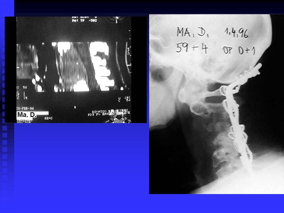
decompression – anterior / posterior posterior fusion and instrumentation Occiput – C2

17
Bone cement with K - wires autograft spacer C 3-7

18
Pelvic autograft and Caspar plate

20
Bone cement with K-wires

21
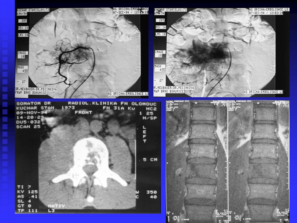
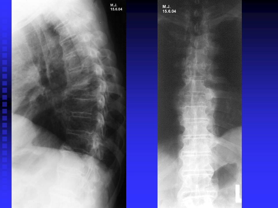
Posterior surgery - decompression + - instrumentation + - fusion T and L spine

22
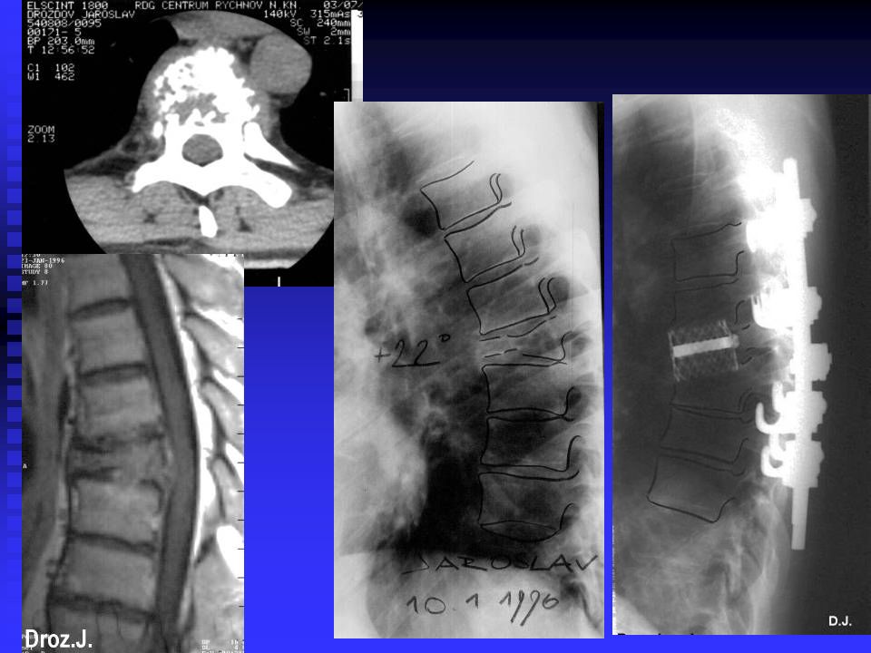
Th and L spine Anterior surgery - decompression - vertebral body replacement - bone cement + K-wires - bone graft - spacer

23
Anterior – vertebral body replacement Posterior - fusion and instrumentation Combined surgeries

25
Harms cage and transpedicular fixator

28
Hartshill system

29
Biopsy - thoracoscopic - lumboscopic - transpedicular Unclear cases

30
spine surgery departments specialized orthopaedic, neurosurgery, traumatology departments Vertebro- and kyfoplasties – radiological departments Surgery in Czech Republic

31
Orthoses – soft collars, Philadelphia collar, three-point body orthoses – Jewett orthosis, belts. Orthotics

32
operated patients: 727 metastases 386 benign 98 malign 175 tumour-like affections 68 1984 – 2005

33
mammar cancer75 Grawitz. tumour54 The most frequent metastases: Malignant tumours: myeloma72 chordoma17 chondrosarcoma12

34
anterior168 posterior350 combined – 1 team 164 - 2 teams 45 Surgeries

35
Frankel scale: A plegia, anesthesia B plegia, some sensory function C usefulles motor function D useful motor (gait) E normal
 E normal")
36
During surgery – heart failure, extensive blood loss Chylothorax Infection Exitus Complications

37
Quick progression of paresis Surgery to 24 hours after plegia onset Conclusion

38
SPINE INJURIES

39
AO classification of T+L injuries A anterior column injury (wedge, split, burst) B both columns – flexion distraction C both columns with rotation
 B both columns – flexion distraction C both columns with rotation")
40
Neural deficit in Th + L spine A type – mainly burst fractures A type – mainly burst fractures B type - seldom B type - seldom C type – majority of patients C type – majority of patients

41
A3.2.1 A3.3.3 A1.3A3.1.1 A3 burst

42
Injuries with neural deficit Transport to spine surgery department Transport to spine surgery department X-rays, CT, (MRI), int., neurol. X-rays, CT, (MRI), int., neurol. Surgery to 6 hours after injury in case of severe neural deficit Surgery to 6 hours after injury in case of severe neural deficit
, int., neurol. Surgery to 6 hours after injury in case of severe neural deficit Surgery to 6 hours after injury in case of severe neural deficit.")
43
Surgery types Posterior – decompression, fusion, instrumentation Posterior – decompression, fusion, instrumentation Anterior – decompression, fusion, instrumentation – spacers, grafts Anterior – decompression, fusion, instrumentation – spacers, grafts Combined – posterior and anterior, anterior – vertebral body replacement in anterior column comminution - destruction Combined – posterior and anterior, anterior – vertebral body replacement in anterior column comminution - destruction

44
Multiple Th fractures – post surg, Hartshill – sublaminar – seldom

45
Posterior TL surgery – transpedicular fixation, fusion and decompression - majority

46
A. Injury of anterior column B. posterior C. both columns C 3-7 INJURIES AO classification 1.Bone injury 2.Bone-ligamentous3.Ligamentous

47
A1 type fracture - bone injury wedge Neural deficit: burst fractures - majority of cases

48
Combined C5-6 surgery C type - dislocation

49
Neural deficit in C spine A type – burst (C5-7) – majority of cases B type – posterior column – seldom C type – both columns – majority of cases Surgery Anterior – decompression, anterior fusion and plates – majority of cases Combined surgery – C type injuries
 – majority of cases B type – posterior column – seldom C type – both columns – majority of cases Surgery Anterior – decompression, anterior fusion and plates – majority of cases Combined surgery – C type injuries")
Similar presentations


 October 29, 2008.>")


