Download presentation
Presentation is loading. Please wait.

1
Nervous Tissue The human nervous system, the most complex system in the human body, the system collect stimuli from the environment, transforms such stimuli into nervous impulses, and passes them to a large highly organized reception and correlation area where they are interpreted, and in turn, issued to effector organs to institute appropriate responses. These functions are performed by highly specialized cells called neurons which together with their supporting or neuroglial cells and associated extracellular material form this integrated communications network.

2
Anatomically, the nervous system can be divided into two parts : The central nervous system (CNS), consisting of the brain and spinal cord, and the peripheral nervous system (PNS), which includes all other nervous tissue. The CNS receives all stimuli originating outside the body (exteroceptive) and all nervous impulses from the body (interoceptive) and act as an integrating and communications center. The PNS serves to interconnect all other tissues and organs with the CNS. Functionally, the nervous system is divided into somatic and autonomic parts,with central & peripheral divisions.
 and all nervous impulses from the body (interoceptive) and act as an integrating and communications center. The PNS serves to interconnect all other tissues and organs with the CNS. Functionally, the nervous system is divided into somatic and autonomic parts,with central & peripheral divisions..")
3
The somatic part is concerned with structures derived from the embryological somites,i.e muscles, bones and skin. The autonomic nervous system innervates smooth & cardiac muscle & the gland of the body, a great extent, its functions are independent of the rest of the nervous system. Structurally, nerve tissue consists of two cell types : nerve cells or neurons, which usually show numerous long processes, and several types of glial cells, which have short processes, support & protect neurons, and participate in neural activity, neural nutrition, defense processes of the CNS. The typical neuron consists of a cell body (perikaryon or soma) with an axon and usually several dendrites
 with an axon and usually several dendrites.")
4
The perikaryon, formed by the nucleus and surrounding cytoplasm, has a receptive function. In most neurons, it receives stimuli generated in other nerve cells, but its main role is as the trophic center of the cells supplying organelles & macromolecules to its processes. Usually the perikaryon is large up to (135) micrometers μm in diameter, although some are only (4) μm. In most nerve cells, the nucleus is large (up to 20 μm) in diameter spherical, and centrally situated in the soma. Chromatin is fine and dispersed, and there is one or more large nucleoli, indicating active synthesis in the cell. The nuclear envelope is distinct and shows numerous pores. Within cytoplasm, the mitochondria are usually small, ovoid or spherical, the Golgi apparatus usually is large, paranuclear in position.Centriols are not prominent in nerve cells (Nerve cells are in capable of cell division).
 micrometers μm in diameter, although some are only (4) μm. In most nerve cells, the nucleus is large (up to 20 μm) in diameter spherical, and centrally situated in the soma. Chromatin is fine and dispersed, and there is one or more large nucleoli, indicating active synthesis in the cell. The nuclear envelope is distinct and shows numerous pores. Within cytoplasm, the mitochondria are usually small, ovoid or spherical, the Golgi apparatus usually is large, paranuclear in position.Centriols are not prominent in nerve cells (Nerve cells are in capable of cell division)..")
5
A characteristic feature of the perikaryon of larger neurons is the presence of Nissl bodies. These represent the basophilic components of the cytoplasm and are stainable by basic aniline dyes. The bodies are formed by stacks of granular endoplasmic reticulum with associated ribosomes, both free and attached. They also are present in dendrites, but are absent in the axon & the axon hillock, a clear conical area at the origin of the axon from the soma. In small soma, only a diffuse basophilia is seen. The reaction of the Nissl bodies to injury or exhaustion after prolonged stimuli is characteristic, the Nissl bodies braking up and diffusing throughout the cytoplasm. This is called chromatolysis. The presence of extensive granular reticulum, prominent nucleoli and numerous mitochondria is concerned with synthesis of new protein that passes to all regions of the neuron, including its axon & dendrites. A second prominent feature of neurons is the presence of neurofibrils visible by light microscopy in silver stains and found throughout the perikaryon, dendrites & axons. These bundles contain both microtubules (neurotubules) and microfilaments (neurofilaments).
 and microfilaments (neurofilaments)..")
6
Nerve cell processes These processes are cytoplasmic extentions of the nerve cell body developed to provide conduction pathways & to provide greater surface area for contact. These processes are of two types : Dendrites : Most nerve cells have numerous dendrites that structurally resemble the perikaryon. Most dendrites show branching, the branches being of smaller diameter than the main stem. Main stem dendrites contain Nissl bodies, mitochondria and small Golgi apparatus, but the content of endoplasmic reticulum & ribosomes is reduced with branching until these are absent from the smaller, more slender twigs. A prominent feature is the present of many neurotubules & neurofilaments,aligned along the axis of the dendrite and in some,extending nearly to the tips. Functionally, there is considerable evidence that the microtubules are involved in transport of organelles such as mitochondria and proteins from the perikaryon to the terminations of the dendrites. In many neurons, the dendrites appear to be beaded, the dendrites covered by numerous, small, spine-like processes called dendritic spines that are specialized for synaptic contact. Generally, spines have a short, slender stem (0.5-.0 ) μm in length with an expanded tip (0.5-2.0) μm in diameter. Spines are prominent, particularly on major dendrites, less so near the perikaryon and at dendritic tips. They represent the main synaptic surface of the dendrites.
 μm in length with an expanded tip ( ) μm in diameter. Spines are prominent, particularly on major dendrites, less so near the perikaryon and at dendritic tips. They represent the main synaptic surface of the dendrites..")
7
Axon The axon is a single, cylindrical process arising from the nerve cell body at a region called the axon hillock. Its initial portion is often the narrowest part. The axon hillock contains no chromophil substance, but contain numerous neurotubules & neurofilaments. Axons generally have a smooth contour and are of uniform diameter, usually more slender, longer and straighter than dendrites varying from a fraction of a millimeter to more than a meter in length. Along its course, an axon may or may not show side branches or collaterals, these leaving the axon at aright angle near the axon hillock. Axons usually terminate in twig-like branchings, the telodendria, which contact the perikaryon, dendrites,or axon of one or more neurons at synapses. At their terminations the axonal twigs show small swellings, called boutons terminaux.

8
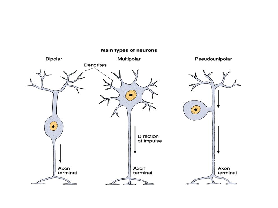
Types of Neurons : According to the size and shape of their processes, there are three types of neurons : Pseudounipolar neurons : which have a single processes that is close to perikaryon and divide form a T- shape with one branch extending to peripheral ending & the other toward the CNS. Pseudounipolar neurons are found in the spinal ganglia & also found in the most cranial ganglia. The nerve cell body is globular in shape. Bipolar neurons : Are found in the retina & olfactory mucosa & inner ear. The nerve cell body is spindle-shaped. Multipolar neurons : have more than two processes, one axon and many (At least two dendrites ). Most neurons in the body (over 99 %) are multipolar.
. Most neurons in the body (over 99 %) are multipolar..")
10
There are great variation in size & shape among the multipolar type. For example : Anterior horn cell or motor neuron cell of the spinal cord, has a large cell body with dendrites radiating in all directions Smaller neurons with dendrites that radiate in all directions are termed stellate Purkinje cells of the cerebellar cortex. The cell body is flask-shaped with a single or more frequently branched dendrites arising from the pointed pole and branching extensively, but in a single plane, a small axon leaves the opposite broader pole of the perikaryon.

11
Pyramidal cells of the cerebral cortex have an apical dendrite and 4 or more branching dendrites passing outward the base of the pyramid, the axon also leaves the base centrally.

12
According to the length of the axons, there are two types of neurons : Golgi type 1 neurons : have a well-developed dendritic tree and along axon that leaves gray matter enters white matter, and run in a major fiber tract of CNS to terminate in other area such as muscle or skin. This type include anterior horn cell ( motor.n ) Purkinje cell & pyramidal cell. Golgi type 11 neurons : have short axons that do not leave the area of their perikarya often passing from their dendrite field for only a short distance > Examples are the interneurons, especially neurons in cerebral & cerebellar cortices. Neurons can also be classified a according to their functional roles : Motor (efferent) neurons control effector organs such as muscle fibers and exocrine & endocrine glands. Sensory (afferent) neurons, are involved in the reception of sensory stimuli from the environment and from within the body. Interneurons, establish relationship among other neurons forming complex functional networks. Interneurons are multipolar neurons between the sensory & motor neurons. the vast majority of neurons are interneuron
 Purkinje cell & pyramidal cell. Golgi type 11 neurons : have short axons that do not leave the area of their perikarya often passing from their dendrite field for only a short distance > Examples are the interneurons, especially neurons in cerebral & cerebellar cortices. Neurons can also be classified a according to their functional roles : Motor (efferent) neurons control effector organs such as muscle fibers and exocrine & endocrine glands. Sensory (afferent) neurons, are involved in the reception of sensory stimuli from the environment and from within the body. Interneurons, establish relationship among other neurons forming complex functional networks. Interneurons are multipolar neurons between the sensory & motor neurons. the vast majority of neurons are interneuron.")
13
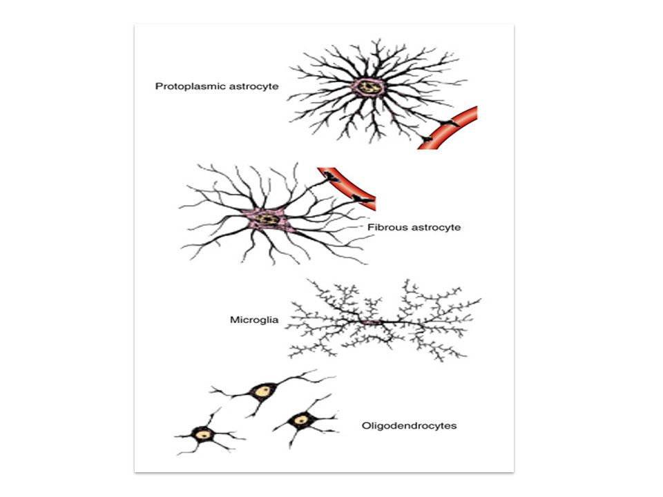
Neuroglia : Although neurons are the principle cells nerve tissue, glial cells play important supporting role. This tissue functions to bind together the nervous tissue of CNS. Neuroglial cells generally are small and only nuclei are seen in routine preparations, thy are studied best by special silver & gold staining that demonstrate the entire cell. Neuroglia include Macroglia, Microglia and Ependymal cells. Macroglia include :- Astrocytes and oligodendrocytes Astrocytes : are star-shaped cells with many branching cytoplasmic processes. Nuclei are large, ovoid or spherical and pale staining. There are bundles of glial filaments, similar to neurofilaments extending into the cytoplasmic processes. Cytoplasmic processes pass between neurons and their processes and extend to blood vessels as pedicle or perivascular feets and to the surface of the brain and spinal cord to the pial surface forming a layer beneath it. Two types of astrocytes are recognized : * Protoplasmic astrocytes, located mainly in gray matter of the brain & spinal cord. They have short thick processes with many branching and lie around neurons, synaptic area, and blood vessels. * Fibrous astrocytes : are found mainly in white matter and have long, slender processes with few or no branches. Glial filaments, are prominent in this type. Astrocytes are important as a supporting or structural elements. In the CNS, after brain damage, astrocytes remove neuronal debris.

14
Oligodendrocytes : ( oligodendroglia ) are smaller than astrocytes with fewer, shorter cell processes. Their nuclei are small ovoid in shape. It occur mainly in two locations : in gray matter closely associated with the perikarya of neurons (perineuronal satellite cells), and among bundles of axon in white matter (interfascicular oligodendrocytes). Others lie in aperivascular position around blood vessels. Oligodendrocytes are responsible for myelin formation thus serving the same function as Schwann cells in the PNS. Unlike Schwann cell, each oligodendrocyte has several processes and thus forms myelin sheaths around several adjacent nerve fibers. Microglia : (Mesoglia) are small, somewhat elongated cells with short spiny processes. Few in number, they are derived from mesoderm, whereas all other types of neuroglia are ectodermal. Microglia lie in both white & gray matter usually near blood vessels. They were believed to be the main source of phagocytic cells in the CNS. ( aperivascular phagocytic cell ). Ependyma : are low columnar epithelial cells lining the ventricles of the brain and central canal of the spinal cord. In some locations the cells are ciliated, which facilitates the movement of cerebrospinal fluid.
, and among bundles of axon in white matter (interfascicular oligodendrocytes). Others lie in aperivascular position around blood vessels. Oligodendrocytes are responsible for myelin formation thus serving the same function as Schwann cells in the PNS. Unlike Schwann cell, each oligodendrocyte has several processes and thus forms myelin sheaths around several adjacent nerve fibers. Microglia : (Mesoglia) are small, somewhat elongated cells with short spiny processes. Few in number, they are derived from mesoderm, whereas all other types of neuroglia are ectodermal. Microglia lie in both white & gray matter usually near blood vessels. They were believed to be the main source of phagocytic cells in the CNS. ( aperivascular phagocytic cell ). Ependyma : are low columnar epithelial cells lining the ventricles of the brain and central canal of the spinal cord. In some locations the cells are ciliated, which facilitates the movement of cerebrospinal fluid..")
16
Ganglia A collection of nerve cell bodies located outside the CNS is called a ganglion. A similar collection in the substance of the CNS is termed a nucleus. Ganglia are of two main types : those of the craniospinal group (sensory ganglia) and those of the autonomic nervous system (Visceral, Motor ganglia). Ganglia vary in size, ranging from very small ones containing only a few nerve cell bodies to very large ones with 50,000 or more cells. Each ganglion has a con. t capsule, which may be quite dense a round large ganglia. Continuous with capsule is a fine con. t network This network, found throughout the substance of the ganglion, is composed of fine collagenous & reticular fibers. Blood vessels run in the con.t. In addition to nerve cell bodies, nerve fibers (axons & dendrites) are present with their supporting sheaths and each ganglionic cell has a capsule, composed of a single layer of small cuboidal cells called satellite cells.
 and those of the autonomic nervous system (Visceral, Motor ganglia). Ganglia vary in size, ranging from very small ones containing only a few nerve cell bodies to very large ones with 50,000 or more cells. Each ganglion has a con. t capsule, which may be quite dense a round large ganglia. Continuous with capsule is a fine con. t network This network, found throughout the substance of the ganglion, is composed of fine collagenous & reticular fibers. Blood vessels run in the con.t. In addition to nerve cell bodies, nerve fibers (axons & dendrites) are present with their supporting sheaths and each ganglionic cell has a capsule, composed of a single layer of small cuboidal cells called satellite cells..")
17
Craniospinal Ganglia : are fusiform or globular swelling of the posterior roots of spinal nerves, and cranial ganglia are similar swelling of some cranial nerves. The ganglionic cells are pseudounipolar, globular. Histologically, both have asimillar structure. The perikarya are arranged in groups at the periphery or cortical zone of the ganglion separated by fiber bundles. The perikarya may be only 15-25μm in diameter with processes that are unmyelinated or up to 100μm with myelinated processes. Each perikarya has a capsule formed by asingle layer of small, low cuboidal cells, the satellite cells or amphicytes around which is a thin layer of vascular con.t. Centrally, located in the ganglion are bundles of nerve fibers, forming amedullary zone, where perikarya are few.

18
Autonomic Ganglia : are swellings located along the sympathetic chain and it branches, and within the walls of organs supplied by the autonomic system, where they may be very small. Like craniospinal ganglia, a con.t capsule envelops the ganglion, except in very small peripheral ganglia. Ganglion cells are multipolar and smaller than those of sensory ganglia being 15- 45μm in diameter. Perikarya do not show a tendency to group, nor are the axons grouped into bundles, perikarya and fibers are intermingled in the ganglion.

19
Nerve Fibers : Nerve fiber consists of axons enveloped by a special sheath derived from cells of ectodermal in origin. Nerve fibers exhibit differences in their enveloping sheaths related to whether the fibers are part of CNS or PNS. Most axons in adult nerve tissue are covered by single or multiple folds of a sheath cell. In PNF, the sheath cell is Schwann cell, and in CNF it is oligodendrocytes. Axons of small diameter are usually unmyelinated n.f. Progressively thicker axons are generally sheathed by increasingly numerous concentric wrappings of the enveloping cell, forming the myelin sheaths. These fibers are known as myeliated n.f. Myelinated Fibers :In myelinated fibers of the peripheral nervous system, the plasmalemma of Schwann cell wind and warp around the axon. The layers of membranes of the sheath cell unite and form myelin a lipoprotein complex whose lipid component removed by histological procedures.. These membrane have a higher proportion of lipids. a lipoprotein complex whose lipid component removed by histological procedures. Myelin consists of many layers of modified cell membranes. These membranes have a higher proportion of lipid than do other cell membranes. Central nervous system myelin contains two major proteins: myelin basic protein & proteolipid protein. Several human demyelinating disease are due to an insufficiency or lack of one or both of these proteins. Each axon is surrounded by myelin formed by a sequential series of Schwann cells. The myelin sheath shows gaps along its path called the nodes of Ranvier, these represent the spaces between adjacent Schwann cells along the length of the axon.

20
The distance between two nodes is called an internode and consists of one Schwann cell. Internode length varies between 1 & 2 mm. In the CNS, the myelin sheath is formed by the processes of the oligodendrocytes. Oligodendrocytes differ from Schwann cells in that different branches of one cell can envelop segments of several axons. Unmyelinated Fibers : In both the CNS & PNS, not all axons are sheathed in myelin. In the PNS, all unmyelinated axons are enveloped within simple clefts of Schwann cells. Each Schwann cell can sheathed many unmyelinated axons. unmyelinated nerve fibers do not have nodes of Ranvier. The CNS is rich in unmyelinated axons, these axons are not sheathed. In brain and spinal cord, unmyelinated axonal processes run free among the other neuronal processes. Schwann cells are essential to the vitality & function of PNF. They form myelin, also are necessary for regeneration of axons, also Schwann cells can become phagocytic after nerve injury, removing cellular debris.

21
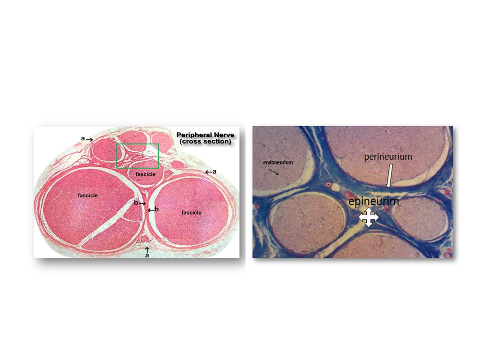
Peripheral Nerve : are composed of bundles of nerve fibers held together by connective tissue and include spinal nerves connected to the spinal cord and cranial nerves connected with the brain. Most nerves are mixed containing both sensory (afferent) and motor (efferent) fibers. Surrounding the entire nerve is a sheath of dense con.t called epineurium. It is composed of fibroblasts and collagenous fibers mainly longitudinal in orientation and few elastic fibers. The epineurium contains the major blood vessels to the nerve. Within the epineurium, nerve fibers are grouped into bundles or fascicles. Each fascicle is surrounded by con.t sheath called perineurium. This sheath is formed by concentric layers or sleeves of flattened fibroblast-like cells, each sleeve being one cell thick. The number of sleeves decreases as the nerve branches becomes smaller. The perineurium provides a barrier to the passages of materials into or out of the nerve fascicle. Within the perineurium are strands of delicate con.t extending around and between individual nerve fibers. This is the endoneurium, composed of delicate collagenous and reticular fibers and flattened, elongated fibroblasts. The peripheral nerves establish communication between brain and spinal cord centers and the sense organs and effectors (muscles, glands, etc).
 and motor (efferent) fibers. Surrounding the entire nerve is a sheath of dense con.t called epineurium. It is composed of fibroblasts and collagenous fibers mainly longitudinal in orientation and few elastic fibers. The epineurium contains the major blood vessels to the nerve. Within the epineurium, nerve fibers are grouped into bundles or fascicles. Each fascicle is surrounded by con.t sheath called perineurium. This sheath is formed by concentric layers or sleeves of flattened fibroblast-like cells, each sleeve being one cell thick. The number of sleeves decreases as the nerve branches becomes smaller. The perineurium provides a barrier to the passages of materials into or out of the nerve fascicle. Within the perineurium are strands of delicate con.t extending around and between individual nerve fibers. This is the endoneurium, composed of delicate collagenous and reticular fibers and flattened, elongated fibroblasts. The peripheral nerves establish communication between brain and spinal cord centers and the sense organs and effectors (muscles, glands, etc)..")
23
Spinal Cord : In cross section the spinal cord is oval in shape covered by the pia mater. Posteriorly is divided into right & left halves by the dorsal median septum, anteriorly there is a deep longitudinal cleft called the anterior ( ventral ) median fissure. The entire cord is surrounded by pia mater which extends into the anterior median fissure. Centrally in the cord in cross section is an H-shaped area of gray matter composed of nerve cells. On each side, the limbs of the H are called the anterior & posterior horns. In addition extending throughout the thoracic and upper one or two lumber segments, there is a lateral horn. The central canal lined by ependyma, is situated in the horizontal bar of the H. Nerve cell bodies lie in groups in the gray matter, the large motor neurons lying in the anterior horn.
 median fissure. The entire cord is surrounded by pia mater which extends into the anterior median fissure. Centrally in the cord in cross section is an H-shaped area of gray matter composed of nerve cells. On each side, the limbs of the H are called the anterior & posterior horns. In addition extending throughout the thoracic and upper one or two lumber segments, there is a lateral horn. The central canal lined by ependyma, is situated in the horizontal bar of the H. Nerve cell bodies lie in groups in the gray matter, the large motor neurons lying in the anterior horn..")
24
The white matter, formed by nerve fibers, surrounded the gray matter, and is divided into longitudinal columns or funiculi. Between the posterior horn and the dorsal median septum is the posterior or dorsal columns. The remainder of white matter is divided by the ventral horn and the anterior median fissure into lateral & ventral columns. Nerve cells in the gray matter are multipolar. The axons of some leave the cord as ventral root fibers; others send axons into the white matter, others have short axons that terminate on neurons near their origin ( Golgi type II ). Generally the white matter contains no nerve cell bodies or dendrites and is formed by myelinated & unmyelinated fibers.
. Generally the white matter contains no nerve cell bodies or dendrites and is formed by myelinated & unmyelinated fibers..")
Similar presentations


 Integration:>")

>")
 1.Microglial cells –Scattered throughout CNS –Support neurons.>")
