Download presentation
Presentation is loading. Please wait.

1
Pharyngeal arches DR N SATYANARAYANA,FOM,DEPARTMENT OF ANATOMY.AIMST UNIVERSITY,MALAYSIA.

2
Pharyngeal arches Cranial most part of foregut is called primitive pharynx Primitive pharynx lies just caudal to stomodeum Primitive pharynx is initially separated from the stomodeum by buccopharyngeal membrane Endoderm of primitive pharynx is separated from the surface ectoderm by thin layer of mesoderm Series of mesodermal thickenings appear on the lateral wall of primitive pharynx These mesodermal thickenings are called pharyngeal (branchial) arches
 arches.")
3
Pharyngeal arches are 6 on each lateral wall of primitive pharynx
Each arch contains central core of mesoderm which is derived from paraxial and lateral plate mesoderm Each arch is covered on the outer surface by the ectoderm and lined on the inner side by the endoderm Pharyngeal arches separated externally from each other by pharyngeal grooves (clefts) Pharyngeal arches separated internally from each other by pharyngeal pouches 5th pharyngeal arch soon degenerates after its formation
 Pharyngeal arches separated internally from each other by pharyngeal pouches. 5th pharyngeal arch soon degenerates after its formation.")
4
Pharyngeal arches

5
Pharyngeal pouch

6
1. Pharyngeal arches 2. Lens placode 3. Pericardial swelling 4
1. Pharyngeal arches 2. Lens placode 3. Pericardial swelling 4. Pharyngeal clefts 5. Hand bud

7
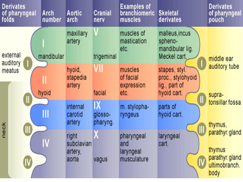
Each arch contains Mesoderm: Cartilage (skeletal element):
which develops into muscles Cartilage (skeletal element): which develops into bones Nerve: supplies muscles developing from the mesoderm of the arch skin developing from the ectoderm of the arch mucous membrane developing from the endoderm of the arch Aortic arch artery: develop into arteries
: which develops into bones. Nerve: supplies. muscles developing from the mesoderm of the arch. skin developing from the ectoderm of the arch. mucous membrane developing from the endoderm of the arch. Aortic arch artery: develop into arteries.")
8
First pharyngeal arch

10
First pharyngeal arch First pharyngeal arch is called the mandibular arch First arch gives 2 process – mandibular process and maxillary process Structures derived from the first arch: Structures developing from the cartilage of 1st arch (Meckel’s cartilage): Incus and malleus bones of middle ear Sphenomandibular ligament and anterior ligament of malleus
: Incus and malleus bones of middle ear. Sphenomandibular ligament and anterior ligament of malleus.")
11
Structures developing from the mesoderm of 1st arch:
Muscles of mastication (masseter, temporalis, medial and lateral pterygoids) Tensor tympani, tensor palati, mylohyoid and anterior belly of digastric Maxilla, mandible, zygomatic bone, palatine bone, part of temporal bone Nerve of the arch: Mandibular nerve is the pretrematic nerve of 1st arch Chorda tympani branch of facial nerve is post trematic nerve of 1st arch 1st aortic arch artery: Forms part of maxillary artery
 Tensor tympani, tensor palati, mylohyoid and anterior belly of digastric. Maxilla, mandible, zygomatic bone, palatine bone, part of temporal bone. Nerve of the arch: Mandibular nerve is the pretrematic nerve of 1st arch. Chorda tympani branch of facial nerve is post trematic nerve of 1st arch. 1st aortic arch artery: Forms part of maxillary artery.")
12
Second pharyngeal arch

13
Second pharyngeal arch
2nd pharyngeal arch is called hyoid arch Structures derived from the 2nd arch: Structures developing from the cartilage of 2nd arch (Reichert’s cartilage): Stapes bone of middle ear Styloid process, stylohyoid ligament Lesser cornu and upper part of body of hyoid bone
: Stapes bone of middle ear. Styloid process, stylohyoid ligament. Lesser cornu and upper part of body of hyoid bone.")
14
Structures developing from the mesoderm of 2nd arch:
Muscles of face Muscles of auricle (auricularis anterior, auricularis superior and auricularis posterior) Muscles of scalp (occipito-frontalis) Platysma, stapedius, posterior belly of digastric and stylohyoid Nerve of the arch: Facial nerve is the nerve of 2nd arch 2nd aortic arch artery: Forms hyoid artery and stapedial artery which degenerates in the adults
 Muscles of scalp (occipito-frontalis) Platysma, stapedius, posterior belly of digastric and stylohyoid. Nerve of the arch: Facial nerve is the nerve of 2nd arch. 2nd aortic arch artery: Forms hyoid artery and stapedial artery which degenerates in the adults.")
15
Third pharyngeal arch

16
Third pharyngeal arch Structures derived from the third arch:
Structures developing from the cartilage of 3rd arch: Greater cornu of hyoid bone Lower part of body of hyoid bone Structures developing from the mesoderm of 3rd arch: Stylopharyngeus muscle Nerve of the arch: Glossopharyngeal nerve is the nerve of 3rd arch 3rd aortic arch artery: Forms common carotid artery and part of internal carotid artery

17
Fourth and sixth pharyngeal arches

18
Fourth and sixth pharyngeal arches
Structures derived from the fourth and sixth arches: Structures developing from the cartilage of 4th and 6th arches: Cartilages of larynx – thyroid, cricoid, arytenoid, epiglottic, cuneiform and corniculate cartilages Structures developing from the mesoderm of 4th arch: Muscles of pharynx, soft palate and cricothyroid Structures developing from the mesoderm of 6th arch: Intrinsic muscles of larynx except cricothyroid

19
Nerve of the 4th arch: superior laryngeal nerve
Nerve of the 6th arch: recurrent laryngeal nerve 4th aortic arch artery: Of the right side forms right subclavian artery Of the left side forms arch of aorta 6th aortic arch artery: On the right side forms right pulmonary artery On the left side forms left pulmonary artery and ductus arteriosus

20
pharyngeal arch derivatives

21
Pharyngeal pouches

22
Pharyngeal pouches

23
Pharyngeal pouches Pharyngeal arches are separated internally from each other by pharyngeal pouches Pharyngeal pouches are lined by endoderm Pharyngeal pouches are 5 in number 5th pharyngeal pouch is rudimentary and considered as part of 4th pouch Endoderm lining the pouch develops into many structures

24
Fate of pharyngeal pouches
First pharyngeal pouch: Dorsal part of 1st pouch along with dorsal part of 2nd pouch forms a stalk like diverticulum called tubotympanic recess Tubotympanic recess comes in contact with first pharyngeal cleft Distal part of tubotympanic recess becomes wide and forms middle ear cavity Proximal part of tubotympanic recess remains narrow and forms auditory tube Tympanic membranic is formed where tubotympanic recess touches the 1st pharyngeal cleft 1. External auditory meatus 2. Auditory tube 3. Primary tympanic cavity 4. Cervical sinus 5. Inferior parathyroid gland 6. Thymus 7. Palatine tonsil 8. Superior parathyroid gland 9. Ultimobranchial body

25
Second pharyngeal pouch:
Dorsal part takes part in the formation of tubotympanic recess along with dorsal part of 1st pouch Endoderm of ventral part forms palatine tonsil Third pharyngeal pouch: Develops into thymus and inferior parathyroid gland Fourth pharyngeal pouch: Forms superior parathyroid gland Forms parafollicular cells thyroid gland along the rudimentary 5th pouch Fifth pharyngeal pouch: Is rudimentary Along with the part of 4th pouch it forms ultimobranchial body which gives origin to parafollicular cells of thyroid gland 1. External auditory meatus 2. Auditory tube 3. Primary tympanic cavity 4. Cervical sinus 5. Inferior parathyroid gland 6. Thymus 7. Palatine tonsil 8. Superior parathyroid gland 9. Ultimobranchial body

26
Fate of pharyngeal clefts

27
Fate of pharyngeal clefts
Pharyngeal arches are separated externally by pharyngeal clefts or grooves Pharyngeal clefts are lined by ectoderm First pharyngeal cleft: Its dorsal part forms external acoustic meatus Series of swellings (auricular hillocks) appear on the 1st and 2nd pharyngeal arch around the dorsal part of 1st cleft These swellings join to form auricle or pinna of the ear Other clefts: Projection arising from the 2nd pharyngeal arch grows down over the lower pharyngeal arches
 appear on the 1st and 2nd pharyngeal arch around the dorsal part of 1st cleft. These swellings join to form auricle or pinna of the ear. Other clefts: Projection arising from the 2nd pharyngeal arch grows down over the lower pharyngeal arches.")
28
The space called cervical sinus separates this projection from the 3rd, 4th and 6th arches
This projection will fuse with the tissues lying caudal to the arches The other arches and clefts become buried by the downward projection of 2nd arch, and side of neck becomes smooth The cavity of cervical sinus normally disappears, sometimes part of it may persist and give rise to swelling in the neck called branchial cysts If such a cyst opens on the surface on the neck, then its called branchial sinus

29
Structures developing the in the floor of primitive pharynx
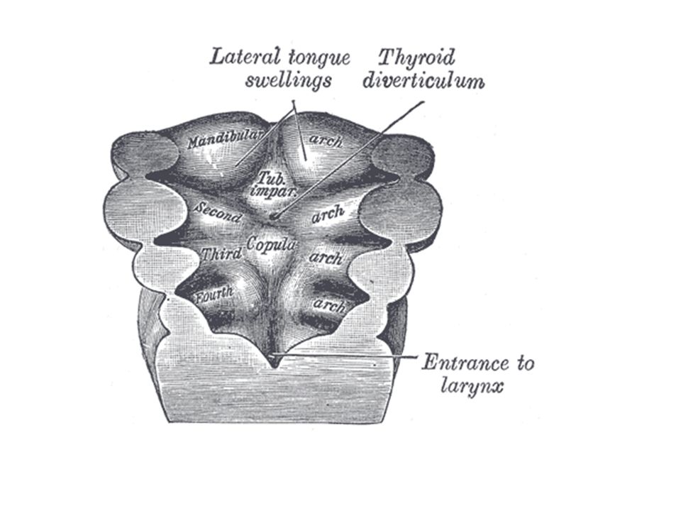
Floor of primitive pharynx at the first pharyngeal arch shows 3 enlargements Median swelling called tuberculum impar 2 lateral lingual sweelings These swellings fuse to form anterior two-third of tongue Just caudal to tuberculum impar floor of primitive pharynx shows thickening in the midline This thickening forms thyroglossal duct which form thyroid gland Floor of primitive pharynx at the region of 3rd and 4th pharyngeal arches shows a bulging called hypobranchial eminence (copula)
")
30
Tongue Development These 3 drawings are viewed from inside the pharynx looking at the floor of the pharynx. Mesodermal swellings in pharynx floor are covered with endoderm. Foramen caecum is the site of initial thyroid cell descent into the hypopharyngeal eminence.

31
In the midline on the surface is a depression called the median sulcus.
Lateral lingual swellings have fused and overgrown medial and 2nd arch components.

32
Oral part is anterior 2/3 Pharyngeal part is posterior 1/3 Circumvillate papilla lie just anterior to terminal sulcus. (Modified from Kaufman and Bard, 1999)
")
34
Floor of primitive pharynx

35
This hypobranchial eminence is divided in to cranial and caudal parts
Cranial part of hyphobranchial eminence grows cranially over the 2nd arch, joins lateral lingual swellings and tuberculum impar and forms posterior one-third of tongue Caudal part of hypobranchial eminence forms epiglottis Tracheobronchial groove appears in the floor of primitive pharynx caudal to hypobranchial eminence This grows downs, elongates to form respiratory diverticulum Respiratory diverticulum forms parts of the respiratory system like larynx, trachea, bronchial tree and lungs

36
Pharyngeal pouches 1. External auditory meatus 2. Auditory tube 3. Primary tympanic cavity 4. Cervical sinus 5. Inferior parathyroid gland 6. Thymus 7. Palatine tonsil 8. Superior parathyroid gland 9. Ultimobranchial body

37
Pharyngeal pouches 1. Auditory tube 2. Foramen cecum 3. Palatine tonsil 4. Ventral syde of pharynx 5. Tympanic cavity 6. Thyroid gland 7. Ultimobranchial body 8. Foregut 9. Thymus 10. Inferior parathyroid gland 11. Superior parathyroid gland 12. External auditory meatus

Similar presentations