Download presentation
Presentation is loading. Please wait.

1
1.Name the cell component that controls entry and exit of substances into and out of the cell? 2.In what part of the cell is chromatin found? 3.What is the function of the ribosomes? 4.What organelle provides energy to the cell through respiration? 5.What do you call the jelly like substance in which chemical reactions in the cell take place?

2
Learning Objectives: 1.Use an optical microscope / bioviewer to observe cells 2.Describe the appearance and function of the main organelles found within cells 3.Understand and explain the principles of cell fractionation Learning Objectives: 1.Use an optical microscope / bioviewer to observe cells 2.Describe the appearance and function of the main organelles found within cells 3.Understand and explain the principles of cell fractionation The structure of Eukaryotic cells

3
In the beginning.. The first cells to exist were really simple cells called ARCHAEA They didn’t have any internal membranes Their DNA was scattered amongst the cytoplasm Some of them are still surviving today, and some have really cool superpowers like being able to withstand extreme temperatures and nasty chemicals

4
Some Archaea evolved into Prokaryotes Prokaryotes are a more complex form of life They are bacteria They still dont have any internal membranes They dont have a nucleus.. PRO – KARYO means before nucleus Their DNA is in a little nucleosome in the cytoplasm There are millions of species of bacteria on the earth today and they are the most prevalent form of life

5
The most complex form of cell Much much BIGGER than prokaryotes They have lots of complex internal membranes Including a nucleus.. EU – KARYO means has a nucleus Their DNA is in a complex organelle called a nucleus which has its own membranes They have mitochondria and other organelles Plant, Animals and Fungi are all Eukaryotes One lucky Prokaryote evolved into a Eukaryote

9
Discuss with your partner, try to figure out the order of procedure

10
To obtain reliable information about the activity of an organelle, it is necessary to isolate it and test it individually. First the cells are broken open or cell fractionation occurs to produce a homogenate or suspension. This is done using a blender with the cells in an isotonic, cold solution. Because the solution is isotonic, the organelles neither gain, or loose water by osmosis and as it is cold, the action of enzymes, which might damage the organelles, is prevented.

11
Differential centrifugation of the suspension is then carried out. A tube containing the suspension is spun in a centrifuge at a speed, which causes the heaviest organelles to be thrown to the bottom, forming a sediment. The other lighter organelles remain floating in the clear supernatant fluid above the sediment. The sediment may be removed and the activity of the heaviest organelles such as the nucleus, determined. The supernatant may then be spun at a faster speed so that lighter organelles like the mitochondria sediment out.

12
Learning Objectives: 1.Use an optical microscope 2.Describe the principles by which transmission and scanning electron microscopes work. 3.Describe the difference between magnification and resolution Learning Objectives: 1.Use an optical microscope 2.Describe the principles by which transmission and scanning electron microscopes work. 3.Describe the difference between magnification and resolution The structure of Eukaryotic cells

13
A telescope must gather large amounts of light from a dim, distant object; therefore, it needs a large objective lens to gather as much light as possible and bring it to a bright focus. Because the objective lens is large, it brings the image of the object to a focus at some distance away, which is why telescopes are much longer than microscopes. The eyepiece of the telescope then magnifies that image as it brings it to your eye.

14
In contrast to a telescope, a microscope must gather light from a tiny area of a thin, well- illuminated specimen that is close-by. So the microscope does not need a large objective lens. Instead, the objective lens of a microscope is small and spherical, which means that it has a much shorter focal length on either side. It brings the image of the object into focus at a short distance within the microscope's tube. The image is then magnified by a second lens, called an ocular lens or eyepiece, as it is brought to your eye.

15
Magnification: How many times an image has been enlarged compared to the original object Resolution: the ability to distinguish between two points that are close together nm = nanometre: there are 1000 nm in 1 µm, or 1000 000 000 nm in 1 m

16
Magnification and Resolution By using more lenses microscopes can magnify by a larger amount, but this doesn't always mean that more detail can be seen. The amount of detail depends on the resolving power of a microscope, which is the smallest separation at which two separate objects can be distinguished (or resolved). The resolving power of a microscope is ultimately limited by the wavelength of light (400- 600nm for visible light). To improve the resolving power a shorter wavelength of light is needed, and sometimes microscopes have blue filters for this purpose (because blue has the shortest wavelength of visible light).
. The resolving power of a microscope is ultimately limited by the wavelength of light ( nm for visible light). To improve the resolving power a shorter wavelength of light is needed, and sometimes microscopes have blue filters for this purpose (because blue has the shortest wavelength of visible light)..")
17
+ electrons Magnification is how much bigger a sample appears to be under the microscope than it is in real life. Resolution is the ability to distinguish between two points on an image i.e. the amount of detail The resolution of an image is limited by the wavelength of radiation used to view the sample. This is because when objects in the specimen are much smaller than the wavelength of the radiation being used, they do not interrupt the waves, and so are not detected. The wavelength of light is much larger than the wavelength of electrons, so the resolution of the light microscope is a lot lower. Using a microscope with a more powerful magnification will not increase this resolution any further. It will increase the size of the image, but objects closer than 200nm will still only be seen as one point.

18
Scanning Electron Microscope

19
Electron vs. Light Microscopes: Basic Differences There are not many things that these two microscope types have in common. Both electron and light microscopes are technical devices which are used for visualizing structures that are too small to see with the unaided eye, and both types have relevant areas of applications in biology and the materials sciences. And this is pretty much it. The method of visualizing the structures is very different. Electron Microscopes use electrons and not photons (light rays) for visualization. The first electron microscope was constructed in 1931, compared to optical microscopes they are a very recent invention.
 for visualization. The first electron microscope was constructed in 1931, compared to optical microscopes they are a very recent invention..")
20
Electron microscopes have certain advantages over optical microscopes: The biggest advantage is that they have a higher resolution and are therefore also able of a higher magnification (up to 2 million times). Light microscopes can show a useful magnification only up to 1000-2000 times. This is a physical limit imposed by the wavelength of the light. Electron microscopes therefore allow for the visualization of structures that would normally be not visible by optical microscopy. Depending on the type of electron microscope, it is possible to view the three dimensional external shape of an object (Scanning Electron Microscope, SEM). In scanning electron microscopy (SEM), due to the nature of electrons, electron microscopes have a greater depth of field compared to light microscopes. The higher resolution may also give the human eye the subjective impression of a higher depth of field.
. In scanning electron microscopy (SEM), due to the nature of electrons, electron microscopes have a greater depth of field compared to light microscopes. The higher resolution may also give the human eye the subjective impression of a higher depth of field..")
21
Electron microscopes have a range of disadvantages as well: They are extremely expensive. Sample preparation is often much more elaborate. It is often necessary to coat the specimen with a very thin layer of metal (such as gold). The metal is able to reflect the electrons. The sample must be completely dry. This makes it impossible to observe living specimens. It is not possible to observe moving specimens (they are dead). It is not possible to observe color. Electrons do not possess a color. The image is only black/white. Sometimes the image is colored artificially to give a better visual impression. They require more training and experience in identifying artifacts that may have been introduced during the sample preparation process. The energy of the electron beam is very high. The sample is therefore exposed to high radiation, and therefore not able to live. The space requirements are high. They may need a whole room. Maintenance costs are high.
. The metal is able to reflect the electrons. The sample must be completely dry. This makes it impossible to observe living specimens. It is not possible to observe moving specimens (they are dead). It is not possible to observe color. Electrons do not possess a color. The image is only black/white. Sometimes the image is colored artificially to give a better visual impression. They require more training and experience in identifying artifacts that may have been introduced during the sample preparation process. The energy of the electron beam is very high. The sample is therefore exposed to high radiation, and therefore not able to live. The space requirements are high. They may need a whole room. Maintenance costs are high..")
22
When should one use optical (light) microscopes? One big advantage of light microscopes is the ability to observe living cells. It is possible to observe a wide range of biological activity, such as the uptake of food, cell division and movement. Additionally, it is possible to use in-vivo staining techniques to observe the uptake of coloured pigments by the cells. These processes can not be observed in real time using electron microscopes, as the specimen has to be fixed, and completely dehydrated (and is therefore dead). The low cost of optical microscopes makes them useful in a wide range of different areas, such as education, the medical sector or for hobbyists. Generally, optical and electron microscopes have different areas of application and they complement each other.
. The low cost of optical microscopes makes them useful in a wide range of different areas, such as education, the medical sector or for hobbyists. Generally, optical and electron microscopes have different areas of application and they complement each other..")
23
. Light MicroscopyLight MicroscopyLight MicroscopyLight Microscopy Electron Microscopy Vs Cheap to purchase (£100 – 500)Expensive to buy (over £ 1 000 000). Cheap to operate.Expensive to produce electron beam. Small and portable.Large and requires special rooms. Simple and easy sample preparation.Lengthy and complex sample prep. Material rarely distorted by preparation.Preparation distorts material. Vacuum is not required.Vacuum is required. Natural colour of sample maintained.All images in black and white. Magnifies objects only up to 2000 timesMagnifies over 500 000 times. Specimens can be living or dead Specimens are dead, as they must be fixed in plastic and viewed in a vacuum Stains are often needed to make the cells visible The electron beam can damage specimens and they must be stained with an electron- dense chemical (usually heavy metals like osmium, lead or gold).
..")
24
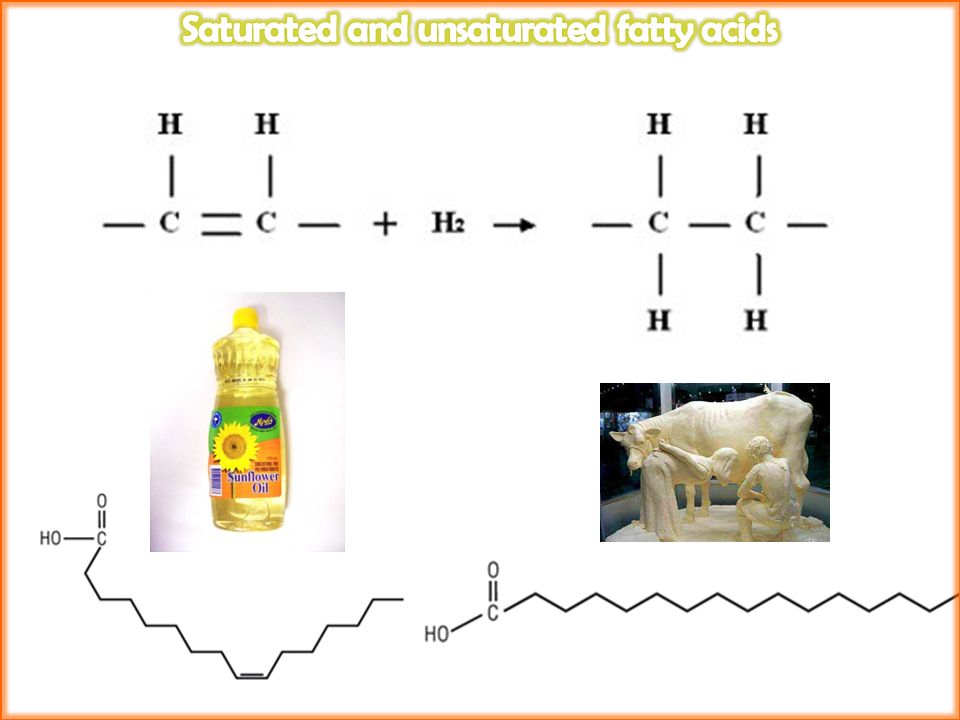
Learning Objectives: 1.Describe the structure of triglycerides and phospholipids 2.Describe the difference between saturated and unsaturated fatty acids. 3.Describe the Fluid mosaic model of membrane structure Learning Objectives: 1.Describe the structure of triglycerides and phospholipids 2.Describe the difference between saturated and unsaturated fatty acids. 3.Describe the Fluid mosaic model of membrane structure Cell Membranes Starter: The term “ Phospholipid bilayer ” is one you’ll hear a lot in AS Biology. How many familiar words can you find in it... And what do they mean

25
A mosaic A picture made of little tiles and pieces of glass gathered and stuck together

26
A Fluid mosaic

27
Appearance of the Cell Membrane Seen using a light microscope, the cell membrane appears as a thin line, but with an electron microscope, it appears as a double line. } 7 – 8 nm This model is referred to as the ‘fluid mosaic model’ because the components are free to move independently of each other.

28
The fluid mosaic model A model of the phospholipid membrane Notice the proteins and glycoproteins stuck in the membrane Some go right through (channel proteins) others are on the inside or the outside of the membrane
 others are on the inside or the outside of the membrane")
29
Adapting your membrane Membranes need to be flexible but stable Cholesterol makes the cell membrane more rigid Plants and Animals who live in extremely hot temperatures are in danger of their membranes rupturing and leaking Plants and Animals who live in extremely cold temperatures are in danger of their membranes being too rigid and crystallising Eukaryotes have adapted their membranes through natural selection to have the right amount of cholesterol, glycoproteins and glycolipids for their environment

30
To calculate the standard deviation of those numbers : 1. Work out the Mean (the simple average of the numbers) 2. Then for each number: subtract the Mean and square the result 3. Then work out the mean of those squared differences. 4. Take the square root of that and you are done!
 2. Then for each number: subtract the Mean and square the result 3. Then work out the mean of those squared differences. 4. Take the square root of that and you are done!.")
31
Lipid Carbohydrate Protein DNA

32
1.What is the function of the cells’ ribosomes? 2.What is a glycoprotein made of? 3.What are phospholipids made of? 4.Why is the term fluid mosaic used to describe the eukaryotic membrane? 5.What is a centrifuge? 6.What is the function of the cells’ cytoplasm? 7.What does a colorimeter measure? 8.How do higher temperatures effect the cells’ membrane? 9.What is the difference between how the light microscope works and how the electron microscope works 10.What are the drawbacks of electron microscopy?

33
. Light MicroscopyLight MicroscopyLight MicroscopyLight Microscopy Electron Microscopy Vs Cheap to purchase (£100 – 500)Expensive to buy (over £ 1 000 000). Cheap to operate.Expensive to produce electron beam. Small and portable.Large and requires special rooms. Simple and easy sample preparation.Lengthy and complex sample prep. Material rarely distorted by preparation.Preparation distorts material. Vacuum is not required.Vacuum is required. Natural colour of sample maintained.All images in black and white. Magnifies objects only up to 2000 timesMagnifies over 500 000 times. Specimens can be living or dead Specimens are dead, as they must be fixed in plastic and viewed in a vacuum Stains are often needed to make the cells visible The electron beam can damage specimens and they must be stained with an electron- dense chemical (usually heavy metals like osmium, lead or gold).
..")
35
Both glycerol and fatty acids are in all fats Some fatty acids are manufactured in the body Others are taken in the diet Essential fatty acids

39
Formation of monoglyceride

40
Formation of triglyceride molecule Triglycerides are hydrophobic -Charges on the molecule are evenly distributed -Hydrogen bonding does not easily occur -They are therefore insoluble in water

41
Note the similar structure to triglycerides Phospholipids have phosphate group instead of 3 rd fatty acid chain The phospholipid is formed by a condensation reaction with the elimination of water

43
The ratio of saturated to unsaturated phospholipids in a membrane defines the fluidity of a membrane Organisms adapted to colder climates have more unsaturated fatty acids (higher melting point) so the membranes remain fluid in low temperatures
 so the membranes remain fluid in low temperatures")
44
The test for Lipids Lipids do not dissolve in water, but do dissolve in ethanol. This characteristic is used in the emulsion test. Do not start by dissolving the sample in water, but instead shake some of the test sample with about 4 cm³ of ethanol. Decant the liquid into a test tube of water, leaving any undissolved substances behind. If there are lipids dissolved in the ethanol, they forming a will precipitate in the water, forming a cloudy white emulsion cloudy white emulsion.

45
Learning Objectives: 1.Define proteins as polymers of amino acids 2.Describe the levels of protein structure Learning Objectives: 1.Define proteins as polymers of amino acids 2.Describe the levels of protein structure Proteins Starter: The term “ Phospholipid bilayer ” is one you’ll hear a lot in AS Biology. How many familiar words can you find in it... And what do they mean

46
© Pearson Education Ltd 2008 This document may have been altered from the original

47
© Pearson Education Ltd 2008 This document may have been altered from the original

49
© Pearson Education Ltd 2008 This document may have been altered from the original

53
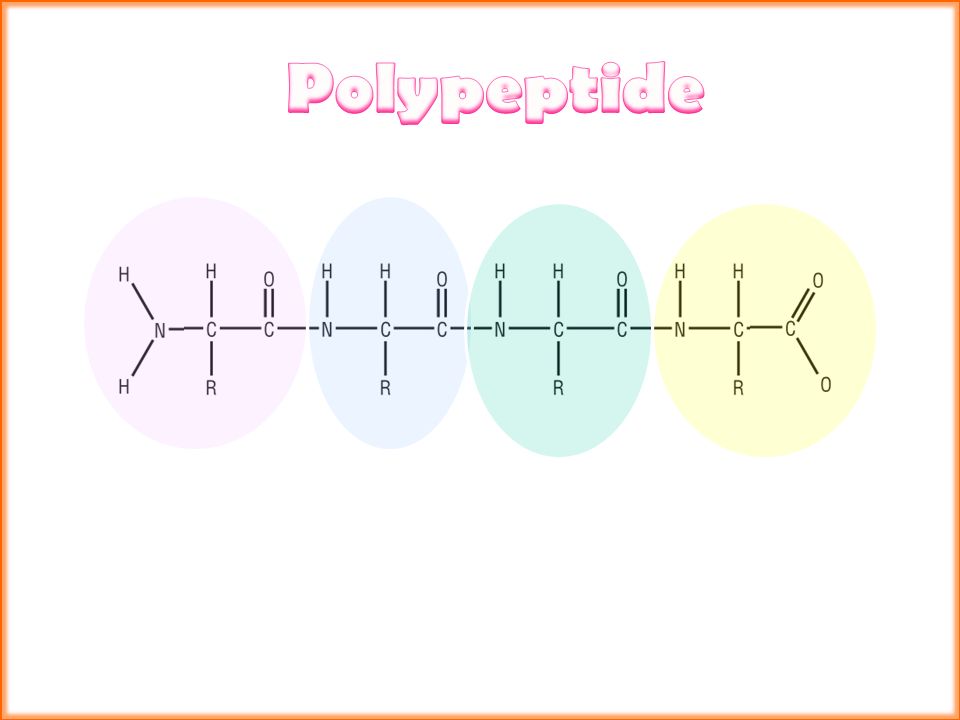
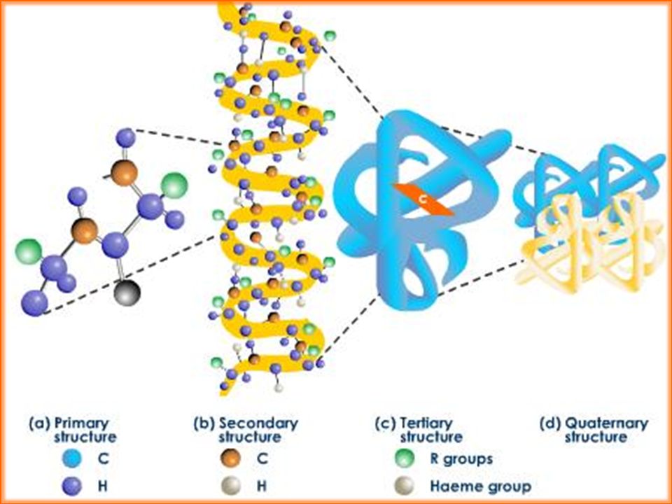
Primary structure is The sequence of amino acids that make up the polypeptide molecule

54
Alpha helix and polypeptide chain Secondary structure is The coils and pleat motifs that occur in the polypeptide molecule

55
The 3D structure of a peptide which is held together by a variety of chemical bonds

56
Ionic bonds Bonds that occur between the positive and negatively charged R groups on amino acids Hydrogen bonds Attraction that occurs between the slightly positive hydrogen atoms and slightly negatively charged oxygen or nitrogen atoms in an amino acid Disulfide bonds Bonds that occur between 2 sulfur containing amino acids. These are very strong and provide a lot of stability to the protein Hydrophobic interactions Strong attraction between oily, water- hating molecules. These regions spontaneously fold in together in a protein Hydrophillic interactions As water-hating molecules group together they push out water-loving molecules to the outside of the globular protein structure

57
Ionic bonds Bonds that occur between the positive and negatively charged R groups on amino acids Hydrogen bonds Attraction that occurs between the slightly positive hydrogen atoms and slightly negatively charged oxygen or nitrogen atoms in an amino acid Disulfide bonds Bonds that occur between 2 sulfur containing amino acids. These are very strong and provide a lot of stability to the protein Hydrophobic interactions Strong attraction between oily, water- hating molecules. These regions spontaneously fold in together in a protein Hydrophillic interactions As water-hating molecules group together they push out water-hating molecules to the outside of the globular protein structure

58
Name the structure and the model Label A - D

59
Draw the generalised structure of an amino acid [3] What part of the amino acid dictates its unique reactivity? What type of bonding holds the secondary structure of proteins? What are triglycerides? Name 2 biomolecules made from cholesterol [2] What are phospholipids made of? What are essential amino acids? What is the R group of an amino acid? What is cytoplasm mostly made of? What type of bonding is common in excreted protein? What reagents are needed for the emulsion test? Name the 4 types of bonds that hold proteins together up to a quarternary structure [4] Draw a triglyceride [3] Draw a phospholipid [3] /24
![Draw the generalised structure of an amino acid [3] What part of the amino acid dictates its unique reactivity.](http://images.slideplayer.com/36/10619048/slides/slide_59.jpg "What type of bonding holds the secondary structure of proteins. What are triglycerides. Name 2 biomolecules made from cholesterol [2] What are phospholipids made of. What are essential amino acids. What is the R group of an amino acid. What is cytoplasm mostly made of. What type of bonding is common in excreted protein. What reagents are needed for the emulsion test. Name the 4 types of bonds that hold proteins together up to a quarternary structure [4] Draw a triglyceride [3] Draw a phospholipid [3] /24.")
60
1.Proteins are made up of glycerol 2.Amino acids contain a carboxylic alkali group 3.Peptide bonds hold amino acids together 4.The secondary structure of an amino acid is the pleats and bends that it has 5.The tertiary structure of a protein is dictated by the sequence of amino acids that it’s made up of

61
The bonds responsible for maintaining tertiary structure © Pearson Education Ltd 2008 This document may have been altered from the original

63
The biuret test is a chemical test used for detecting the presence of peptide bonds. In the presence of peptides, a copper(II) ion forms a violet-coloured complex in an alkaline solution 1.Using a syringe, add 2 cm 3 of protein suspension to a test tube. 2.Using a clean syringe, add 2 cm 3 of sodium hydroxide and shake to mix. 3.Add a small amount of copper sulfate drop by drop, shaking between each addition. 4.Check for any colour change. Do not get confused with the blue colour of the copper sulfate. 5.Repeat the test using a clean test tube and syringes and substituting distilled water for the protein suspension. 6.Check for any colour change.
 ion forms a violet-coloured complex in an alkaline solution 1.Using a syringe, add 2 cm 3 of protein suspension to a test tube. 2.Using a clean syringe, add 2 cm 3 of sodium hydroxide and shake to mix. 3.Add a small amount of copper sulfate drop by drop, shaking between each addition. 4.Check for any colour change. Do not get confused with the blue colour of the copper sulfate. 5.Repeat the test using a clean test tube and syringes and substituting distilled water for the protein suspension. 6.Check for any colour change..")
64
Polypeptide Monosaccharide Peptide bond Carboxylic acid Copper sulfate Hydrophobic interactions Maltose hydrolysis Condensation Alpha Structural Glycogen Glycosidic bond Polypeptide Secondary 3-Dimensional Turgid Cellulose Dipeptide Disulphide Ionic Benedicts Carbon Hydrophillic interactions

65
Learning Objectives: 1.Define diffusion is the passive movement down a concentration gradient. 2.Explain that osmosis is special type of diffusion defined in terms of water potential Learning Objectives: 1.Define diffusion is the passive movement down a concentration gradient. 2.Explain that osmosis is special type of diffusion defined in terms of water potential Diffusion and Osmosis

66
(a) Simple diffusion © Pearson Education Ltd 2008 This document may have been altered from the original
 Simple diffusion © Pearson Education Ltd 2008 This document may have been altered from the original")
67
© Pearson Education Ltd 2008 This document may have been altered from the original (b) Facilitated diffusion using a channel protein
 Facilitated diffusion using a channel protein")
68
© Pearson Education Ltd 2008 This document may have been altered from the original (c) Facilitated diffusion using a carrier protein
 Facilitated diffusion using a carrier protein")
69
Molecules will move from where they are at a high concentration to where they are at a lower concentration. i.e. they diffuse down a concentration gradient. The blood system in humans continually brings more oxygen to the cell and takes carbon dioxide away. This maintains a high concentration gradient. Since the movement is always down the concentration gradient, it requires no energy. The small molecules pass from one side of the membrane to the other by moving between the lipid molecules.

70
Factors affecting the rate of diffusion Fick’s law notes that the rate of diffusion is in direct proportion to:

71
Factors affecting the rate of diffusion The higher the surface area to volume ratio, the faster diffusion occurs. Vs By maintaining a steep concentration gradient, diffusion rate increases Vs

72
The difference in concentration is maintained by breathing, which brings in air with a high oxygen concentration and removes the air with a high carbon dioxide concentration and by a good blood supply. The capillaries surrounding the alveoli take away the oxygenated blood and replace it with blood with a high carbon dioxide concentration. The walls of the alveoli are only one cell thick, so the surface across which diffusion occurs is thin and the rate is high In a nutshell.. The larger the area and difference in concentration and the thinner the surface, the quicker the rate In a nutshell.. The larger the area and difference in concentration and the thinner the surface, the quicker the rate For example.. in the lung the surface area is made very large by the presence of many alveoli.

73
Define DIFFUSION: The movement of particles from an area of high concentration to low concentration Water potential describes the tendency of water to move away from dissolved solvents Water Loves solute The more solute dissolved in a volume of water the water wants to go and join them Water moves towards an area with a high concentration of solute and away from other water molecules (to “even out”) Substitute in water potential for solute concentration in your definition of diffusion... Water potential describes the tendency of water to move away from dissolved solvents Water Loves solute The more solute dissolved in a volume of water the water wants to go and join them Water moves towards an area with a high concentration of solute and away from other water molecules (to “even out”) Substitute in water potential for solute concentration in your definition of diffusion... Define DIFFUSION using water potential : The movement of water from an area of high water potential (high H 2 O concentration) low water potential (Low H 2 O concentration) Define DIFFUSION using water potential : The movement of water from an area of high water potential (high H 2 O concentration) low water potential (Low H 2 O concentration)
 Substitute in water potential for solute concentration in your definition of diffusion... Define DIFFUSION using water potential : The movement of water from an area of high water potential (high H 2 O concentration) low water potential (Low H 2 O concentration) Define DIFFUSION using water potential : The movement of water from an area of high water potential (high H 2 O concentration) low water potential (Low H 2 O concentration).")
74
Osmosis is the process used by cells to exchange water with their environment. It is a passive process similar to diffusion but it is water molecules that move.

75
© Pearson Education Ltd 2008 This document may have been altered from the original Osmosis is the process used by cells to exchange water with their environment. It is a passive process similar to diffusion but it is water molecules that move. A standard definition of osmosis is: The net movement of water molecules from a region of high water potential to a region where their water potential is low, through a partially permeable membrane (a membrane permeable to water and specific solutes).
..")
76
Pure water has a water potential of 0 Cells have a much lower water potential than pure water.. Why?

77
Hypotonic vs. Hypertonic solutions Hypotonic One of two solutions with MORE water and less solute One of two solutions with less water and MORE solute Which solution will have the most negative water potential? Write a definition for Isotonic What will happen to animal and plants cells placed in a)Hypertonic solutions? b) Hypotonic solutions? Hypertonic
Hypertonic solutions. b) Hypotonic solutions. Hypertonic.")
78
What happens in pure water? Animal cell will eventually burst-HAEMOLYSED Plant cells-swelling cytoplasm & vacuole will push against the cell wall which will stop the cell getting any larger-TURGID Solutions of high water potential - before and after

79
What happens in concentrated Sugar? Animal cell: cell contents shrink & membrane wrinkles- FLACCID Plant cell-cytoplasm & vacuole shrink and PM pulls away from cell wall-PLASMOLYSIS Solutions of low water potential - before and after

80
A solution’s water potential will fall as solutes are added because water molecules will cluster around the solute molecules. Water molecule Solute molecule

81
Osmosis in an animal cell

82
If this cell is placed in a solution that’s hypotonic to its cytosol, then water will move into the cell causing it to expand. 4.9

83
Osmosis in an animal cell If this cell is placed in a solution that’s isotonic to its cytosol, then the same amount of water enters the cell as moves out of it, so the cell is not damaged. 4.9

84
Osmosis in a plant cell Plant cells behave in the same way as animal cells when placed in an isotonic solution: they don’t gain or lose water. But the cell wall is inflexible and causes plant cells to behave differently in a hypertonic and hypotonic solution.

85
In a hypotonic solution water will enter the cell and fill the vacuole. The plasma membrane will push against the cell wall making the cell very inflexible. It is said that cells in this state are turgid. In a hypertonic solution the cell loses water and goes flaccid because the vacuole becomes flaccid and the cytoplasm stops pushing against the cell wall. This state is called plasmolysis. A cell at this stage is said to be in plasmolysis.

86
Skills: 1.Producing an appropriate dilution series when provided with stock solutions of reagents. 2.Collection of reliable quantitative data where there are changes in mass or length. 3.Collect and present raw data in a suitable table conforming to conventions (IOB). 4.Take all measurements to an appropriate level of accuracy and precision. 5.Using a standard scientific calculator to calculate mean, standard deviations and percentage change. Plotting data as line graphs.
. 4.Take all measurements to an appropriate level of accuracy and precision. 5.Using a standard scientific calculator to calculate mean, standard deviations and percentage change. Plotting data as line graphs..")
87
Instructions Read through all this before you start. 1. Choose a recorder from within your group to record all the measurements and recordings. Use the table provided to record your group’s results. You only need to initially record one copy per group, but you will all need a table of results to submit with your report. 2. Check that you have 6X 250ml labelled beakers. Pour 50mls of the particular molar sucrose solution, into the correct beaker. 3. You need to carry out steps 4 to 10 quickly and carefully to avoid the bores drying out, as this may affect your results. 4. Take 18 prepared potato bores; divide them into 6 sets of 3. 5. Place 3 of them side by side on a white tile. Lay a ruler down the edge of the bores and trim the ends with a scalpel, so that they are all the same length. This is clearly shown on the attached diagram. Use this as guidance. 6. Repeat this with the remaining 5 sets of 3 bores, keeping each set of 3 separate. It may be an idea to place each set of 3 bores on a separate scrap of paper to prevent confusion. Call these sets 0.2, 0.4, 0.6, 0.8, 1.0 and distilled water. 7. Gently roll the bores on a piece of paper towel or filter paper to lightly dry the surface. 8. Measure and record the length of all the bores, accurate to 0.5mm. Do this keeping them in their sets of 3. 9. Weigh and record the total mass of the 3 bores of each set. 10. Quickly place each set of bores in their respective beakers of solution.

88
Instructions 11. Leave the apparatus for 20 minutes. 12. Whilst you are waiting you can calculate and record the average initial length and average initial mass for each set of potato bores. You can also start to write up your experiment. Use the separate sheet as a guide. 13. After 20 minutes remove the bores using forceps and place each set on the appropriately labelled paper. 14. Gently roll each set of bores, re-measure and record the final lengths of the bores. 15. Weigh and record the final mass for each set of 3 potato bores. 16. You can now calculate and record the average final length and average final mass for each set of potato bores. 17. For each of the 6 separate solutions, calculate and record the following ratio for each set of bores: final average length ÷ Initial average length 18. Repeat step 17 for mass, so that you calculate the ratio for each solution. 19. Plot 2 graphs. The first one should be the concentration of sugar solutions (molarity), against: Final average length ÷ Initial average length (ratio) The second graph should be the concentration of sugar solutions (molarity), against: Final average mass ÷ Initial average mass (ratio) 20. From your graphs state the concentrations of sucrose solution, which are isotonic with the potato bores, from the length and mass results. This will be where the ratio is 1:1.
, against: Final average length ÷ Initial average length (ratio) The second graph should be the concentration of sugar solutions (molarity), against: Final average mass ÷ Initial average mass (ratio) 20. From your graphs state the concentrations of sucrose solution, which are isotonic with the potato bores, from the length and mass results. This will be where the ratio is 1:1..")
89
Place these features in the correct part of the Venn Diagram Involves water only Requires energy Is passive Movement of particles Needs a semi-permeable membrane High to low concentration Against a concentration gradient Occurs in nature How minerals get into root hair cells How oxygen leaves a leaf How water keeps plant cells turgid Involves transport of solutes DIFFUSION

90
A cell cannot get everything it needs via diffusion and osmosis. Sometimes a cell will need more of a particular substance than there is outside of the cell; or in other cases, it may just be that the cell needs to get a particular substance inside the cell quicker than simple diffusion allows. This would obviously require energy to drive the process What is Active transport? Unjumble the definition or molecules across a cell The movement of ions membrane from a region of higher concentration, requiring energy assisted by protein pumps and. concentration to a region of low What is Active transport? Unjumble the definition or molecules across a cell The movement of ions membrane from a region of higher concentration, requiring energy assisted by protein pumps and. concentration to a region of low What is Active transport? The movement of ions or molecules across a cell membrane from a region of higher concentration to a region of low concentration, assisted by protein pumps and requiring energy. What is Active transport? The movement of ions or molecules across a cell membrane from a region of higher concentration to a region of low concentration, assisted by protein pumps and requiring energy.

91
Some of the carrier proteins found in membranes act as “pumps.” These proteins are similar to the carrier proteins used for facilitated diffusion. They are shaped in a way that is complementary to the molecules they carry. They carry larger or charged ions through membranes. These are the molecules that cannot pass through the lipid bilayer using diffusion. These protein pumps differ significantly from the proteins used in facilitated diffusion:

92
What do protein pumps do? 1.they carry specific molecules one way across the membrane 2.in carrying molecules across the membrane, they use energy in the form of ATP 3.they can carry molecules against the concentration gradient (from low to high) 4.they can carry molecules at a much faster and more efficient rate than diffusion
 4.they can carry molecules at a much faster and more efficient rate than diffusion.")
93
DIFFUSION OSMOSIS ACTIVE TRANSPORT Involves water only Requires energy Is passive Movement of particles Needs a semi- permeable membrane High to low concentration Against a concentration gradient Occurs in nature How minerals get into root hair cells How oxygen leaves a leaf How water keeps plant cells turgid Involves transport of solutes

94
Find the right box below for each of these words CELL TRANSPORT ACTIVE TRANSPORT FACILITATED DIFFUSION GAS EXCHANGE IN THE LUNGS GATED CHANNELS HELPS CELL TURGOR IN PLANTS HIGH Ψ TO LOW Ψ MEMBRANE PROTEINS INVOLVED OSMOSIS PASSIVE PROCESSES REQUIRES ATP SIMPLE DIFFUSION STEROID ABSORPTION THROUGH PHOSPHOLIPID BILAYER UPTAKE OF GLUCOSE IN SMALL INTESTINE UPTAKE OF MINERAL IONS UPTAKE OF SOIL MINERALS IN PLANT ROOTS

96
Learning Objectives: 1.Compare and contrast the structures of plant and animal cells 2.Describe how some plant cells are adapted to carry out photosynthesis Learning Objectives: 1.Compare and contrast the structures of plant and animal cells 2.Describe how some plant cells are adapted to carry out photosynthesis Ref p 158 in books

98
Perhaps not! If alien humanoid like creatures had chloroplasts in their cells, they could carry out photosynthesis and become producers in their own food chain.

99
o The cell wall is rigid and made of cellulose fibres running through a mixture of other polysaccharides (more complex sugars) o The sticky middle lamella holds next-door cells together o The cell wall is fully permeable unless a substance called lignin is deposited in the cellulose layers. Lignin makes the cell wall very strong and resistant to strain but it also makes it impermeable. If all the gaps between the fibres are filled in, the wall becomes completely impermeable and the cell will die.

100
Unjumble the functions of the cell wall: mechanical To strength provide bursting To cell under osmotic prevent the pressure through the plant pass along it and To allow water to move mechanical To strength provide bursting To cell under osmotic prevent the pressure through the plant pass along it and To allow water to move To provide mechanical strength To prevent the cell bursting under osmotic pressure To allow water to pass along it and move through the plant To provide mechanical strength To prevent the cell bursting under osmotic pressure To allow water to pass along it and move through the plant

101
o Like the mitochondria - they have an envelope of two membranes making up the outer "wall". o They have pairs of membranes called thylakoids arranged in stacks, each stack being called a granum. o Connecting different grana together are inter-granal thylakoids. o Surrounding the internal membranes, inside the envelope is the stroma. o The reactions of photosynthesis take place in the membranes and stroma of the chloroplast.

Similar presentations
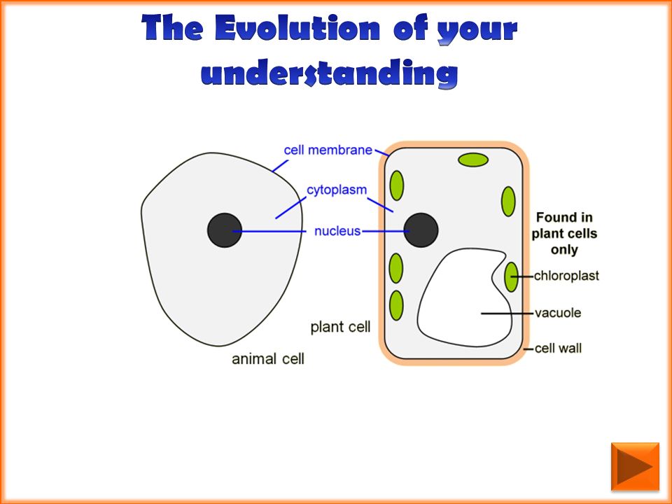


. Regulates what enters and leaves.>")


