Download presentation
Presentation is loading. Please wait.

3
NORMAL AND DYSPLASTIC EPITHELIUM

4
DYSPLASTIC EPITHELIUM

5
NORMAL AND CARCINOMA IN SITU

6
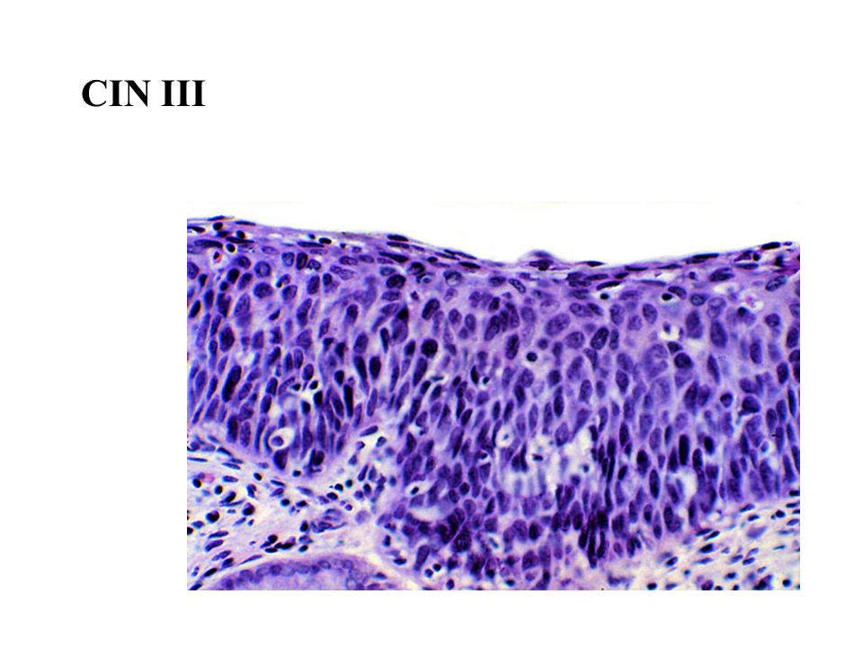
CIN I, II, and III CIN III

8
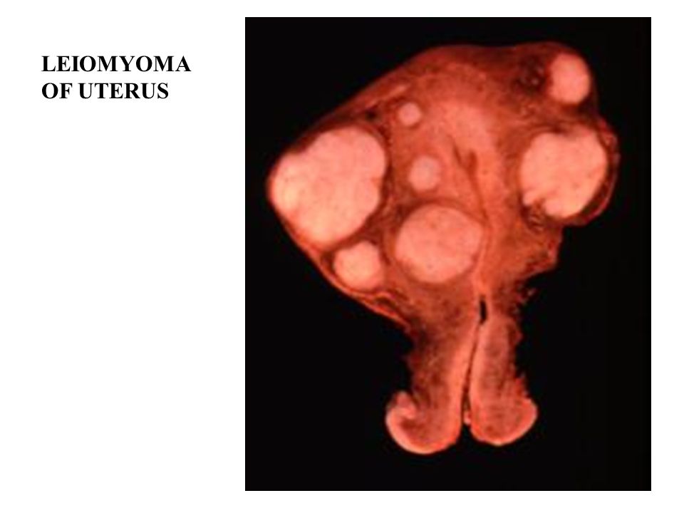
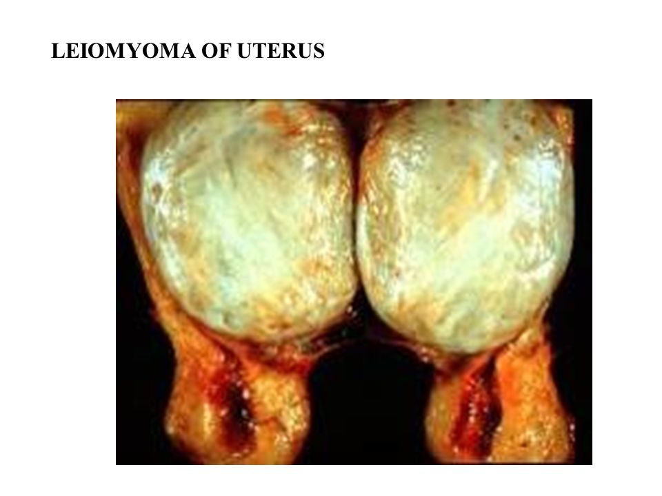
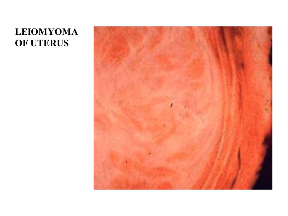
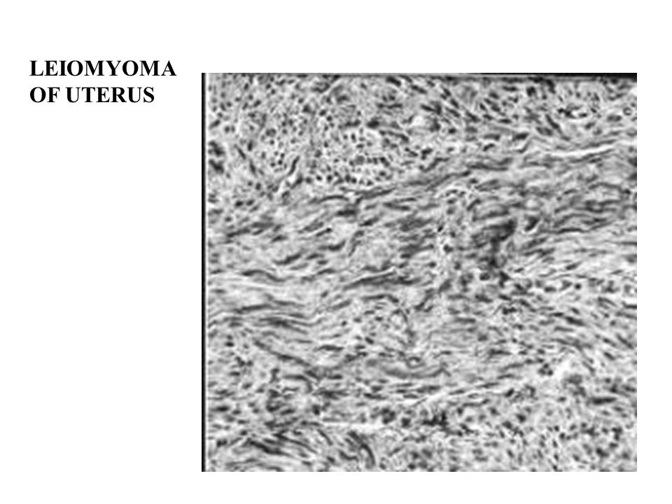
LEIOMYOMA OF UTERUS

13
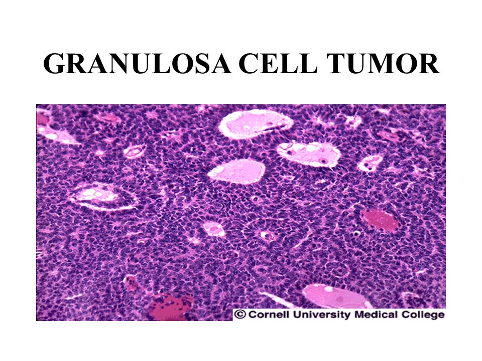
GRANULOSA CELL TUMOR

15
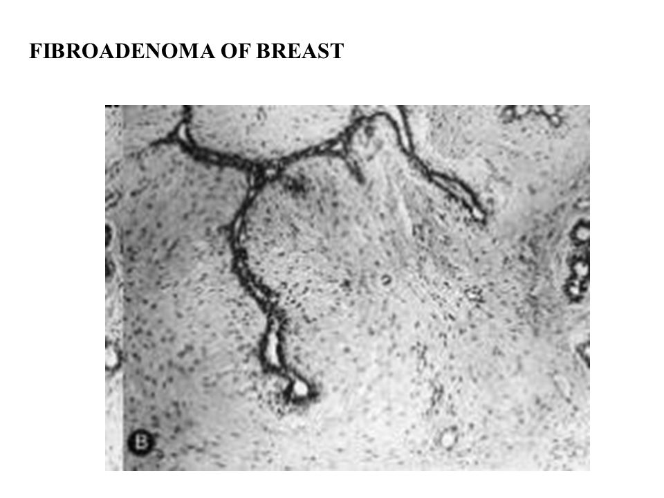
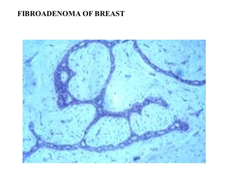
FIBROADENOMA OF BREAST

18
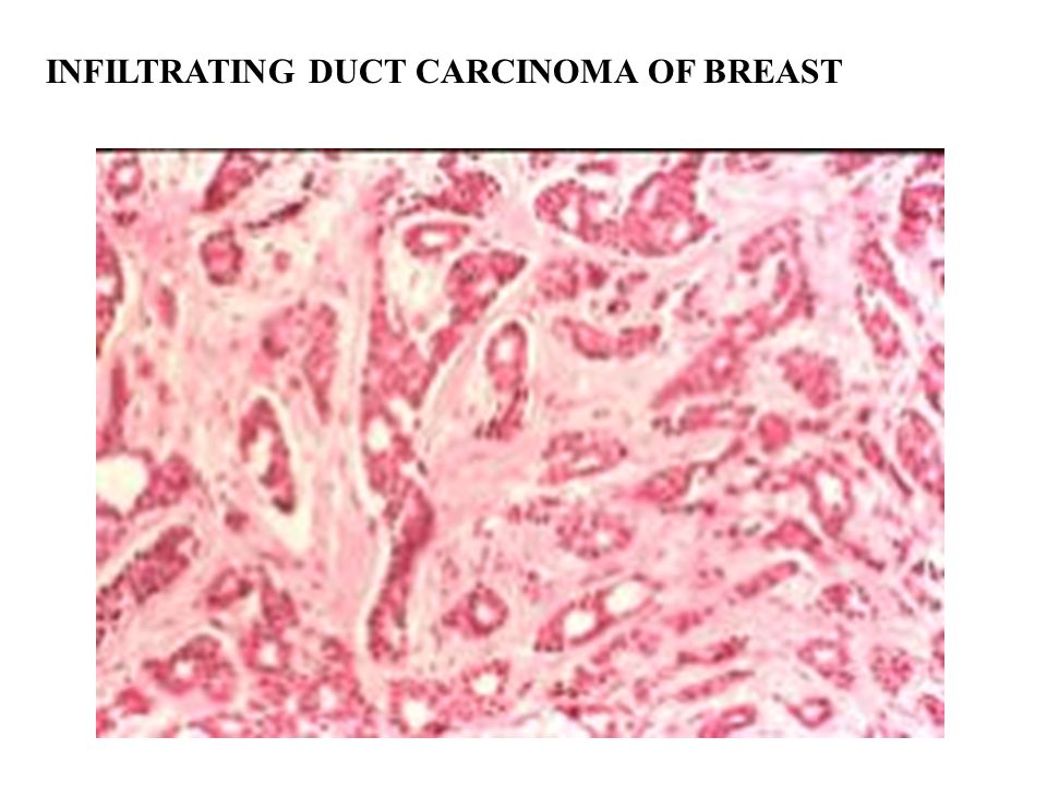
INFILTRATING DUCT CARCINOMA OF BREAST

20
INFILTRATING LOBULAR CARCINOMA OF BREAST Note “Indian file” pattern

21
Infiltrating lobular carcinoma Note “Indian file” pattern

22
GRAVES DISEASE

23
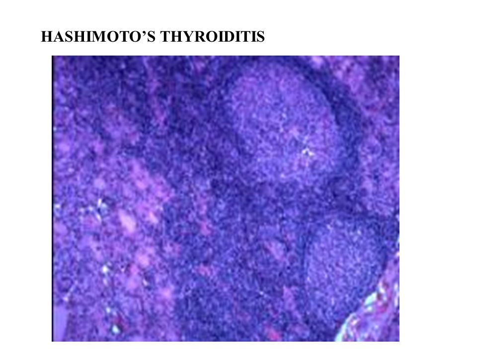
HASHIMOTO’S THYROIDITIS

25
PAPILLARY CARCINOMA OF THYROID

26
PAPILLARY CARCINOMA OF THE THYROID

27
PSAMMOMA BODIES

28
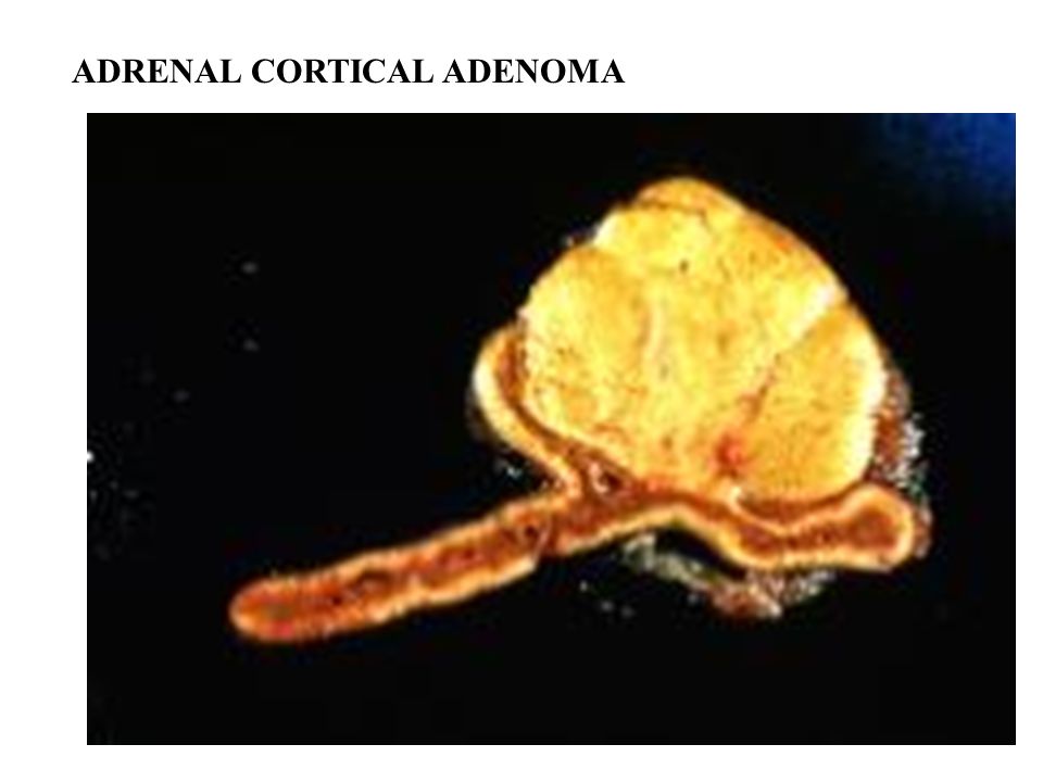
ADRENAL CORTICAL ADENOMA

30
SQUAMOUS CELL CARCINOMA

31
BASAL CELL CARCINOMA

32
Basal Cell Carcinoma

33
Duchenne’s Muscular Dystrophy Common sex-linked recessive trait. Fundamental lesion is a lack of dystrophin, an inner-sarcolemmal cytoskeletal component. Affected boys and their carrier mothers have markedly elevated creatine kinase.

34
Duchenne’s Muscular Dystrophy The muscle is abnormal at birth, and problems usually begin obvious in early childhood. These boys have difficulty must resort to unusual methods to standing up and become wheelchair-bound by their early teens. Fatty ingrowth produces characteristic "pseudohypertrophy of the calves".

35
Duchenne’s Muscular Dystrophy Pathology changes include: degeneration with phagocytosis and regeneration attempts along the muscle fibers. Eventually the fibers are replaced by fat and scar tissue. The healthier fibers undergo hypertrophy.

36
DUCHENNE’S MUSCULAR DYSTROPHY

37
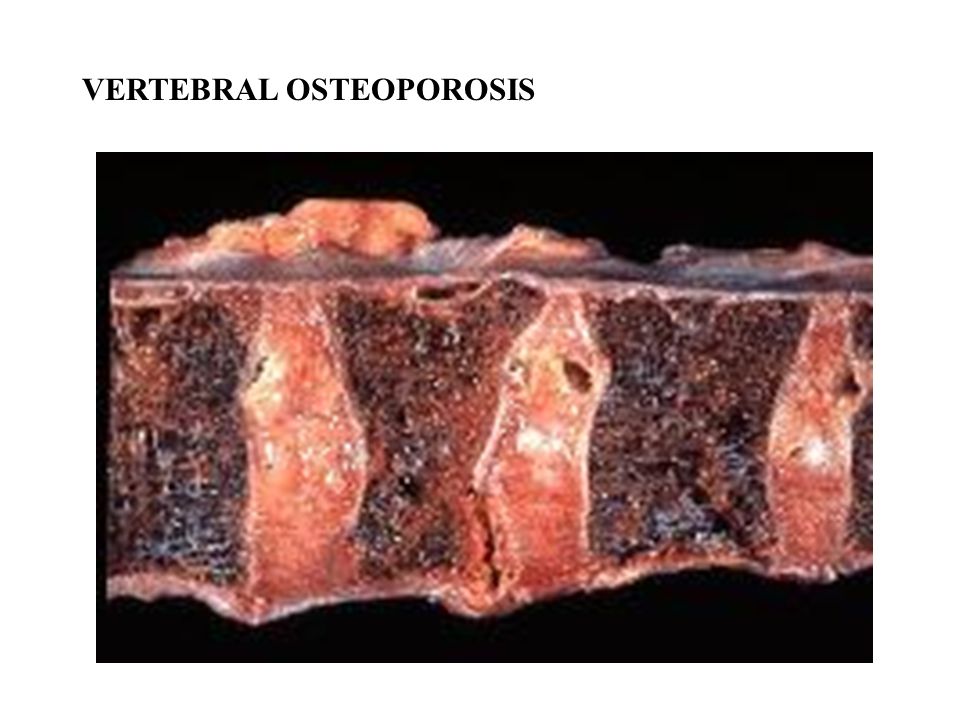
VERTEBRAL OSTEOPOROSIS

39
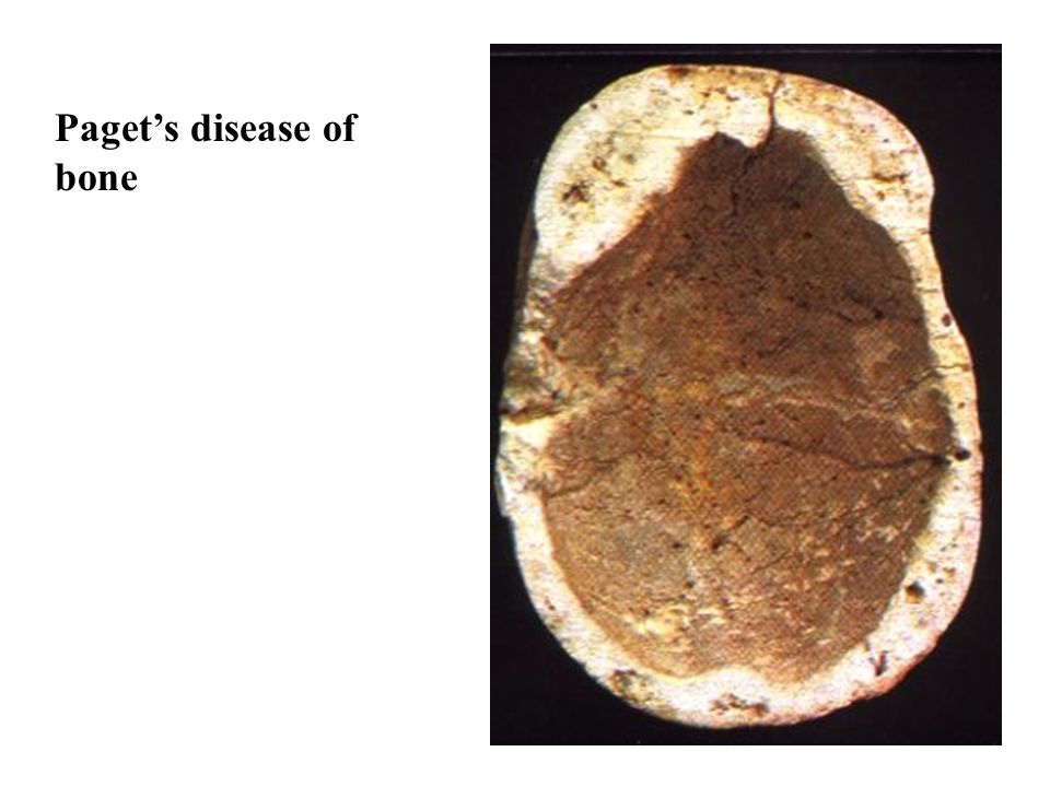
Paget’s disease of bone

40
Complications of Paget’s Disease Fractures Bone sarcomas (osteosarcoma, fibrosarcoma,) High-output cardiac failure
 High-output cardiac failure")
41
PAGET’S DISEASE

42
Paget’s disease of bone

44
Osteogenesis Imperfecta Group of disorders caused by deficiencies in synthesis of type I collagen resulting in bones that fracture easily. Other findings include blue sclera.

45
Blue Sclera in Osteogenesis Imperfecta

46
Osteopetrosis Osteopetrosis is a group of diseases characterized by progressive obliteration of the marrow cavity by bone (due to a defect in osteoclast function) preventing formation of bone marrow (resulting in anemia) and resulting in abnormal bone development and fragile bones.
 preventing formation of bone marrow (resulting in anemia) and resulting in abnormal bone development and fragile bones.")
47
Fibrous Dysplasia is a condition in which there is a localized replacement of bone by fibrous tissue with poorly-formed spicules of bone. Simple fibrous dysplasia can be monostotic (involvement of a single bone) or polyostotic (involvement of several bones). Polyostotic Fibrous Dysplasia associated with melanotic pigmentation of the skin and endocrine disorders is known as the McCune-Albright syndrome. Fibrous Dysplasia
 or polyostotic (involvement of several bones). Polyostotic Fibrous Dysplasia associated with melanotic pigmentation of the skin and endocrine disorders is known as the McCune-Albright syndrome. Fibrous Dysplasia.")
48
McCune-Albright Syndrome McCune-Albright syndrome is a condition featuring café-au-lait spots (with irregular borders compared to those seen in neurofibromatosis), precocious puberty, and frequently other endocrine disorders such as Cushing syndrome. Associated with mutation of G- proteins. “Coast of maine” Café au lait spot

49
OSTEOSARCOMA

50
Codman’s triangle

51
Osteosarcoma

52
Most common primary malignant tumor of bone. More commonly seen in men (about 2:1). Most frequently arise near the knee joint.

53
OSTEOSARCOMA

54
SUBARACHNOID HEMORRHAGE

55
Epidurual Hematoma Epidural hematoma develops between the bone and dura. It develops when a fracture of the vault of skull, usually the temporal bone, transects a blood vessels of the dura, usually a branch of the middle meningeal artery.

56
EPIDURAL HEMATOMA Occurs when blood accumulates within the space between the dura and the skull. Such a location for hemorrhage is almost always the result of trauma that causes a tear in the middle meningeal artery.

57
Subdural Hematoma Usually venous in origin, with the bridging veins being a common source. Usually, blood seeps from the injured veins slowly and causes a gradual deterioration over a period of several hours or a day.

58
SUBDURAL HEMATOMA is usually the result of trauma with tearing of the bridging veins.

59
BRIDGING VEINS

60
EXTRADURAL HEMORRHAGE

61
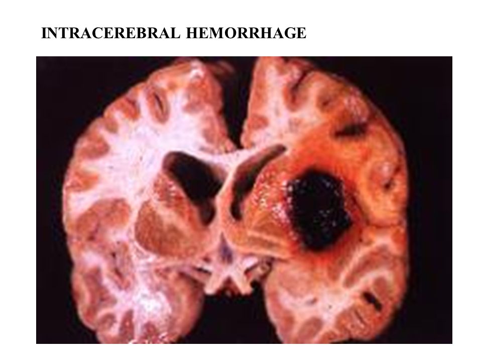
INTRACEREBRAL HEMORRHAGE

63
PYOGENIC MENINGITIS

64
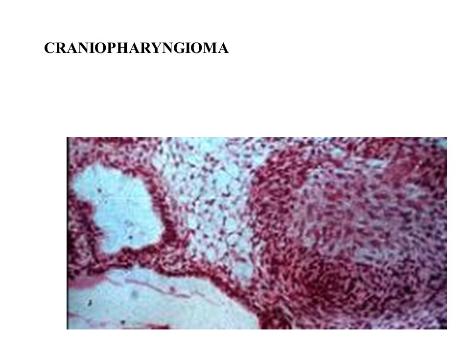
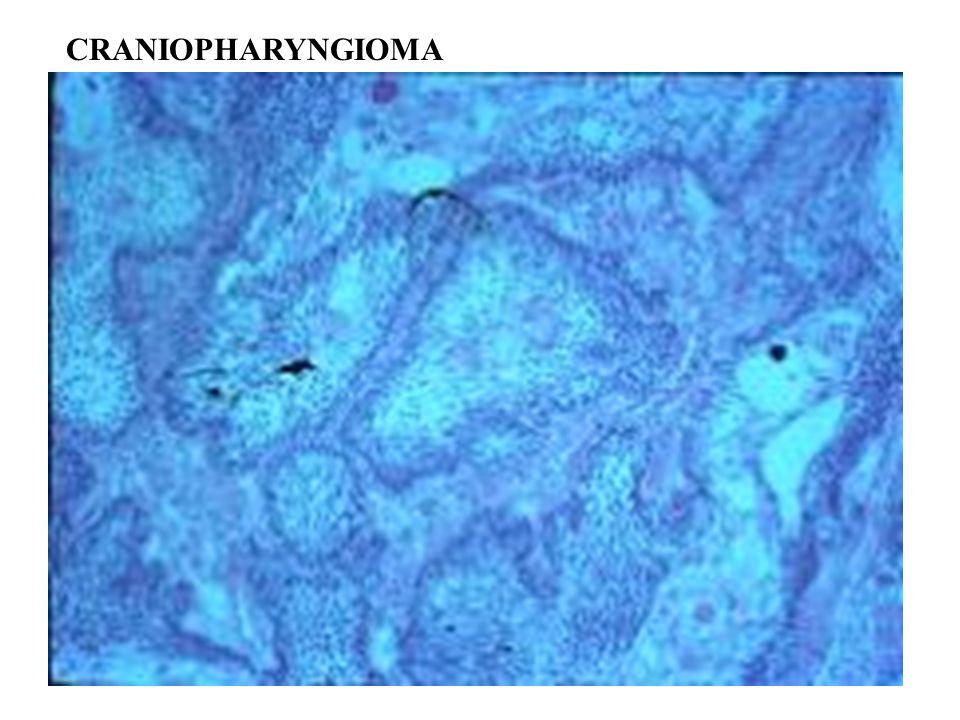
CRANIOPHARYNGIOMA develops in the hypothalamus, close to the pituitary gland.

65
CRANIOPHARYNGIOMA Histologically, these lesions vary in pattern and may recapitulate the enamel organ of the tooth, and are hence known as adamatinomas or ameloblastomas.

66
CRANIOPHARYNGIOMA

70
MENINGIOMA Meningiomas make up nearly 20% of all primary brain tumors. Meningiomas are more common in women than men.

71
MENINGIOMA

72
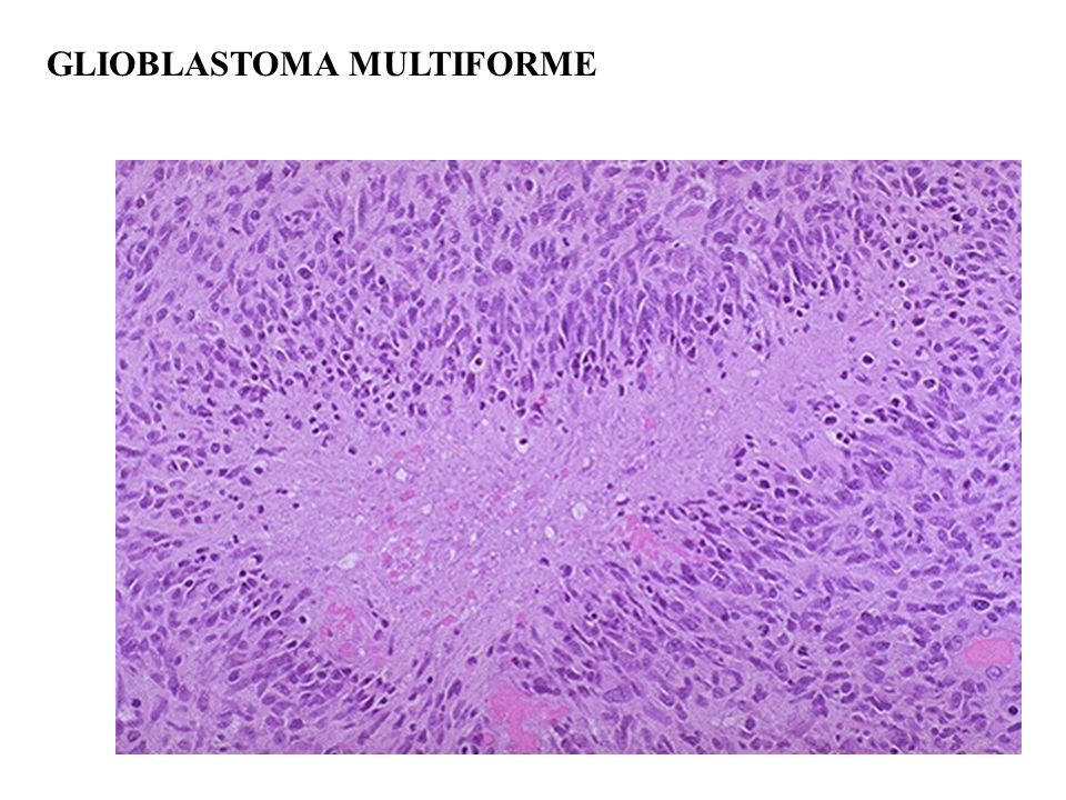
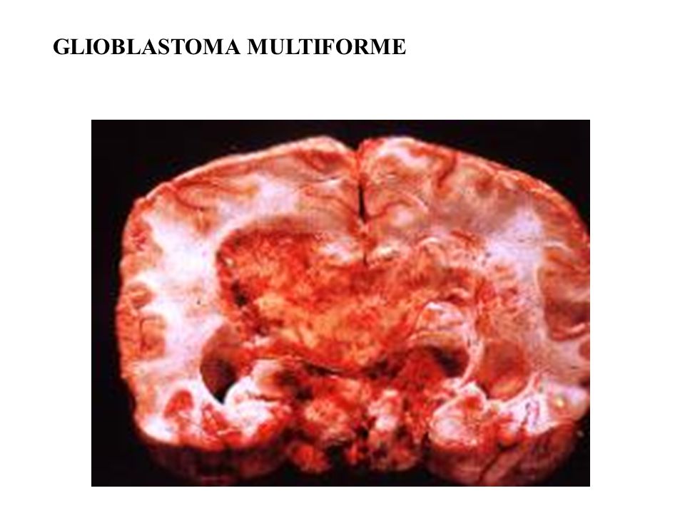
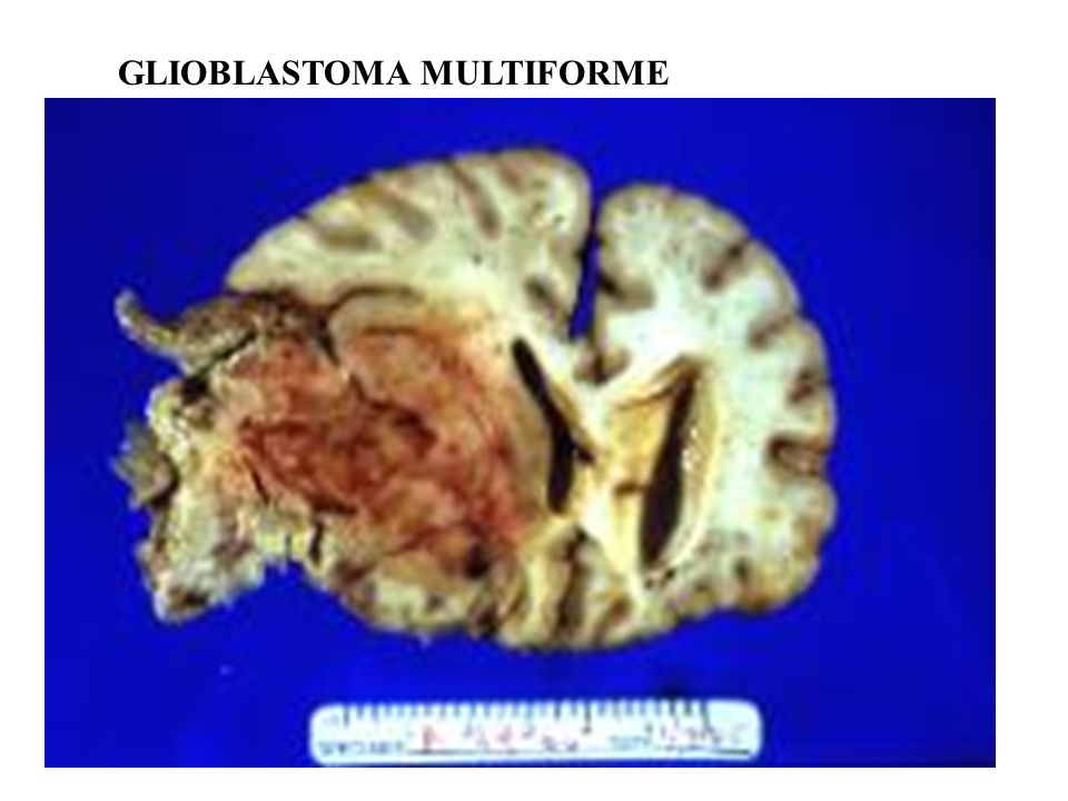
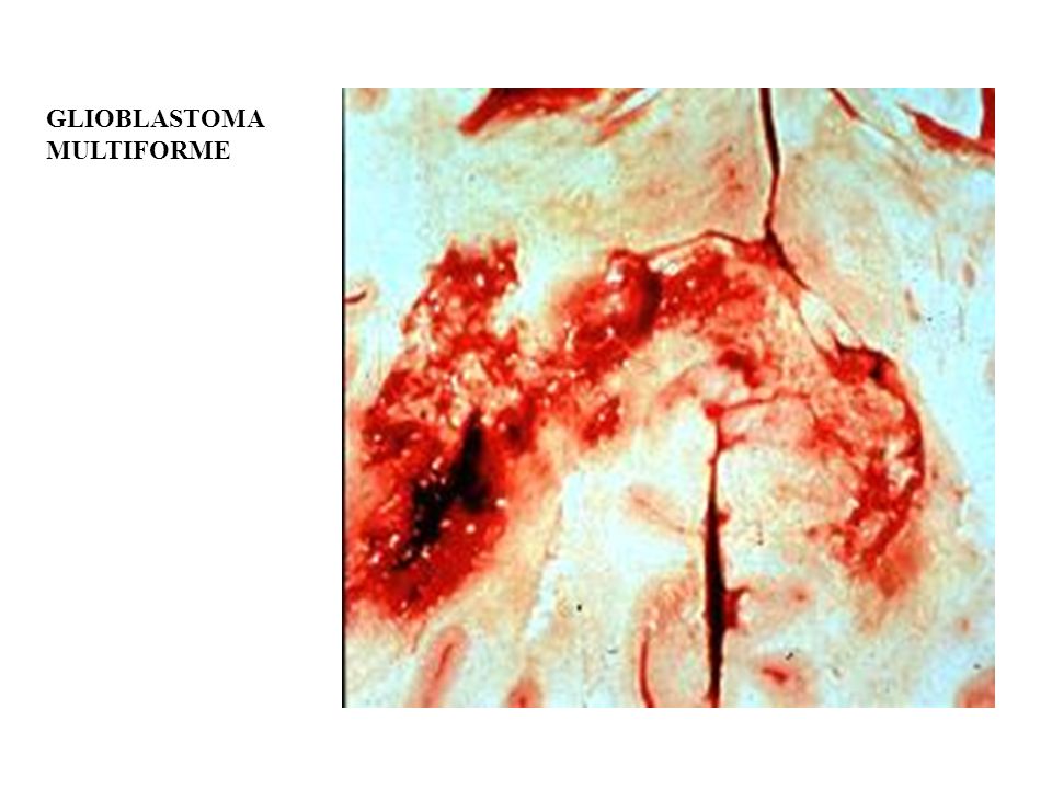
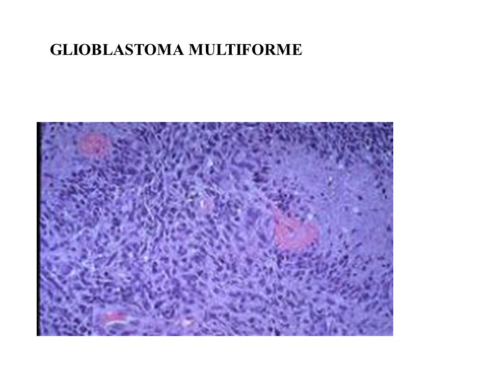
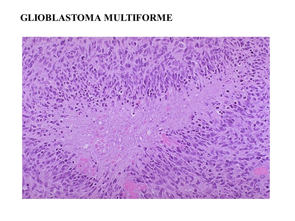
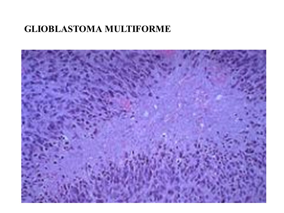
GLIOBLASTOMA MULTIFORME

82
MULTIPLE SCLEROSIS

83
Amyotrophic Lateral Sclerosis (ALS) Lou Gehrig’s disease – neuromuscular condition involving degeneration of motor nerve cells in the brain (upper motor neurons) and spinal cord (lower motor neurons). Symptoms – loss of the ability to speak, swallow, and breathe Cause unknown, but disease may be linked to malfunctioning genes for superoxide dismutase enzyme Death often occurs within five years…

84
AMYOTROPIC LATERAL SCLEROSIS ALS is characterized by a progressive degeneration of motor nerve cells in the brain (upper motor neurons) and spinal cord (lower motor neurons).
 and spinal cord (lower motor neurons).")
85
PERNICIOUS ANEMIA

86
Amyotrophic lateral sclerosis Subacute combined degeneration Pyramidal tracts Ventral horn Posterior funiculus Pyramidal tracts

87
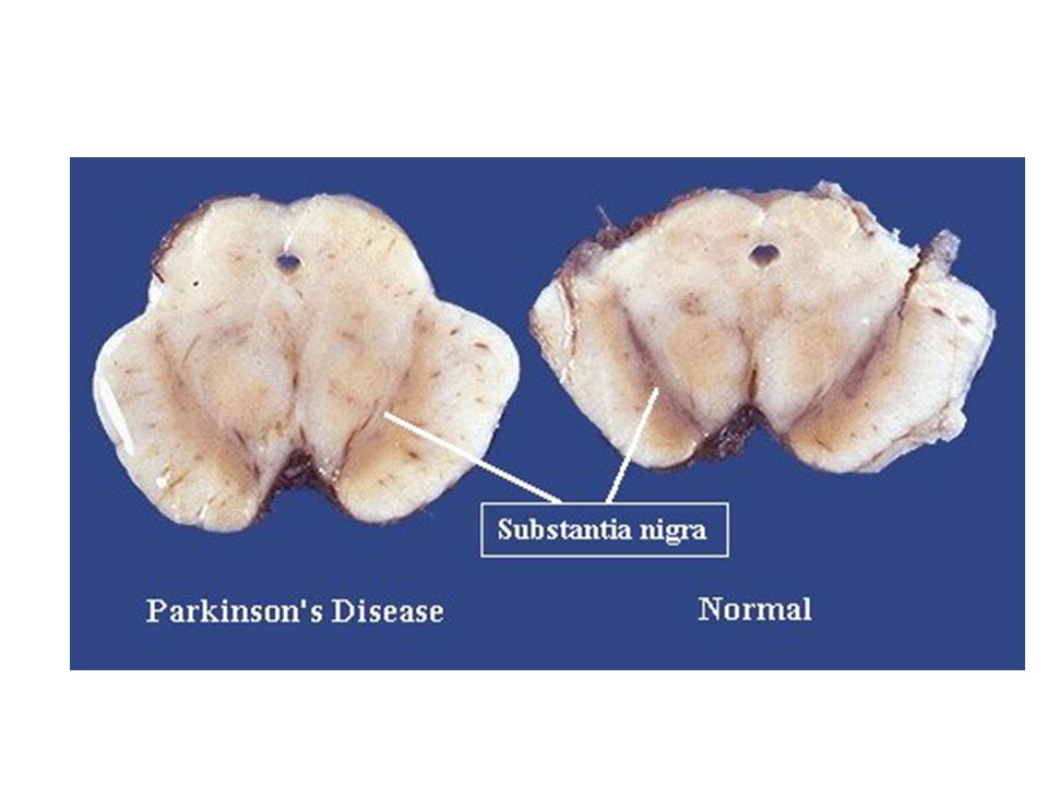
IDIOPATHIC PARKINSON’S DISEASE LOSS OF PIGMENT IN SUBSTANTIA NIGRA

88
Idiopathic Parkinson disease Note loss of pigment in substantia nigra in section on left versus normal appearance of section on right.

90
LEWY BODIES are cytoplasmic inclusion bodies which contain accumulations of normal neurofilament and stain positively with ubiquitin.

91
MITRAL STENOSIS

92
BUTTERFLY RASH IN SLE

Similar presentations