Download presentation
Presentation is loading. Please wait.

1
Learning Objectives Describe the location , external features, relations, lobes, segments & applied anatomy of Liver. Describe parts, relations, & clinical importance of Biliary apparatus (gall bladder, cystic duct, hepatic ducts & bile duct).
.")
2
Learning outcomes At the end of the lecture,student should be able to
Describe the gross anatomy ,blood supply,nerve supply and lymphatic drainge of the liver Explain its various peritoneal ligaments . Discuss its applied anatomy and vacsular segments Explain the gross anatomy of the different parts of the extrahepatic biliary apparatus Describe the blood supply,nerve supply and lymphatic drainage of its different parts Discuss the clinical significance of its different parts .

3
LIVER & BILIARY APPARATUS
Dr.chandraleha

4
Liver-Introduction Also called ‘hepar’. Largest gland in the body.
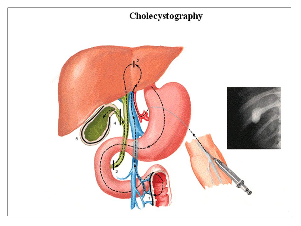
Weighs about 1600 gm in males,1300 gm in females. Occupies the right hypochondrium, epigastrium & left hypochondrium. Most part of the liver is covered by ribs & costal cartilages. The bile secreted by liver carried to the gall-bladder by the cystic duct or poured directly into the duodenum by the common bile duct to aid digestion.

5
Liver- Position

6
External features Wedge shaped, resembles four sided pyramid.
3 surfaces - - Superior, posterior & inferior. Inferior surface is well defined -also called visceral surface. Inferior border is well defined & the other borders are rounded.

7
External features Superior surface is attached to the diaphragm and anterior abdominal wall by falciform ligament and the free margin contains ligamentum teres (obliterated umbilical vein). Inferior and posterior surfaces are divided into four lobes namely right lobe, left lobe, quadrate lobe & caudate lobe.
. Inferior and posterior surfaces are divided into four lobes namely right lobe, left lobe, quadrate lobe & caudate lobe.")
8
External features Inferior and posterior surfaces Superior surface

9
Ligaments of Liver Ligaments are two layered peritoneal folds.
Falciform ligament connecting the anterior abdominal wall to the anterosuperior surface of the liver to become the coronary ligament and triangular ligaments. Left triangular ligament connecting the superior surface of left lobe of liver to the diaphragm. Right triangular ligament connecting the lateral part of posterior surface of right lobe of liver to the diaphragm. Coronary ligament having anterior & posterior layers, to enclose the bare area of liver. Lesser omentum connecting the stomach & first part of duodenum to the fissure for the ligamentum venosum & margins of porta hepatis.

10
Ligaments of Liver

11
Bare Area of Liver Area of liver not covered by peritoneum, directly comes in contact with diaphragm. Triangular area bounded by the anterior and posterior layers of coronary ligament and the inferior vena cava. One of the site of porto caval anastomosis.

12
Bare Area of Liver Inferior vena cava Bare area Coronary ligament

13
Lobes of Liver Liver is divided into right & left lobes by falciform ligament ,fissure for ligamentum teres & fissure for ligamentum venosum. Right lobe is larger than left and has caudate & quadrate lobes.

14
Lobes of Liver Quadrate lobe Left lobe Right lobe Caudate lobe

15
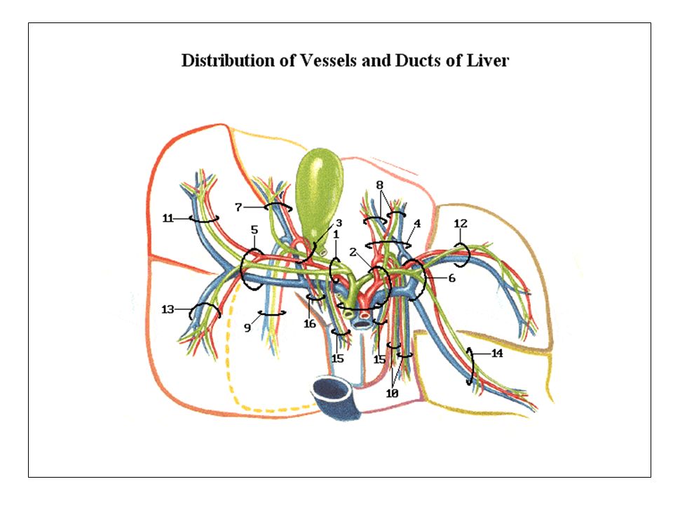
Porta Hepatis Portahepatis, is the hilum of the liver transmitting the hepatic ducts, hepatic artery and portal vein in that order from before backwards. Lesser omentum extends between porta hepatis and the lesser curvature of stomach. Porta hepatis

16
Liver-Relations All the surfaces except visceral surface are related to the diaphragm. Visceral surface is related to oesophagus, stomach, first part of duodenum, the gall bladder, right kidney and suprarenal gland.

17
Visceral surface-Relations

18
Hepatic Segments Medial segment Lateral segment
Posterior medial segment Left anterior lateral segment Posterior lateral segment Medial segment Right anterior lateral segment Anterior medial segment

20
Liver-Blood supply Arterial supply is by portal vein & hepatic artery.
Venous drainage is by hepatic veins which drain into inferior vena cava.

21
Lymphatic Drainage Lymphatics from upper surface drain into nodes in the posterior mediastinum. Lymphatics from lower surface drain into hepatic nodes and celiac nodes.

22
Nerve Supply of Liver Parasympathetic supply is by the preganglionic fibers of the vagus nerve. Sympathetic innervation is by the postganglionic fibers from the coeliac plexus.

23
CLINICAL ANATOMY Inflammation of liver – hepatitis.
Cirrhosis- the liver is replaced by fibrous tissue & shrinks in size.

24
Cirrhosis of Liver

26
Variations-Liver

27
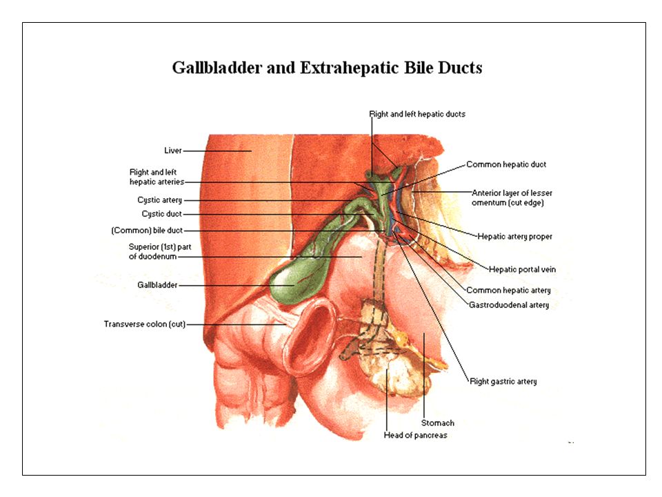
Biliary Apparatus Gall bladder. Cystic duct.
Right and left hepatic ducts which unite to form common hepatic duct. Bile duct formed by the union of cystic duct and common hepatic duct.

30
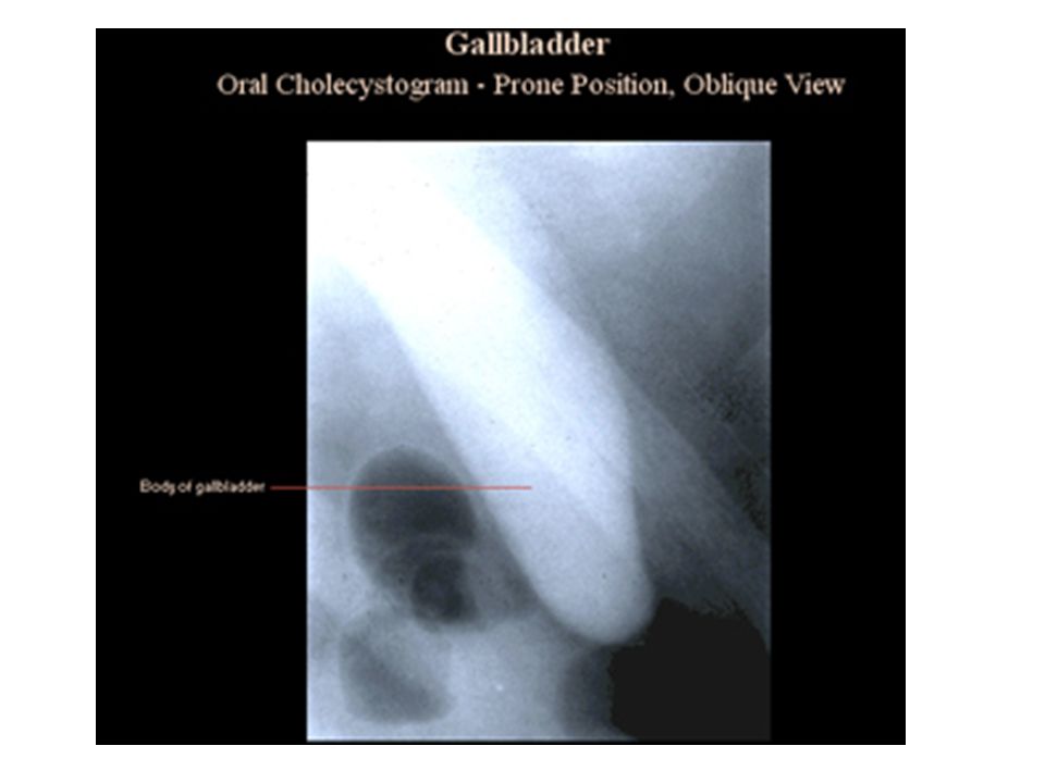
Gall Bladder Gall bladder is a store house for bile and has a capacity of 50 cc. It lies in a fossa on the visceral surface of liver . Covered by peritoneum.

31
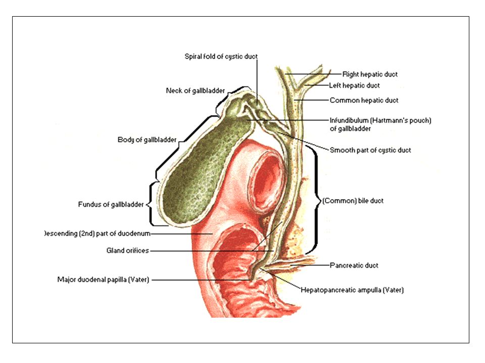
Parts of Gall Bladder Fundus is the blind end which projects beyond the lower margin of the liver Body is the upward continuation of fundus Neck is the narrow part of gall bladder Cystic duct is the S shaped continuation of the neck

32
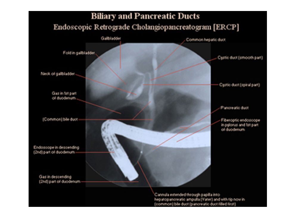
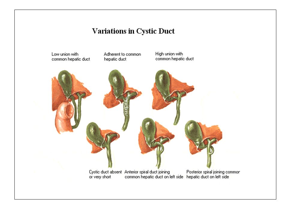
Cystic and Hepatic Ducts
Cystic duct joins the common hepatic duct to form the common bile duct.

33
Common Bile Duct Common bile duct is 9 cm long and has three parts.
Supraduodenal part in the free border of lesser omentum with hepatic artery to its left and portal vein behind.

34
Common Bile Duct Retro duodenal part behind the first part of duodenum. Pancreatic part lying in between the pancreas and the 2nd part of duodenum.

35
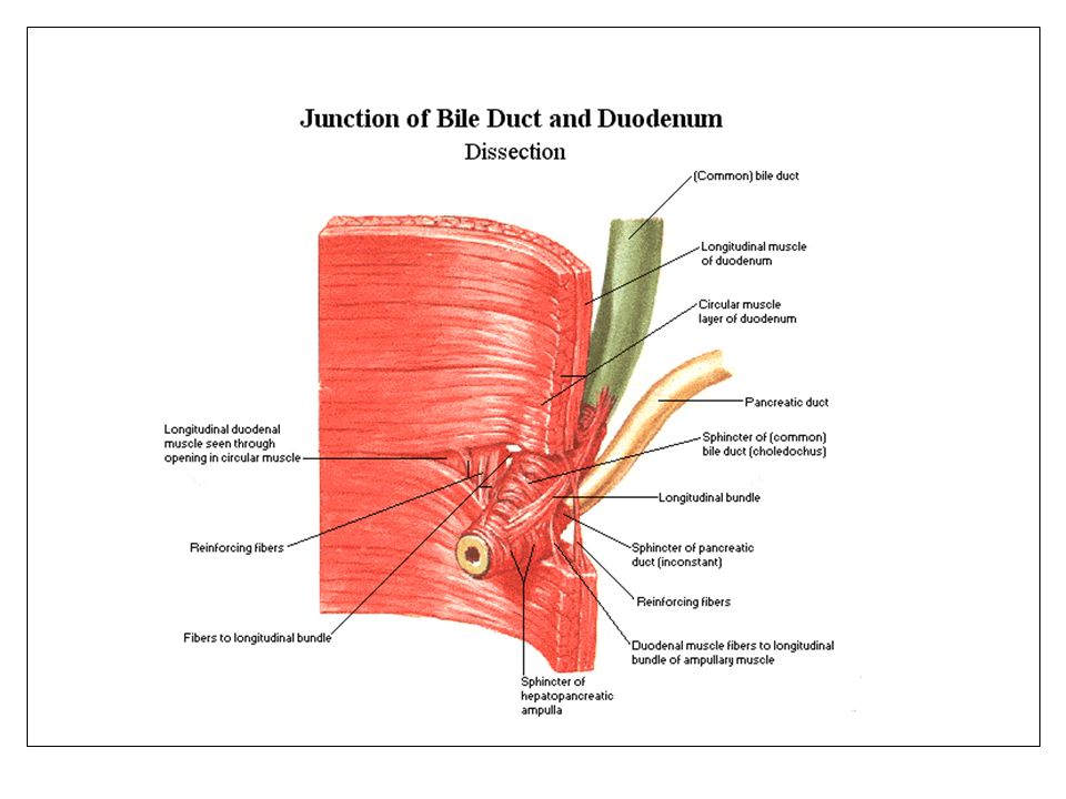
Ampula of Vater Common bile duct is joined by the main pancreatic duct before it opens into the duodenal papilla (Ampula of Vater). Smooth muscle fibers surrounding terminal part of the duct thickens to form the sphincter Oddi. .

36
Major & Minor duodenal Papillae

38
Blood Supply Arterial supply is by cystic artery, a branch of the right hepatic artery. Venous drainage is into the portal vein.

39
Lymphatic Drainage Lymphatics drain into cystic nodes, hepatic nodes and coeliac nodes.

40
Nerve Supply Parasympathetic supply is by pre-ganglionic fibers from the vagus nerve. Sympathetic innervation is by post-ganglionic fibers from the coeliac plexus.

41
Surface Marking Fundus of the gall bladder - Angle between the right costal margin and lateral border of rectus abdominis muscle.

42
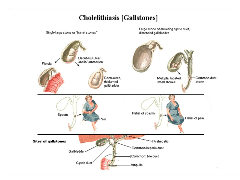
Clinical Anatomy Cholicystitis, Gall stones.

47
Bile duct - Variations

49
Any questions?

Similar presentations