Download presentation
Presentation is loading. Please wait.

1
이근욱 분당서울대학교병원/서울의대 혈액종양내과 부교수
제12회 분당서울대학교병원 내과 연수강좌 면역증강 표적항암제란? 이근욱 분당서울대학교병원/서울의대 혈액종양내과 부교수

2
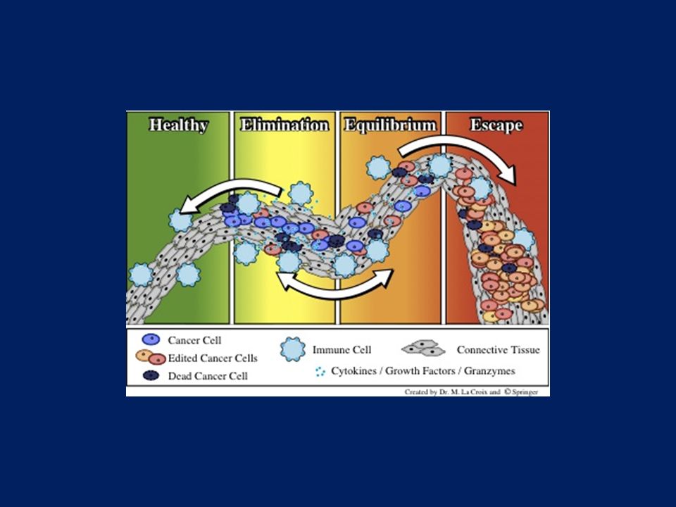
Immunotherapy of cancer
ACTIVE Acts directly on immune system PASSIVE (Adoptive) Targets the tumor; may utilize immune system Enhancing immune cell function Therapeutic vaccines Immune checkpoint inhibitors Cytokines Inhibition of CTLA-4 PD-1 PDL-1 Anti-tumor mAbs Adoptive Cetuximab Rituximab Bevacizumab … Adoptive cell transfer
 Targets the tumor; may utilize immune system. Enhancing. immune cell function. Therapeutic. vaccines. Immune checkpoint inhibitors. Cytokines. Inhibition of CTLA-4. PD-1. PDL-1. Anti-tumor. mAbs. Adoptive. Cetuximab. Rituximab. Bevacizumab. … Adoptive cell transfer.")
3
19 patients (7%) remain disease-free
RR 20%, including 23 [9%] CR 19 patients (7%) remain disease-free 2 patients with treatment-related mortality Overall Survival Klapper JA et al. Cancer 2008
 remain disease-free. 2 patients with treatment-related mortality. Overall Survival. Klapper JA et al. Cancer")
4
MAGRIT a double-blind, randomized, placebo-controlled phase III study to assess the efficacy of the recMAGE-A3 + AS15 as adjuvant therapy in resected MAGE-A3-positive NSCLC Melanoma-associated antigen 3 (MAGE-A3) is a protein that in humans is encoded by the MAGEA3 gene. The normal function of MAGE-A3 in healthy cells is unknown.[4] The presence of the antigen on tumor cells has been associated with worse prognosis. In one study, high levels of MAGE-A3 in lung adenocarcinoma were associated with shorter survival.[5] MAGE-A3 is a tumor-specific protein, and has been identified on many tumors including melanoma, non-small cell lung cancer, hematologic malignancies, among others Vansteenkiste et al. ESMO 2014
 is a protein that in humans is encoded by the MAGEA3 gene. The normal function of MAGE-A3 in healthy cells is unknown.[4] The presence of the antigen on tumor cells has been associated with worse prognosis. In one study, high levels of MAGE-A3 in lung adenocarcinoma were associated with shorter survival.[5] MAGE-A3 is a tumor-specific protein, and has been identified on many tumors including melanoma, non-small cell lung cancer, hematologic malignancies, among others. Vansteenkiste et al. ESMO")
5
TOGA : Trastuzumab for GC (Phase 3)
HER2 overexpression in GC 1:1 Bang YJ et al, Lancet 2010 5

6
Monoclonal Antibodies
Murine Mab : - omab Chimeric Mab : - ximab Humanized Mab : - zumab Basic antibody structure and the different types of therapeutic antibody. (a) Basic antibody structure. (b) Basic structure of a murine, chimeric, humanised, and human monoclonal antibody. Red indicates murine sequence and black indicates human sequence. CDR, complementarity-determining region Murine monoclonal antibodies (suffix -omab) Initially, murine antibodies were obtained by hybridoma technology, for which Kohler and Milstein received a Nobel prize. However the dissimilarity between murine and human immune systems led to the clinical failure of these antibodies, except in some specific circumstances. Major problems associated with murine antibodies included reduced stimulation of cytotoxicity and the formation complexes after repeated administration, which resulted in mild allergic reactions and sometimes anaphylactic shock.[8] [edit] Chimeric and humanized monoclonal antibodies (suffixes -ximab, -zumab respectively) To reduce murine antibody immunogenicity, murine molecules were engineered to remove immunogenic content and to increase their immunologic efficiency.[8] This was initially achieved by the production of chimeric and humanized antibodies. Chimeric antibodies are composed of murine variable regions fused onto human constant regions. Human gene sequences, taken from the kappa light chain and the IgG1 heavy chain, results in antibodies that are approximately 65% human. This reduces immunogenicity, and thus increases serum half-life. Humanised antibodies are produced by grafting murine hypervariable[disambiguation needed ] amino acid domains into human antibodies. This results in a molecule of approximately 95% human origin. However it has been shown in several studies that humanised antibodies bind antigen much more weakly than the parent murine monoclonal antibody, with reported decreases in affinity of up to several hundredfold.[10][11] Increases in antibody-antigen binding strength have been achieved by introducing mutations into the complementarity determining regions (CDR),[12] using techniques such as chain-shuffling, randomization of complementarity determining regions and generation of antibody libraries with mutations within the variable regions by error-prone PCR, E. coli mutator strains, and site-specific mutagenesis.[1] [edit] Human monoclonal antibodies (suffix -umab) Human monoclonal antibodies are produced using transgenic mice or phage display libraries. Human monoclonal antibodies are produced by transferring human immunoglobulin genes into the murine genome, after which the transgenic mouse is vaccinated against the desired antigen, leading to the production of monoclonal antibodies.,[9] allowing the transformation of murine antibodies in vitro into fully human antibodies.[3] The heavy and light chains of human IgG proteins are expressed in structural polymorphic (allotypic) forms. Human IgG allotype has been considered as one of the many factors that can contribute to immunogenecity.[13] The general scheme of a monoclonal antibody development program is described in.[14] [edit] Targeted conditions Human Mab : - umab CDR, complementarity-determining region
 Basic antibody structure. (b) Basic structure of a murine, chimeric, humanised, and human monoclonal antibody. Red indicates murine sequence and black indicates human sequence. CDR, complementarity-determining region Murine monoclonal antibodies (suffix -omab) Initially, murine antibodies were obtained by hybridoma technology, for which Kohler and Milstein received a Nobel prize. However the dissimilarity between murine and human immune systems led to the clinical failure of these antibodies, except in some specific circumstances. Major problems associated with murine antibodies included reduced stimulation of cytotoxicity and the formation complexes after repeated administration, which resulted in mild allergic reactions and sometimes anaphylactic shock.[8] [edit] Chimeric and humanized monoclonal antibodies (suffixes -ximab, -zumab respectively) To reduce murine antibody immunogenicity, murine molecules were engineered to remove immunogenic content and to increase their immunologic efficiency.[8] This was initially achieved by the production of chimeric and humanized antibodies. Chimeric antibodies are composed of murine variable regions fused onto human constant regions. Human gene sequences, taken from the kappa light chain and the IgG1 heavy chain, results in antibodies that are approximately 65% human. This reduces immunogenicity, and thus increases serum half-life. Humanised antibodies are produced by grafting murine hypervariable[disambiguation needed ] amino acid domains into human antibodies. This results in a molecule of approximately 95% human origin. However it has been shown in several studies that humanised antibodies bind antigen much more weakly than the parent murine monoclonal antibody, with reported decreases in affinity of up to several hundredfold.[10][11] Increases in antibody-antigen binding strength have been achieved by introducing mutations into the complementarity determining regions (CDR),[12] using techniques such as chain-shuffling, randomization of complementarity determining regions and generation of antibody libraries with mutations within the variable regions by error-prone PCR, E. coli mutator strains, and site-specific mutagenesis.[1] [edit] Human monoclonal antibodies (suffix -umab) Human monoclonal antibodies are produced using transgenic mice or phage display libraries. Human monoclonal antibodies are produced by transferring human immunoglobulin genes into the murine genome, after which the transgenic mouse is vaccinated against the desired antigen, leading to the production of monoclonal antibodies.,[9] allowing the transformation of murine antibodies in vitro into fully human antibodies.[3] The heavy and light chains of human IgG proteins are expressed in structural polymorphic (allotypic) forms. Human IgG allotype has been considered as one of the many factors that can contribute to immunogenecity.[13] The general scheme of a monoclonal antibody development program is described in.[14] [edit] Targeted conditions. Human Mab. : - umab. CDR, complementarity-determining region.")
10
Immune checkpoints Co-stimulatory pathways. Co-stimulations either enhance or down-regulate T cell activation following the initial TCR and peptide-MHC ligation. Positive co-stimulatory pathways include B7–CD28, CD40L–CD40, ICOS–ICOS-L, and OX40–OX40L. Negative co-stimulatory pathways include B7–CTLA-4 and PD-1–PD-L. Immune checkpoints are molecules in the immune system that either turn up a signal (co-stimulatory molecules) or turn down a signal. Many cancers protect themselves from the immune system by inhibiting the T cell signal. Since around 2010[citation needed] inhibitory checkpoint molecules have been increasingly considered as new targets for cancer immunotherapies due to the effectiveness of two checkpoint inhibitor drugs that were initially indicated for advanced melanoma - Yervoy, from Bristol-Myers Squibb, and Keytruda, from Merck.
 or turn down a signal. Many cancers protect themselves from the immune system by inhibiting the T cell signal. Since around 2010[citation needed] inhibitory checkpoint molecules have been increasingly considered as new targets for cancer immunotherapies due to the effectiveness of two checkpoint inhibitor drugs that were initially indicated for advanced melanoma - Yervoy, from Bristol-Myers Squibb, and Keytruda, from Merck.")
11
CTLA vs. PD-1 Ribas A et al. N Engl J Med 2012
CTLA-4 antibodies release an immune checkpoint at the activation step of an immune response to cancer PD-1 and PD-L1 antibodies release an immune checkpoint at the effector step of n immune response to cancer. Ribas A et al. N Engl J Med 2012

12
Blockade of CTLA Ribas A et al. N Engl J Med 2015
CTLA-4 antibodies release an immune checkpoint at the activation step of an immune response to cancer PD-1 and PD-L1 antibodies release an immune checkpoint at the effector step of n immune response to cancer. Ribas A et al. N Engl J Med 2015

13
Improved survival of melanoma with ipilimumab
626 patients with previously treated melanoma CTLA-4 antibodies release an immune checkpoint at the activation step of an immune response to cancer PD-1 and PD-L1 antibodies release an immune checkpoint at the effector step of n immune response to cancer. Hodi et al. N Engl J Med 2010

14
Pooled survival analysis from all phase I-III, including BMS EAP
CTLA-4 antibodies release an immune checkpoint at the activation step of an immune response to cancer PD-1 and PD-L1 antibodies release an immune checkpoint at the effector step of n immune response to cancer.

15
Blockade of PD-1 pathway
Ribas A et al. N Engl J Med 2015

16
PD1/PD-L1 inhibitors in clinical development
Target Agent (Sponsor) Type of mAb PD-1 Nivolumab (BMS-ONO; OPDIVO) IgG4 fully human Pembrolizumab (MK-3475; Merck Sharp & Dohme; KEYTRUDA) IgG4 humanized Pidilizumab (CureTech-Teva) IgG1 humanized AMP-514 (AZ/MedImmune) IgG PDL-1 Atezolizumab (Genetech/Roche) IgG1; fully human engineered Durvalumab (MED4736; AZ/MedImmune) Avelumab (MSB C; Merck Serno) IgG1; fully human BMS (BMS-ONO) IgG4; fully human Antibody-dependent cell-mediated cytotoxicity (ADCC)
 Type of mAb. PD-1. Nivolumab (BMS-ONO; OPDIVO) IgG4 fully human. Pembrolizumab (MK-3475; Merck Sharp & Dohme; KEYTRUDA) IgG4 humanized. Pidilizumab (CureTech-Teva) IgG1 humanized. AMP-514 (AZ/MedImmune) IgG. PDL-1. Atezolizumab (Genetech/Roche) IgG1; fully human engineered. Durvalumab (MED4736; AZ/MedImmune) Avelumab (MSB C; Merck Serno) IgG1; fully human. BMS (BMS-ONO) IgG4; fully human. Antibody-dependent cell-mediated cytotoxicity (ADCC)")
17
Previously untreated melanoma without BRAF mutation: Nivolumab vs
Previously untreated melanoma without BRAF mutation: Nivolumab vs. Dacarbacine OS Robert C et al. N Engl J Med 2015

18
Pembrolizumab versus ipilimumab in advanced melanoma
Statistically significant improvement in overall survival with KEYTRUDA® (pembrolizumab) 10 mg/kg Q3W vs ipilimumab (hazard ratio [HR]=0.69; 95% confidence interval [CI], 0.52–0.90; P=0.004) and with KEYTRUDA 10 mg/kg Q2W vs ipilimumab (HR=0.63; 95% CI, 0.47–0.83; P<0.001). Robert C et al. N Engl J Med 2015
 10 mg/kg Q3W vs ipilimumab. (hazard ratio [HR]=0.69; 95% confidence interval [CI], 0.52–0.90; P=0.004) and with KEYTRUDA 10 mg/kg Q2W vs ipilimumab. (HR=0.63; 95% CI, 0.47–0.83; P<0.001). Robert C et al. N Engl J Med")
19
Blockade of PD-1 pathway in other tumors
Alley et al. AACR2015

20
Phase 3 trials of nivolumab in NSCLC

21
Phase 3 trials of nivolumab in NSCLC
Primary endpoint: Overall survival (OS) CheckMate 017 Squamous NSCLC1 CheckMate 057 Non-squamous NSCLC2 1Reckamp K et al., WCLC 2015; 2Horn L et al., ESMO 2015
 CheckMate 017. Squamous NSCLC1. CheckMate 057. Non-squamous NSCLC2. 1Reckamp K et al., WCLC 2015; 2Horn L et al., ESMO")
22
Phase 3 trials of nivolumab in NSCLC
CheckMate 017 Squamous NSCLC1 CheckMate 057 Non-squamous NSCLC2 1Reckamp K et al., WCLC 2015; 2Horn L et al., ESMO 2015

23
Immune related adverse events (AEs) may be associate with PD-1/PD-L1 blockade
Confirmed immune-related AEs Flu like febrile sense Autoimmune hepatitis, elevated transaminases Colitis / Duodenitis Rash Myositis/myasthenia gravis Pneumonitis Hypothyroidism / Hyperthyroidism Pan hypopituitarism (endocrinopathy) Pancreatitis Type 1 diabetes mellitus
 Pancreatitis. Type 1 diabetes mellitus.")
24
Incidence of Immune-related AEs
( ; Benjamin A. Teply & Evan J. Lipson)
")
25
Safety Profile of Nivolumab in Patients with Advanced Melanoma
: A Pooled Analysis Weber JS et al., ASCO 2015 Abstract 9018

26
Pneumonitis ( ; Benjamin A. Teply & Evan J. Lipson)
")
27
General Management Algorithm of IrAEs

28
Issues & future directions
Cost Biomarkers predictive of treatment response Combination of immune checkpoint inhibitors (or with other chemotherapeutic agents)
")
30
OS with pembrolizumab in NSCLC by PD-L1 expression
Garon et al. N Engl J Med 2015

Similar presentations


:311-9.>")


>")
>")


