Download presentation
Presentation is loading. Please wait.

1
LOGO 미생물학실험 Microbiology Laboratory 생물환경학과 김정호

2
현미경의 3 주요 부분 Light source ( 광원 ) Lens Condenser ( 집광기 ) 현미경의 3 주요 기능 Magnification ( 배율 ) Resolution ( 해상도, 분해능 ) Contrast ( 대비력 ) Light microscope (LM) Electron microscope (EM) Scanning probe microscope ( 주사탐침현미경 ) Microscopy
 Lens Condenser ( 집광기 ) 현미경의 3 주요 기능 Magnification ( 배율 ) Resolution ( 해상도, 분해능 ) Contrast ( 대비력 ) Light microscope (LM) Electron microscope (EM) Scanning probe microscope ( 주사탐침현미경 ) Microscopy")
3
Light (Optical) Microscope ( 광학현미경 ) Visible light, UV Stereomicroscope ( 실체 현미경 ) Biological microscope ( 생물 현미경 ) Bright field microscope ( 명시야 현미경 ) Dark field microscope ( 암시야 현미경 ) Phase contrast microscope ( 상대비 현미경 ) Fluorescence microscope ( 형광 현미경 ) Confocal scanning laser microsc ope ( 공초점 현미경 ) Differential interference contrast (DIC) microscope : 3D ( 분별간섭대비현미경 ) Light Microscope (LM)
 Microscope ( 광학현미경 ) Visible light, UV Stereomicroscope ( 실체 현미경 ) Biological microscope ( 생물 현미경 ) Bright field microscope ( 명시야 현미경 ) Dark field microscope ( 암시야 현미경 ) Phase contrast microscope ( 상대비 현미경 ) Fluorescence microscope ( 형광 현미경 ) Confocal scanning laser microsc ope ( 공초점 현미경 ) Differential interference contrast (DIC) microscope : 3D ( 분별간섭대비현미경 ) Light Microscope (LM)")
4
Electron microscope Electron beam, magnet Scanning electron microscope (SEM) : 주사전자현미경 표면 관찰 Transmission electron microscope (TEM) : 투과전자현미경 세포 내부 구조 관찰 Scanning probe microscope ( 주사탐침현미경 ) Scanning tunneling microscope Atomic force microscope (AFM) Electron Microscope (EM)
 : 주사전자현미경 표면 관찰 Transmission electron microscope (TEM) : 투과전자현미경 세포 내부 구조 관찰 Scanning probe microscope ( 주사탐침현미경 ) Scanning tunneling microscope Atomic force microscope (AFM) Electron Microscope (EM)")
5
Resolution ( 해상도, 분해능 ) Abbé equation d = 0.5λ / n sinθ d : minimum distance between two objects that reveals them as separate entities λ : wavelength n sinθ : numerical aperture (NA, 조리개수 ) λ↓ - light source NA ↑ - lens, immersion oil ( 유침유 ) Contrast ( 대비력 ) Instrumental : phase contrast Staining
 Abbé equation d = 0.5λ / n sinθ d : minimum distance between two objects that reveals them as separate entities λ : wavelength n sinθ : numerical aperture (NA, 조리개수 ) λ↓ - light source NA ↑ - lens, immersion oil ( 유침유 ) Contrast ( 대비력 ) Instrumental : phase contrast Staining")
8
현미경 취급시 주의 사항 충격, 습기, 열, 먼지 !!!

9
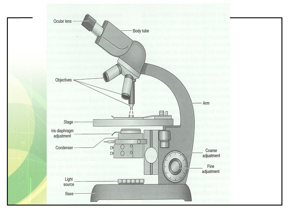
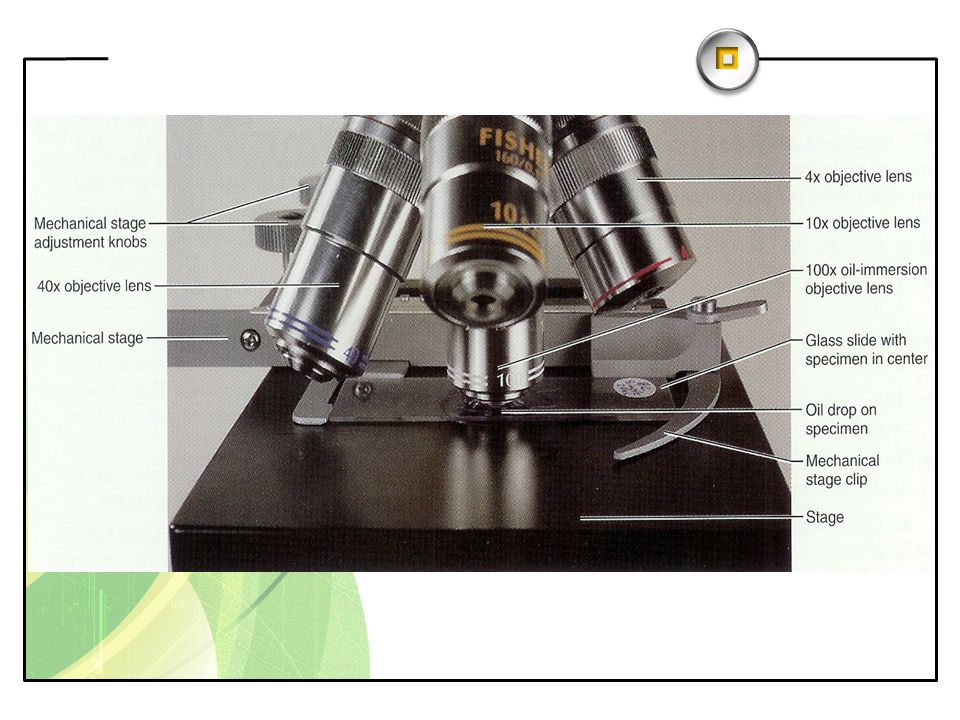
현미경 사용법

10
Micrometer Micrometer ( 측미계 ) Ocular micrometer Stage (objective) micrometer : 0.01 mm (10 µm) 길이 측정 방법
 Ocular micrometer Stage (objective) micrometer : 0.01 mm (10 µm) 길이 측정 방법")
11
Contrast ( 대비력 ) Fixation ( 고정 ) Heat fixation Chemical fixation : chemical fixatives ethanol, acetic acid, mercuric chloride, formaldehyde, glutaraldehyde Staining ( 염색 ) Dye : chromophore ( 발색단 ) Basic (cationic) dye ( 염기성 ) : 세포 염색 ( 세포 표면 : 음성 ) Acidic (anionic) dye ( 산성 ) : 바탕 염색 (negative staining) Neutral dye ( 중성 ) : 지질 등 염색 Mordant ( 매염제 ) iodine, ferrous sulfate, phenol, tannic acid Staining
 Fixation ( 고정 ) Heat fixation Chemical fixation : chemical fixatives ethanol, acetic acid, mercuric chloride, formaldehyde, glutaraldehyde Staining ( 염색 ) Dye : chromophore ( 발색단 ) Basic (cationic) dye ( 염기성 ) : 세포 염색 ( 세포 표면 : 음성 ) Acidic (anionic) dye ( 산성 ) : 바탕 염색 (negative staining) Neutral dye ( 중성 ) : 지질 등 염색 Mordant ( 매염제 ) iodine, ferrous sulfate, phenol, tannic acid Staining")
12
Basic (cationic) dye ( 염기성 염료 ) methylene blue, crystal violet, safranin, basic fuchsin, gentian violet, methyl violet, malachite green, rosaniline, hematoxylin Acidic (anionic) dye ( 산성 염료 ) nigrosin, eosin, rose bengal, erythrosin, picric acid, acidic fuchsin, orange G, India ink Neutral dye ( 중성 염료 ) Combination of acidic & basic dyes Sudan III (Sudan Black) : lipid Feulgen stain : sugar (DNA) Giemsa stain Wright stain Leishman stain Dyes
 dye ( 염기성 염료 ) methylene blue, crystal violet, safranin, basic fuchsin, gentian violet, methyl violet, malachite green, rosaniline, hematoxylin Acidic (anionic) dye ( 산성 염료 ) nigrosin, eosin, rose bengal, erythrosin, picric acid, acidic fuchsin, orange G, India ink Neutral dye ( 중성 염료 ) Combination of acidic & basic dyes Sudan III (Sudan Black) : lipid Feulgen stain : sugar (DNA) Giemsa stain Wright stain Leishman stain Dyes")
13
Staining Simple staining ( 단순염색 ). size, shape, arrangement. crystal violet, basic fuchsin, methylene blue nigrosin (negative staining) Differential staining ( 분별염색 ). stain specific cells :. Gram stain : Crystal violet & Safranin Acid-fast stain : Basic fuchsin & Methylene blue Mycobacterium. stain specific structures flagella, endospore, capsule, nuclear stain
 Differential staining ( 분별염색 ). stain specific cells :. Gram stain : Crystal violet & Safranin Acid-fast stain : Basic fuchsin & Methylene blue Mycobacterium. stain specific structures flagella, endospore, capsule, nuclear stain.")
14
Nigrosin acidic dye : negatively charged cell envelope : negatively charged negative stain no heat or chemical fixation Nigrosin stain

15
Gram stain

16
Mycobacterium tuberculosis ( 결핵균 ) mycolic acid in cell wall impenetrable by aqueous stain solutions Koch & Ehrlich simultaneously introduced a stain for M. tuberculosis Ziehl & Neelsen method modifications carbol fuchsin stain, acid alcohol decolorizer, counter stain Muller & Chermock method addition of a surfactant (wetting agent) Kinyoun method Fluorescence microscopy : auramine-rhodamine stain Preliminary diagnosis of active tuberculosis : sputum samples Acid-fast stain
 Kinyoun method Fluorescence microscopy : auramine-rhodamine stain Preliminary diagnosis of active tuberculosis : sputum samples Acid-fast stain.")
Similar presentations

 iris diaphragm.>")


 or 1000000000.>")

