Download presentation
Presentation is loading. Please wait.

1
Mammography

2
Introduction and History Breast cancer is 2nd only to lung cancer as cause of death in women –Very treatable with early detection! 1st innovation since radical mastectomy introduction in 1898 –In 1913, radiographic appearance of breast cancers was first reported Mammography became a reliable diagnostic tool in 1950s when industrial grade x-ray film introduced

3
History Of Mammography (cont’d) 1960’s – Xerography introduced – much lower dose Research conducted in 1970s clearly showed mammography to be essential part of early diagnosis 1975 – High speed/resolution film introduced by DuPont 1992 – MQSA implemented (Mammography Quality Standards Act)
 1960’s – Xerography introduced – much lower dose Research conducted in 1970s clearly showed mammography to be essential part of early diagnosis 1975 – High speed/resolution film introduced by DuPont 1992 – MQSA implemented (Mammography Quality Standards Act)")
4
Cancer that forms in tissues of breast, usually ducts (tubes that carry milk to nipple) and lobules (glands that make milk). Occurs in both men and women (male breast cancer is rare) Definition of breast cancer:
 Definition of breast cancer:.")
5
MQSA Mammography was 1st and only federally regulated imaging exam with implementation of Mammography Quality Standards Act (MQSA) Mandated following: –Formal training and continuing education –Required regular inspection of equipment –Documentation of quality assurance –Reporting results, follow-up, tracking pts, and monitoring outcomes
 Mandated following: –Formal training and continuing education –Required regular inspection of equipment –Documentation of quality assurance –Reporting results, follow-up, tracking pts, and monitoring outcomes")
6
Principles Of Breast Cancer Pt.s in early stages respond well to treatment Patients with advanced disease do poorly Earlier diagnosis, better chance of survival Mammography is tool for early detection

7
Risk v. Benefit Breast cancer in United States in 2009 (estimated): New cases: 192,370 (female); 1,910 (male) Deaths: 40,170 (female); 440 (male) Us population 306 million in 2007- 133 deaths /million Mortality risk from mammography induced radiation is 5 deaths/ million pts. using screen film mammography
: New cases: 192,370 (female); 1,910 (male) Deaths: 40,170 (female); 440 (male) Us population 306 million in deaths /million Mortality risk from mammography induced radiation is 5 deaths/ million pts. using screen film mammography.")
8
Breast Cancer Screening Very 1 st Mammogram is Baseline (or first mammo. after surgery) There after: screening mammogram pt. must be asymptomatic – no known breast problems American Cancer Society and American College of Radiology recommend screening annually for women over age 40
 There after: screening mammogram pt. must be asymptomatic – no known breast problems American Cancer Society and American College of Radiology recommend screening annually for women over age 40.")
9
Diagnostic Mammogram For woman presenting with clinical evidence of breast disease, palpable mass or other symptom Uses specific projections to –Rule out cancer –Demonstrate suspicious area seen on screening mammogram

10
Tissue Variations Breasts -glandular and connective Ability to visualize depends upon amount of fat within and around breast lobules- provides contrast Postpuberty breasts contain primarily dense connective tissue

11
During pregnancy, breasts undergo hypertrophy

12
Fatty tissue replaces glandular tissue after lactation and advancing age After menopause, glandular tissue begins to atrophy

13
Typical Mammography Unit Equipment is C-arm SID is fixed at 24 – 26”

14
Mammography Equipment Dedicated units have high-frequency generators Provide more precise control of kVp, mA, and exposure time Specially designed to produce high- contrast and high-resolution images

15
Mammography uses Low kVp : 25 – 28 AEC Anode material made of molybdenum, with rhodium target Grid with ratio: 4:1, or 5:1 200 lines/inch

16
Magnification Increases visibility of small structures Increase OID Uses air gap Radiation dose increases with magnification

17
Compression Device Compression decreases thickness of breast, magnification and scatter Increases contrast Reduces motion unsharpness Reduces dosage

18
Compression Device Made of firm plastic Amount of compression: between 25 and 40 pounds pressure Compression may be uncomfortable!

19
Screen-Film Systems Mammography cassettes contain a single screen Film is single emulsion Occasionally, extended time processing is used –(reduces dose and increases contrast)
")
20
Digital Mammography State of the art! No film or chemical processing Images easily sent over internet Much better definition Possible downside-if 1st digital compared to previous film mammo., can give false positives due to increased sensitivity! - Slightly higher dosage

21
Procedure Complete, careful history and physical assessment –Take notes on location of scars, palpable masses, skin abnormalities, and nipple alterations Examine previous mammograms for positioning, compression, and exposure factors

22
Procedure (con’t) Patients dress in open-front gown Breasts must be bared for imaging –Cloth will cause image artifact Remove deodorant and powder from axilla and breast –Can mimic calcifications on image
 Patients dress in open-front gown Breasts must be bared for imaging –Cloth will cause image artifact Remove deodorant and powder from axilla and breast –Can mimic calcifications on image")
23
Procedure (cont’d) Explain procedure to pt., including possibility for additional projections Consider natural mobility of breast before positioning Support breast firmly so that nipple is directed forward Profile nipple, if possible
 Explain procedure to pt., including possibility for additional projections Consider natural mobility of breast before positioning Support breast firmly so that nipple is directed forward Profile nipple, if possible")
24
Procedure Apply proper compression to produce uniform breast thickness –Essential to high-quality mammograms Place ID markers according to standard convention

25
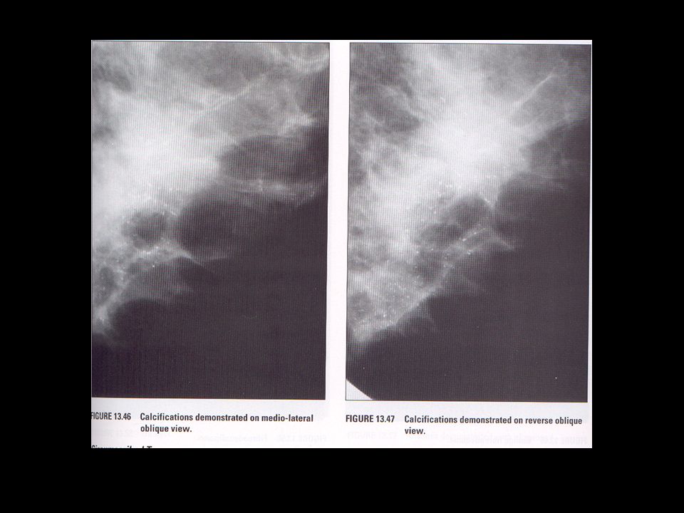
Routine mammography projections Craniocaudal (CC) Mediolateral oblique (MLO)
 Mediolateral oblique (MLO)")
26
Radiography Of Augmented Breast (implants) 8 projections must be obtained (2x4) MRI and sonography can help determine rupture or leakage Four standard images with implant displaced posteriorly into chest wall are obtained
 8 projections must be obtained (2x4) MRI and sonography can help determine rupture or leakage Four standard images with implant displaced posteriorly into chest wall are obtained")
27
Breast Implants

28
Saline vs Silicone Some surgeons feel silicone implants have a more natural look and feel because silicone gel texture similar to breast tissue. Silicone implant ruptures are harder to detect. When saline implants rupture, they deflate -results are seen almost immediately. When silicone implants rupture, breast often looks and feels same because silicone gel may leak into surrounding areas of breast without a visible difference. Replacing a ruptured silicone gel implant is more difficult than repairing saline implant. Silicone implants have higher rate of capsular contracture (scarring and hardening around implant). Saline implants inflated to desired size with saline, then valve is sealed by surgeon
. Saline implants inflated to desired size with saline, then valve is sealed by surgeon.")
29
Radiography Of Augmented Breast (implants) Complications: Increased fibrous tissue surrounding implant (contracture) Shrinking Hardening Leakage Pain
 Complications: Increased fibrous tissue surrounding implant (contracture) Shrinking Hardening Leakage Pain")
30
Male Mammography Approximately 1000 males develop breast cancer every year Standard CC and MLO are obtained Males not screened- mammogram only if lump discovered

31
Treatment For Breast Cancer Lumpectomy Partial or radical mastectomy Radiation Chemotherapy

32
CC view ( lesion)
")
33
Needle Localizations Used to localize breast lesions before surgery Special, open-hole plate may be used for ease of localization –Plate contains grid to plot coordinates –Operative stereotactic surgery may be used Two offset images are obtained to create a 3- dimensional image

34
Needle Localization

35
Breast Specimen Radiography Performed after surgery once lump has been excised Determines extent of calcifications within specimen Magnification technique may be used

36
Breast Specimen Radiograph

38
19 yr. old (never pregnant) 24 yr. old (has children)
 24 yr. old (has children)")
39
Calcified Milk Ducts

41
Various abnormal mammograms

42
The End

Similar presentations


 cells are found in the breast tissue. This can happen to males and females. Worldwide, breast cancer is.>")


