Download presentation
Presentation is loading. Please wait.

1
Helicobacter pylori and gastric ulcers

2
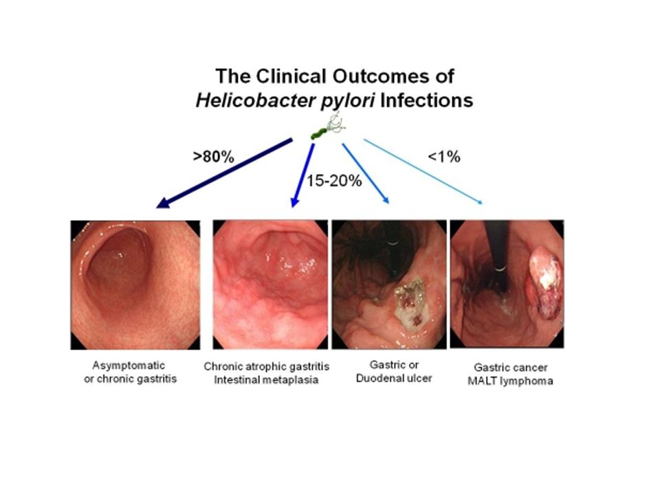
Helicobacter pylori (H. pylori) is a spiral shaped bacterium that lives on the lining of the stomach; inflammation in lining called GASTRITIS, extended to inflammation in epithelial cells of stomach causing ULCER.
 is a spiral shaped bacterium that lives on the lining of the stomach; inflammation in lining called GASTRITIS, extended to inflammation in epithelial cells of stomach causing ULCER..")
3
Genome 1.7 mbp genome Four regulatory sites in genome

4
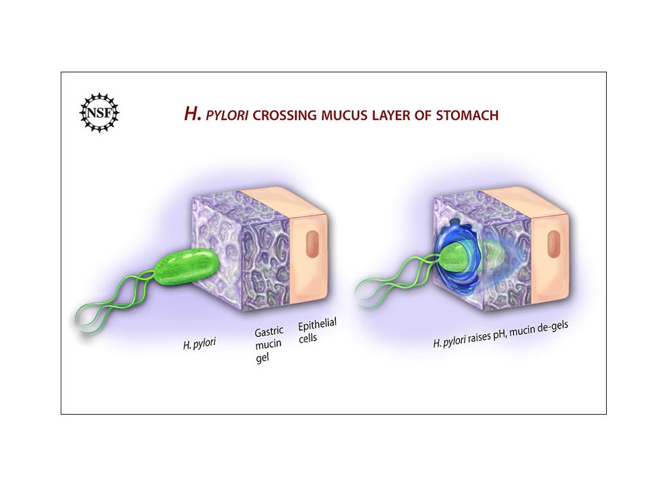
1. Colonization in Stomach MOTILITY Is sensitive to acidic condition so doesn’t colonize in lumen but rather colonize in mucin layer of gastric mucosa Mucus resists the diffusion of protons because it is composed of negatively charged sulfated polysacchrides Mucus act as buffer to maintain the slightly alkaline condition on mucosal surface thus protecting the mucosal cells from extensive acids Bacteria use this advantage of the property of mucin

6
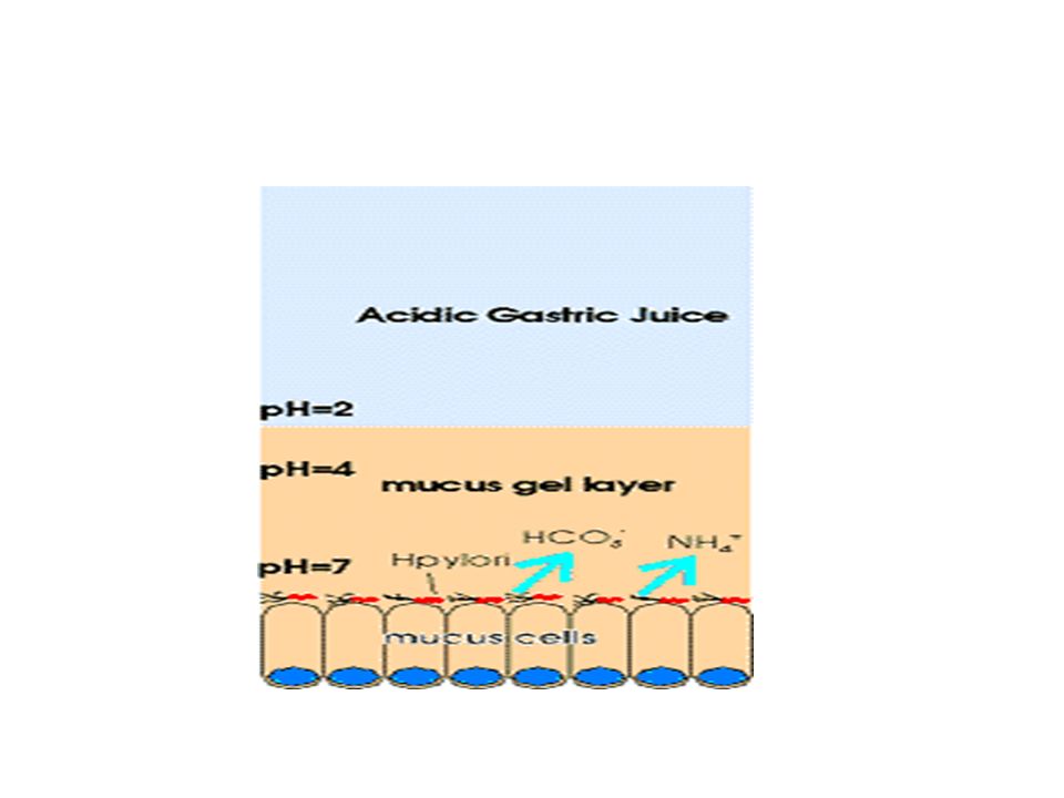
Urease, temporary protection H. pylori is able to fight off the stomach acid that does reach it with the enzyme urease. Urease converts urea secreted by gastric cells into bicarbonate and ammonia, which are strong bases. These acid neutralizing chemicals around the H. pylori protect if from the acid in the stomach. Ammonia is toxic to eukaryotic cells having inflammatory effect on gastric mucosa

9
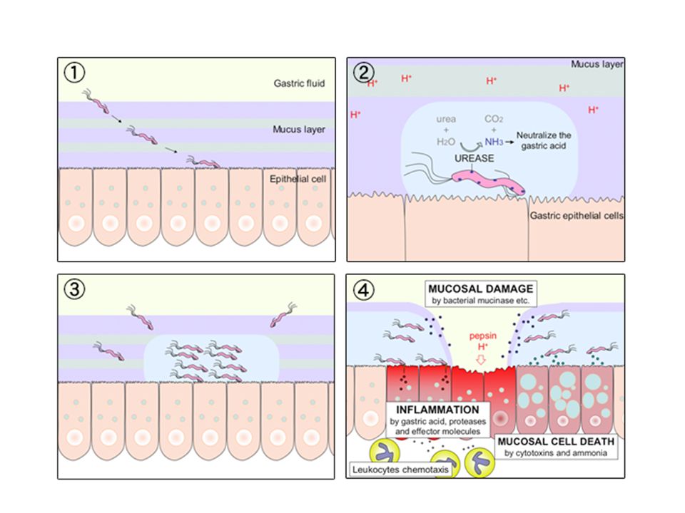
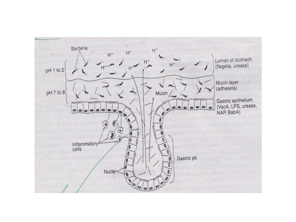
2. Persistence in Mucin Layer Mucus is continuously produced and sloughed into lumen of stomach, flagella burrows in viscous mucin layer Specimen from infected person showed bacteria adherent to muciin layer as they have an adhesin that binds to sulfated mucin sugars. H. pylori also produces adhesins that help them to attach to epithelial cells BabA BabA recognizes the lewis B Ag a carbohydrate Ag found on the surface of epithelial cells Although most of the bacteria don't attach to epithelial cells, few that do elicit inflammatory response tends to ULCER Urease and flagella are found in all spp but BabA is found in few

10
To avoid the acidic environment of the interior of the stomach (lumen), H. pylori uses its flagella to burrow into the mucus lining the stomach to reach the epithelial cells underneath, where there is a more neutral pH. H. pylori is able to sense the pH gradient in the mucus and move towards the less acidic region (chemotaxis).lumenepithelial cellschemotaxis H. pylori is found in the mucus, on the inner surface of the epithelium, and occasionally inside the epithelial cells themselves. It adheres to the epithelial cells by producing adhesins, which bind to lipids and carbohydrates in the epithelial cell membrane.adhesinscell membrane
.lumenepithelial cellschemotaxis H. pylori is found in the mucus, on the inner surface of the epithelium, and occasionally inside the epithelial cells themselves. It adheres to the epithelial cells by producing adhesins, which bind to lipids and carbohydrates in the epithelial cell membrane.adhesinscell membrane.")
12
Immune response H. pylori can also survive because the body’s natural defenses cannot reach the bacterium in the mucus lining of the stomach. Inflammation in epithelium results in infiltraition of PMNs, T cells and B cells. (VacA) and Neutrophil activation protein (NAP) released by Helicobacter pylori activates neutrophils. All strains of Helicobacter pylori express the secreted virulence factors vacuolating cytotoxin (VacA) to directly inhibit T cell activation, proliferation and effector functions. The immune system responds to an H. pylori infection by sending white cells, killer T cells, and other infection fighting agents, but they cannot reach the infection because they cannot easily get through the stomach lining.
 and Neutrophil activation protein (NAP) released by Helicobacter pylori activates neutrophils. All strains of Helicobacter pylori express the secreted virulence factors vacuolating cytotoxin (VacA) to directly inhibit T cell activation, proliferation and effector functions. The immune system responds to an H. pylori infection by sending white cells, killer T cells, and other infection fighting agents, but they cannot reach the infection because they cannot easily get through the stomach lining..")
13
Transmission It is thought that H. pylori is transmitted orally by ingestion of food or water contaminated with fecal matter. It is possible that H. pylori could be transmitted from the stomach to the mouth through gastro-esophagal reflux, or burping.

14
Diagnosis There are three diagnostic tests that can be done to test for the bacteria. *Breath tests *Blood tests *Endoscopy

15
Breath Tests There are two forms of the diagnostic breath test, the Carbon-14 and the Carbon-13 urea breath tests. Patients swallow urea labelled with an uncommon isotope either radioactive carbon-14 or non-radioactive carbon-13. In the subsequent 10–30 minutes, the detection of isotope- labelled carbondioxide in exhaled breath indicates that the urea was split; this indicates that urease (the enzyme that H. pylori uses to metabolize urea) is present in the stomach, and hence that H. pyloribacteria are present. The reaction of urea Hydrolysis is important for diagnosis of H. pylori by the breath test.
 is present in the stomach, and hence that H. pyloribacteria are present. The reaction of urea Hydrolysis is important for diagnosis of H. pylori by the breath test..")
16
Blood test & Endoscopy A blood test can also be done to test for antibodies which stick to H. pylori. An endoscopy is another diagnostic test for H. pylori where a biopsy is taken and tested for the bacterium using a gram stain.

17
Symptoms The main symptom is a burning or gnawing pain in the epigastrum, usually when the stomach is empty, between meals, or in the early morning. These symptoms can last for minutes to hours and may be relieved by eating or taking antacids.

19
Treatment Antibiotics such as amoxicillin or tetracycline are used to cure ulcers. Therapy is 1-2 weeks of one or two antibiotics and a medicine that will reduce the acid in the stomach. The acid suppressor is paired with the antibiotics because it helps alleviate ulcer- related symptoms, and helps heal gastric mucosal inflammation.

20
Vaccines Vaccines are being tested on animals and humans, but currently there are no vaccines for the prevention of H. pylori.

Similar presentations


![Peptic Ulcer Disease Dr Maha Arafah. Objectives Upon completion of this lecture the students will : A] Understand the Pathophysiology of acute and chronic.](/13/3809458/big_thumb.jpg "Peptic Ulcer Disease Dr Maha Arafah. Objectives Upon completion of this lecture the students will : A] Understand the Pathophysiology of acute and chronic.>")


![BY RANJEET RAMAN GRAM NEGATIVE BACILLI- MICRO {ST1]](/15/4636473/big_thumb.jpg "BY RANJEET RAMAN GRAM NEGATIVE BACILLI- MICRO {ST1]>")

>")

 Stomach ulcer or peptic ulcer is the damage of the protective layer (lining) of stomach or gastrointestinal tract It may be.>")


