Download presentation
Presentation is loading. Please wait.

1
* Failure of laboratory personnel to document the time a semen sample is collected primarily affects the interpretation of semen: * Appearance * Volume * Ph * Viscosity * A semen specimen delivered to the laboratory in a condom has a normal sperm count and markedly decreased sperm motility. This is indicative of: * Decreased fructose * Antispermicide in the condom * Increased semen viscosity * Increased semen alkalinity * Photolytic enzymes may be added to semen specimens to: * Increase the viscosity * Dilute the specimen * Decrease the viscosity * Neutralize the specimen

2
Lab 11, 12

3
Serous fluids are fluids within the closed cavities of the body. These cavities are lined by an adjacent membrane, which forms a double layer of mesothelial cells, called the serous membrane. The cavities are the pleural (around the lungs), pericardial (around the heart), and peritoneal (around the abdominal and pelvic organs). A small amount of serous fluid fills the space between the two layers and lubricate the surfaces of these membranes as they move against each other. The fluids are ultrafiltrate of plasma, which continuously formed and reabsorbed at a constant rate, leaving only a very small volume within the cavities. An increased volume of any of these fluids is referred to as an effusion. Effusions may be either transudates or exudates. Exudates are usually effusions, which result from conditions that directly affect the membranes lining the serous cavity.
, pericardial (around the heart), and peritoneal (around the abdominal and pelvic organs). A small amount of serous fluid fills the space between the two layers and lubricate the surfaces of these membranes as they move against each other. The fluids are ultrafiltrate of plasma, which continuously formed and reabsorbed at a constant rate, leaving only a very small volume within the cavities. An increased volume of any of these fluids is referred to as an effusion. Effusions may be either transudates or exudates. Exudates are usually effusions, which result from conditions that directly affect the membranes lining the serous cavity..")
4
Serous fluids are formed as ultrafiltrate of plasma, with no additional material contributed by the membrane cells. The small amount of protein is removed by the lymphatic system. Production and reabsorption are subject to hydrostatic & colloidal (oncotic) pressures from the capillaries serving the cavities. Under normal conditions, colloidal pressure from serum proteins is the same in the capillaries on both sides of the membrane. Therefore, the greater hydrostatic in the systemic capillaries on the parietal side favors fluid production through the parietal membrane and reabsorption through the visceral membrane.
 pressures from the capillaries serving the cavities. Under normal conditions, colloidal pressure from serum proteins is the same in the capillaries on both sides of the membrane. Therefore, the greater hydrostatic in the systemic capillaries on the parietal side favors fluid production through the parietal membrane and reabsorption through the visceral membrane..")
7
1. Increased capillary hydrostatic pressure Congestive heart failure Salt and fluid retention 2. Decreased oncotic pressure Nephrotic syndrome Hepatic cirrhosis Malnutrition Protein-losing enteropathy 3. Increased capillary permeability Microbial infections Membrane inflammations Malignancy 4. Lymphatic obstruction Malignant tumors, lymphomas Infection and inflammation Thoracic duct injury

8
Fluids for laboratory examination are collected by needle aspiration from the respective cavities. These aspiration procedures are referred to as thoracentesis (pleural), pericardiocentesis (pericardial), and paracentesis (peritoneal). Abundant fluid (greater than 100 mL) is usually collected; therefore, suitable specimens are available for each section of the laboratory. An ethylenediaminetetraacetic acid (EDTA) tube is used for cell counts and the differential. Sterile heparinized evacuated tubes are used for microbiology and cytology.
, pericardiocentesis (pericardial), and paracentesis (peritoneal). Abundant fluid (greater than 100 mL) is usually collected; therefore, suitable specimens are available for each section of the laboratory. An ethylenediaminetetraacetic acid (EDTA) tube is used for cell counts and the differential. Sterile heparinized evacuated tubes are used for microbiology and cytology..")
10
For better recovery of microorganisms and abnormal cells, concentration of large amounts of fluid is performed by centrifugation. Chemistry tests can be run on clotted specimens in plain tubes or on heparinized tubes. Specimens for pH must be maintained anaerobically in ice. Chemical tests performed on serous fluids are frequently compared with plasma chemical concentrations because the fluids are essentially plasma ultrafiltrates. Therefore, blood specimens should be obtained at the time of collection.

11
In human anatomy, the pleural cavity is a body cavity containing the lungs; the lungs are surrounded by two serous membranes, the pleurae. The outer pleura (parietal pleura) covers and is attached to the chest wall. The inner pleura (visceral pleura) covers and is attached to the lung and other structures, i.e. blood vessels, bronchi and nerves. Between the two is a thin space known as the pleural space, which normally contains a small amount of pleural fluid
 covers and is attached to the chest wall. The inner pleura (visceral pleura) covers and is attached to the lung and other structures, i.e. blood vessels, bronchi and nerves. Between the two is a thin space known as the pleural space, which normally contains a small amount of pleural fluid.")
14
When there is an excess fluid accumulation in the pleural cavity, this is called pleural effusion, which may be transudates, exudates or fluid from extra pleural origin such as: 1.Ruptured esophagus which is characterized by increase fluid amylase and decrease of PH. 2.Pancreatitis which is characterized by increase amylase.

15
Effusion that forms because of systemic disorder that disrupts the balance in the regulation of fluid filtration and reabsorption such as: 1. The changes in the hydrostatic pressure (increasing) created by a mechanical process such as congestive heart failure (CHF) or by pulmonary embolism. 2. Decrease the plasma oncotic pressure such as nephrotic syndrome or hepatic cirrhosis
 created by a mechanical process such as congestive heart failure (CHF) or by pulmonary embolism. 2. Decrease the plasma oncotic pressure such as nephrotic syndrome or hepatic cirrhosis.")
16
Effusions that are produced by conditions that directly involve the membranes of the particular cavity (from an inflammatory process which including infections and malignancies) that leads to: 1. Increased capillary permeability. 2. Decreased lymphatic resorption.

18
Volume: 1-15 ml Color and Appearance: 1. Transudates, Clear, Pale Yellow. 2. Exudates, cloudy, opaque appearance indicates more cell components. 3. Bloody fluid Hemothorax, Hemorrhagic effusion, Pulmonary embolis, Tuberculosis, and Malignancy

19
To differentiate between a hemothorax and hemorrhagic exudate, a hematocrit can be run on the fluid. If the blood is from a hemothorax, the fluid hematocrit is more than 50% of the whole blood hematocrit, because the effusion is actually occurring from the inpouring of blood from the injury. A chronic membrane disease effusion contains both blood and increased pleural fluid, resulting in a much lower hematocrit.

20
4. Milky Chylous Pseudochylous Differentiation Between Chylous and Pseudochylous Pleural

21
5. Black fluid: Aspergillus niger (fungi) infection 6. Purulent fluid: Indicates infection 7. Turbid and greenish yellow : Rheumatoid effusion 8. Viscous Malignant mesothelioma (increased hyaluronic acid)
.")
23
RBC’s Little value WBC’s Total lower than 1000/µl LE cells Macrophages Mesothelial cells Total RBCs count RBCs (5000-6000) are needed to give red appearance to pleural fluid RBCs > 100.000 is grossly hemorrhagic and suggests malignancy, pulmonary infarct, or trauma but occasionally seen in congestive heart failure alone. Hemothorax suggests trauma, bleeding from a vessel, bleeding disorder, or malignancy.

24
Total WBC count Transudates are usually > 1000/µl WBC’s >10.000 /µl indicates inflammation, most commonly with pneumonia, pulmonary infarct, Pancreatitis. WBC’s > 50.000 /µl is typical only in Para pneumonic effusions, usually empyema In malignancy & tuberculosis are usually < 5000 /µl.

25
Mononuclear cells predominate in transudates and early effusions and chronic exudates. PMNs predominate in early inflammatory effusion neutrophil: 90% in the following Acute inflammation due to pneumonia pulmonary infection Pancreatitis After several days, mesothelial cells, macrophage, lymphocytes may be predominating.

26
Lymphocyte (80- 90%) increased in the following cases: Tuberculosis pneumonia True Chylous S.L.E Uremic effusion Subacut inflammation Eosinophilia : Eosinophilie in pleural fluid( > 10% of total WBC) is of diagnostically significant Pneumothorax. Post pneumonia effusion. Chest trauma. Pulmonary infection. Congestive heart failure. S.L.E.

27
LE cells: occasionally LE cells make the diagnosis of SLE. Mesothelial cells: Normal and reactive forms have no clinical significance Decreased mesothelial cells are associated with tuberculosis Plasma cells: Tuberculosis Malignant cells: Primary adenocarcinoma Small cell carcinoma

29
1. Protein and LDH To differentiate transudates from exudates. Protein electrophoresis shows an elevation of albumin & absence of fibrinogen in comparison to that of plasma. 2. Glucose Same as serum value in transudates. Usually normal, but if it lowers than 60 mg\dl may be found in: 1. Rheumatoid arthritis 2. Empyema 3. Malignancy 4. TB 5. Esophageal rupture 6. SLE

30
3. Amylase Increase in acute pancreatitis (may reach 2 times plasma amylase) Perforated peptic ulcer. Necrosis of small intestine. Some times in metastatic cancer and esophageal ruptured. 4. Lipids Triglycerides Lipoproteins Cholesterol.
 Perforated peptic ulcer. Necrosis of small intestine. Some times in metastatic cancer and esophageal ruptured. 4. Lipids Triglycerides Lipoproteins Cholesterol..")
31
5. PH Pleural fluid pH lower than 7.0 may indicate the need for chest-tube drainage, in addition to administration of antibiotics in cases of pneumonia. In cases of acidosis, the pleural fluid pH should be compared to the blood pH. Pleural fluid pH at least 0.30 degrees lower than the blood pH is considered significant. The finding of a pH as low as 6.0 indicates an esophageal rupture that is allowing the influx of gastric fluid. 6. ADA (adenosine deaminase) levels over 40 U/L are highly indicative of tuberculosis. They are also frequently elevated with malignancy.
 levels over 40 U/L are highly indicative of tuberculosis. They are also frequently elevated with malignancy..")
32
Used to differentiate effusions of immunologic and malignant origin from those of non inflammatory and non malignant origin. The tests includes: Tumor Marker : CEA (60-70% of lung cancer), 40-50% of other malignancies. The CEA test measures the level of carcinoembryonic antigen (CEA) in the blood. CEA is a protein normally found in the tissue of a developing baby in the womb. The blood level of this protein disappears or becomes very low after birth. In adults, an abnormal level of CEA may be a sign of cancer. RF, complement, ANF, immunoglobulin Increased levels of immunoglobulins and CEA or decreased complement is indicative of inflammatory and neoplastic reaction.
, 40-50% of other malignancies. The CEA test measures the level of carcinoembryonic antigen (CEA) in the blood. CEA is a protein normally found in the tissue of a developing baby in the womb. The blood level of this protein disappears or becomes very low after birth. In adults, an abnormal level of CEA may be a sign of cancer. RF, complement, ANF, immunoglobulin Increased levels of immunoglobulins and CEA or decreased complement is indicative of inflammatory and neoplastic reaction..")
33
Gram stain, acid-fast stain, cultures.

35
The pericardial space enclosing the heart normally contains about 25 to 50 mL of a clear, straw colored ultrafiltrate of plasma, called pericardial fluid. When an abnormal accumulation of pericardial fluid occurs, it fills up the space around the heart and can mechanically inhibit the normal action of the heart., In this case, immediate aspiration of the excess fluid is indicated.

36
Pericardial effusion is usually caused by: 1.Infection: Which may be bacterial, tuberculosis, fungal or viral. 2.Neoplasm: Which may be due to metastatic carcinoma or lymphoma. 3.Myocardial infarction. 4.Hemorrhage due to trauma. 5.SLE. Sample collection called pericardiocentesis

37
Volume 10-50ml Appearanceclear pale yellow. Bloody due to T.B., or other wide variety of diseases Milky (chylous and pseudochylous). Laboratory tests Tests performed on pericardial fluid are primarily directed at determining if the fluid is a transudate or an exudate
. Laboratory tests Tests performed on pericardial fluid are primarily directed at determining if the fluid is a transudate or an exudate.")
38
WBCs: Little clinical value, although a count of greater than 1000 WBCs/mm 3 with a high percentage of neutrophils can be indicative of bacterial endocarditis. LE cells Cytological examination of pericardial exudates for the presence of malignant cells is an important part of the fluid analysis. Cells most frequently encountered are the result of metastatic lung or breast carcinoma.

39
Protein (little value in differential diagnosis. Glucose. Lipids Triglycerides Lipoproteins Cholesterol Serology ANA, CEA Microbiology Gram stain, acid fast stain and cultures.

41
Accumulation of fluid between the peritoneal membranes is called ascites, and the fluid is commonly referred to as ascetic fluid rather than peritoneal fluid.

42
Normal saline is sometimes introduced into the peritoneal cavity to act as a lavage for the detection of abdominal injuries that have not yet resulted in the accumulation of fluid. Peritoneal lavage is a sensitive test for the detection of intra-abdominal bleeding in blunt trauma cases, and results of the RBC count can be used along with radiographic procedures to aid in determining the need for surgery. RBC counts > 100,000/µL are indicative of blunt trauma injuries.

43
Accumulation of peritoneal is a common complication in many diseases which may be: Transudate due to: 1. Congestive heart failure 2. Constrictive pericarditis 3. Hypoproteinemia 4. Nephrotic syndrome 5. Liver cirrhosis Exudate due to: 1. Peritoneal malignancy 2. Tuberculous peritonitis. 3. Pancreatic ascites. 4. Billie peritonitis. 5. Trauma.

44
Volume: lower than 50 ml. Appearance:clear pale yellow. Turbidity Appendicitis Pancreatitis Strangulated intestine Ruptured bovel Bacterial peritonitis Milky Chylous Pseudochylous. Greenish Perforated duodenal ulcer Perforated intestine Chlocystitis Perforated gall bladder Acute pancreatitis

45
Normal WBC counts are less than 350 cells/µL, and the count increases with bacterial peritonitis and cirrhosis. To distinguish between those two conditions, an absolute neutrophil count should be performed. An absolute neutrophil count greater than 250 cells/µL or greater than 50% of the total WBC count is indicative of infection. Lymphocytes are the predominant cell in tuberculosis. Examination of ascitic exudates for the presence of malignant cells is important for the detection of tumors of primary and metastatic origin. Malignancies are most frequently of gastrointestinal, prostate, or ovarian origin. Cells present in ascitic fluid include leukocytes, abundant mesothelial cells, and macrophages.

46
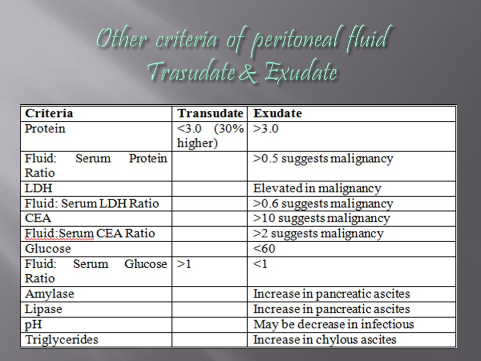
1. Protein 2. Glucose Decreased in tubercular peritonitis and malignancy 3. Amylase Increased in pancreatitis, gastrointestinal perforation 4. ALP An elevated alkaline phosphatase level is also highly diagnostic of intestinal perforation.

47
5. CEA and CA 125 Measurement of the tumor markers CEA and CA 125 is a valuable procedure for identifying the primary source of tumors producing ascitic exudates. The presence of CA 125 antigen with a negative CEA suggests the source is from the ovaries, fallopian tubes, or endometrium 6. Urea nitrogen, ammonia and creatinine in the fluid are requested when a ruptured bladder or accidental puncture of the bladder during the paracentesis is of concern.

48
Differentiation between ascitic fluid transudates and exudates is more difficult than for pleural and pericardial effusions. The serum-ascites albumin gradient (SAAG) is recommended over the fluid: serum total protein and LD ratios for the detection of transudates of hepatic origin Fluid and serum albumin levels are measured concurrently, and the fluid albumin level is then subtracted from the serum albumin level.
 is recommended over the fluid: serum total protein and LD ratios for the detection of transudates of hepatic origin Fluid and serum albumin levels are measured concurrently, and the fluid albumin level is then subtracted from the serum albumin level..")
49
A difference (gradient) of 1.1 or greater suggests a transudate effusion of hepatic origin, and lower gradients are associated with exudative effusions. Serum albumin- Fluid albumin = 3.8 mg/dL -1.2 mg/dL Gradient = 2.6 in transudate Serum albumin- Fluid albumin = 3.8 mg/dL-3 mg/dL Gradient = 0.8 in exudate

51
Microbiology Gram stain, acid fast stain, culture

Similar presentations


 Interstitial fluid (surrounds the.>")


 LECTURE TWO Dr. Essam H. Jiffri.>")
 LECTURE ONE Dr. Essam H. Jiffri.>")
 Dr. Essam H. Jiffri.>")


. -Cytological tests (>")

 The fluid is a plasma filtrate.>")


