Download presentation
Presentation is loading. Please wait.

2
Dr. Seyed Amir Farzam Associated prof. of Qazvin Univercity, Medical Faculty

3
Small-bowel tumors comprise <3% of gastrointestinal neoplasms. Because of their rarity, a correct diagnosis is often delayed

4
SMALL INTESTINE TUMORS Benign Epithelial Tumors Malignant Epithelial Tumors Lympho- proliferative disorders Mesenchymal Tumors Brunner Gland Lesions Benign Intestinal polyp T cell B cell GIST Fatty tumors Neural tumors Para gangl. Smooth Ms tumors Vasc. tumors Lipoma Liposarcoma Schwannoma Neurofibroma Granular cell tumor Leiomyoma Leiomyosarcoma Haemangioma Angiosarcoma Lymphangioma Kaposi sarcoma Benign Malignant Enteropathy associated T-cell lymphoma Diffuse large cell lymphoma. Small non cleaved cell lymphoma. MALT cell lymphoma. Mantle cell lymphoma. Immuonoproliferative small intestine disease Prim. Adca Metastasis. Carcinoid. Adenomas Hamartomas

5
Adenoma Leiomyoma Angioma Lipomas other s

6
Adenocarcinoma Lymphomas Leiomyosarcomas others Carcinoid Tumors

7
not truly neoplastic a hypertrophy or hyperplasia of submucosal duodenal glands appear as small nodules in the duodenal mucosa that secrete a highly viscous alkaline mucus an incidental radiographic finding not associated with any specific clinical disorder

8
Polypoid Adenomas About 25% of benign small-bowel tumors Gardner's syndromeFAP the sessile or papillary form of the tumor is sometimes associated with a coexisting carcinoma Hamartomatous polyps In Peutz-Jeghers (not adenoma)
")
9
they frequently cause intestinal bleeding. They may take the form of telangiectasia or hemangiomas Multiple intestinal telangiectasias occur in a nonhereditary form confined to the gastrointestinal tract or as part of the hereditary Osler-Rendu-Weber syndrome of isolated hemangiomas, most commonly in the jejunum

10
Adenoma in duodenum

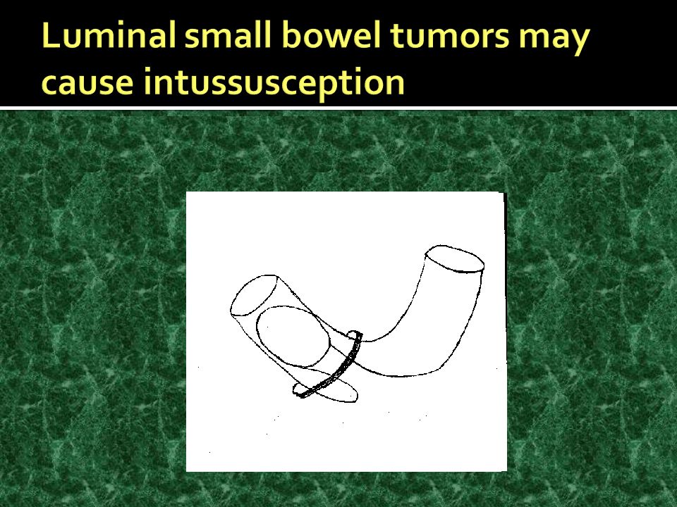
11
A, Film from an enteroclysis demonstrating a smooth, submucosal lesion that was found to be lipoma (arrow). B, Surgical resection specimen of a lipoma from another patient who presented with intussusception and bleeding

12
Small bowel follow through examination demonstrates a smooth, well-circumscribed mass arising from the wall of the terminal ileum. The appearance is consistent with a benign mesenchymal tumor, such as a lipoma or a carcinoid tumor

13
Small bowel follow through examination shows a polypoid eccentric mass arising from the wall of the terminal ileum (arrow).
.")
14
long-standing regional enteritis celiac sprue AIDS

15
The most common primary cancers of the small bowel are adenocarcinomas, accounting for ~50% of malignant tumors most often in the distal duodenum and proximal jejunum, where they tend to ulcerate and cause hemorrhage or obstruction Radiologically, they may be confused with chronic duodenal ulcer disease or with Crohn's disease

16
S.I. ADENOCARCINOMA

17
Film from a small bowel follow through demonstrating an “apple-core” appearance caused by a metastatic lesion to the small intestine from a scirrhous gastric cancer.

18
Upper gastrointestinal endoscopy shows a duodenal adenocarcinoma in the second portion of the duodenum in a patient who presented with heme positive stool. The mass occupied approximately 50 percent of the diameter of the duodenum. The thick erythematous folds in the upper half of the image distinguish the lesion from the pale, thin folds of the normal tissue in the lower half.

19
Histologic confirmation of lymphoma Normal peripheral blood smear or on bone marrow aspiration and biopsy No palpable adenopathy No hepatosplenomegaly No evidence of lymphoma is seen on: Chest radiograph CT scan primary intestinal lymphoma

20
Involvement of the intestine by a lymphoid malignancy extending from involved retroperitoneal or mesenteric lymph nodes

21
suspected from the appearance on contrast radiographs of patterns such as infiltration and thickening of mucosal folds, mucosal nodules, areas of irregular ulceration, or stasis of contrast materia Intestinal lymphoma can occasionally be diagnosed by peroral intestinal mucosal biopsy, but since the disease mainly involves the lamina propria, full-thickness surgical biopsies are usually required. The diagnosis can be confirmed by surgical exploration and resection of involved segments

23
Barium enema shows a large soft tissue mass in the cecum (arrows) caused by intussusception of a lymphoma arising in the terminal ileum
 caused by intussusception of a lymphoma arising in the terminal ileum")
24
diffusely involves the entire intestine B cell tumor chronic diarrhea and steatorrhea associated with vomiting and abdominal cramps; clubbing of the digits presence in the blood and intestinal secretions of an abnormal IgA that contains a shortened - heavy chain and is devoid of light chains clinical course of exacerbations and remissions

25
The use of oral antibiotics such as tetracycline appears to be beneficial in the early phases of the disorder, suggesting a possible infectious etiology. Combination chemotherapy has been administered during later stages of the disease, with variable results

26
Carcinoid tumors Carcinoid tumors arise from argentaffin cells of the crypts of Lieberkühn and are found from the distal duodenum to the ascending colon, areas embryologically derived from the midgut. More than 50% of intestinal carcinoids are found in the distal ileum, with most congregating close to the ileocecal valve. Most intestinal carcinoids are asymptomatic and of low malignant potential, but invasion and metastases may occur, leading to the carcinoid syndrome

27
S.I. CARCINIOD

28
Leiomyosarcomas often are >5 cm in diameter and may be palpable on abdominal examination Bleeding, obstruction, and perforation are common Such tumors should be analyzed for the expression of mutant c- kit receptor (defining GIST

29
Capsule endoscopy view of an ulcerated mass in a patient who presented with gastrointestinal bleeding. Four ulcerated, bleeding masses were found throughout the small bowel; these were confirmed at surgery and found to be sarcomas

Similar presentations


 bowel habit change (-) bearing down sensation PMHx. hemorrhoidectomy,>")


>")


