Download presentation
Presentation is loading. Please wait.

1
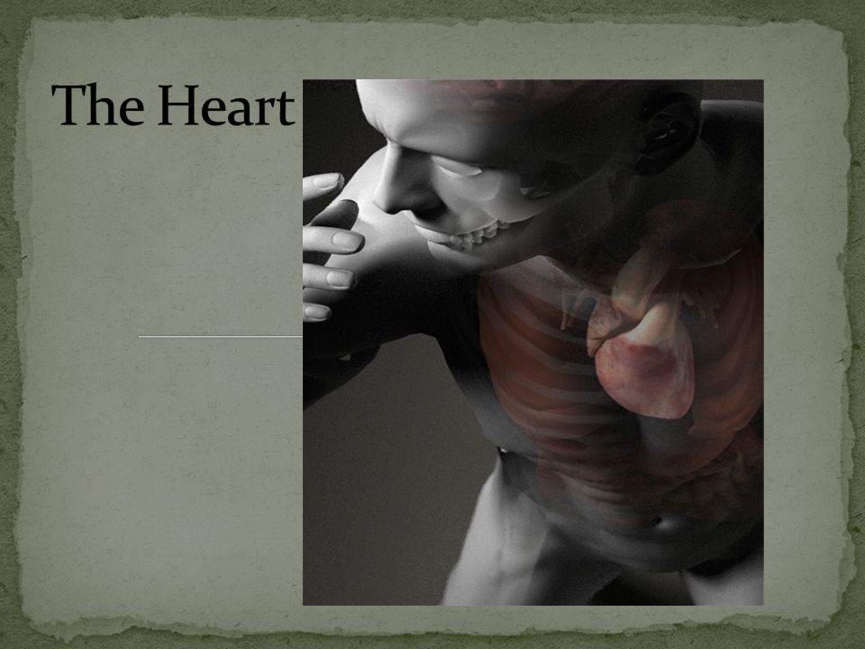
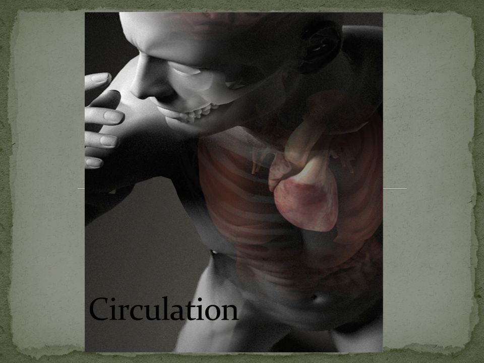
Unit 2 Seminar: Heart & Circulation

3
Chambers 2 upper chambers: R/L Atria 2 Lower Chambers: R/L Ventricles Wall Cardiac Muscle: Myocardium Lining Epithelial lining: Endocardium, includes cardiac valves

5
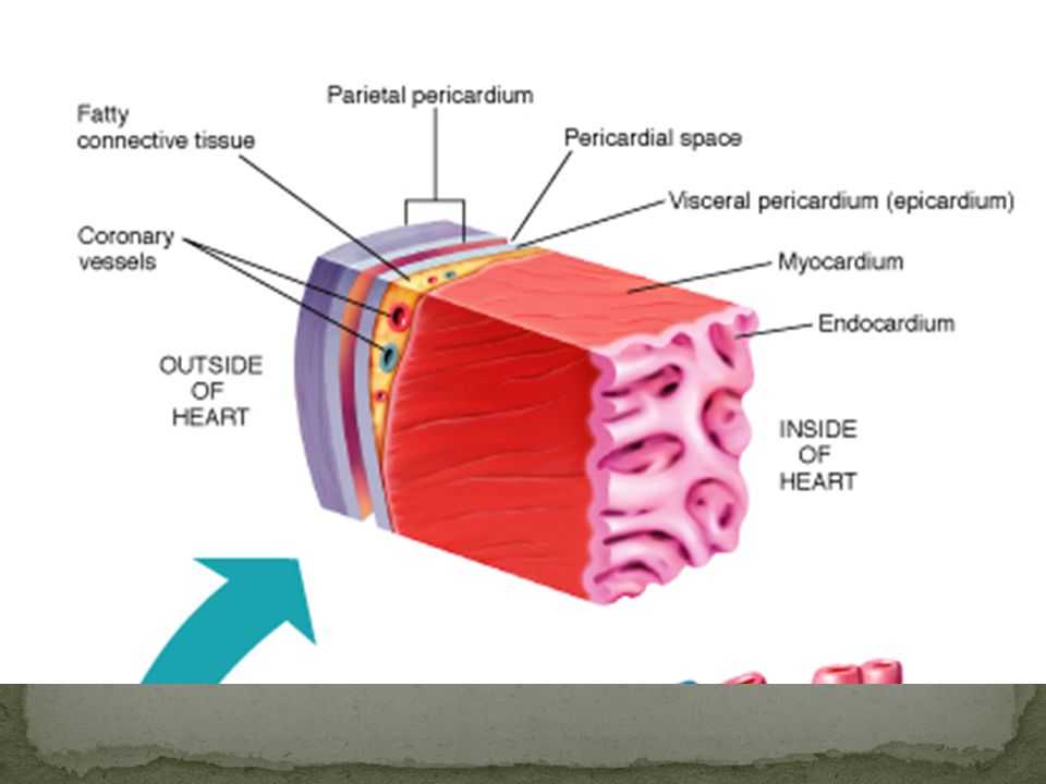
Pericardium 2-layered fibrous sac Lubricated space between layers Inner layer: Visceral Pericardium (Epicardium) Outer layer: Parietal Pericardium Inflammation: Pericarditis Fluid buildup in pericardial space: Pericardial effusion —may lead to Tamponade
 Outer layer: Parietal Pericardium Inflammation: Pericarditis Fluid buildup in pericardial space: Pericardial effusion —may lead to Tamponade")
7
Relaxation: Diastole Contraction: Systole

8
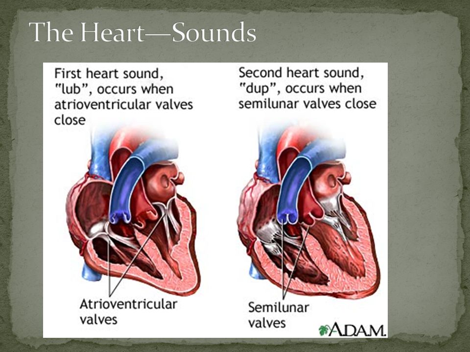
Four valves Keep blood flowing; prevent backflow Right atrioventricular: Tricuspid Between R atrium and ventricle Left atrioventricular: Mitral (Bicuspid) Between L atrium and ventricle Right Semilunar: Pulmonic At beginning of Pulmonary Artery Left Semilunar: Aortic At beginning of Aorta
 Between L atrium and ventricle Right Semilunar: Pulmonic At beginning of Pulmonary Artery Left Semilunar: Aortic At beginning of Aorta")
9
Valves don’t fully close—cause leakage: Regurgitation Narrowed valves prevent normal flow: Stenosis Mitral Valve pops open backwards—allows reverse flow: Mitral Valve Prolapse (MVP)
")
12
http://depts.washington.edu/phys dx/heart/demo.html http://www.dundee.ac.uk/medther /Cardiology/hsmur.html

13
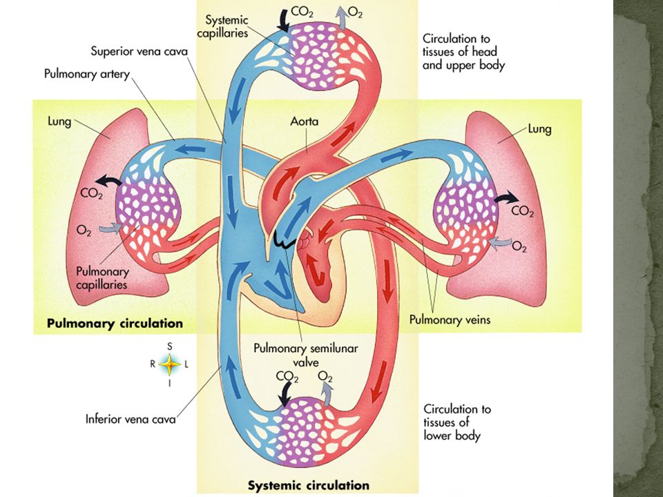
Heart is 2 separate pumps: Right side—pumps deoxygenated blood to lungs; low pressure side Left side—pumps oxygenated blood to body; high pressure side

14
Venous blood enters R Atrium from Sup. and Inf. Vena Cavae Passes from RA through Tricuspid valve to R Ventricle From RV through Pulmonic (semilunar) valve to Pulmonary artery to lungs
 valve to Pulmonary artery to lungs.")
15
Blood from lungs enters L Atrium from Pulmonary Veins From LA, passes through Mitral Valve (bicuspid) to L Ventricle From LV, passes through Aortic (semilunar) valve to Aorta and out to body
 to L Ventricle From LV, passes through Aortic (semilunar) valve to Aorta and out to body")
18
Blood supplying oxygen and nutrients to cardiac muscle flows through R and L Coronary Arteries Blockage of flow through Coronary AA causes heart attack: Myocardial Infarction Buildup of fatty deposits on the inner wall of arteries can cause flow blockage: Atheroscleosis Chest pain due to heart hypoxia: Angina Pectoris

22
Heart Failure Right-sided Left-sided (Congestive Heart Failure-CHF) Ischemic Heart Disease Pericarditis Cardiomyopathy
 Ischemic Heart Disease Pericarditis Cardiomyopathy")
24
Arteries Arterioles Veins Venules Capillaries

25
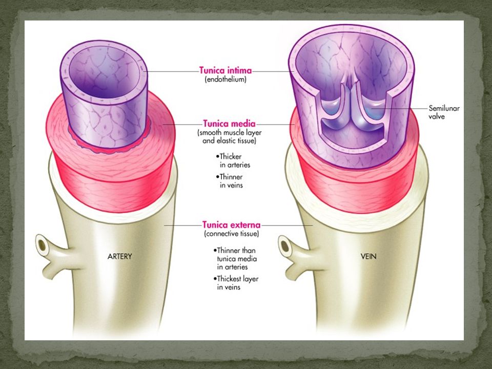
Arteries Tunica Intima Tunica Media Tunica Externa Capillaries Tunica Intima Veins Tunica Intima Tunica Media Tunica Externa

27
Arteriosclerosis Hardening

28
Aneurysm Ballooning of wall

29
Varicose veins (varices) Enlarged veins allow blood to pool
 Enlarged veins allow blood to pool")
30
Venous Stasis Leads to skin breakdown and ulceration

31
Rate at which your heart beats

33
Ask ME or ask your classmates!!

34
See you in discussion!

Similar presentations