Download presentation
Presentation is loading. Please wait.

1
CNS Cases 2 OSPE stations

2
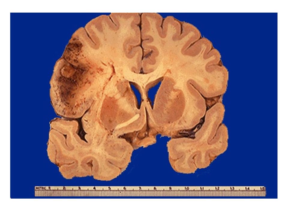
Case1 A 43-year-old, previously healthy woman presented with headache and fever for the past two weeks. She had a history of severe respiratory tract infection two weeks back. On physical examination, her temperature is 38.5̊ C. There is LEFT hemiparesis. CT scan of the head shows a sharply demarcated, 3 cm, ring- enhancing lesion in the right frontal region. While undergoing treatment the patient went into respiratory failure because of unknown cause and died. Autopsy was carried out as per hospital protocol. The slice of brain and microscopic image is provide for interpretation.

3
Can bring 2 pics together and focus on the triad of abscess in the case

4
What is the most likely diagnosis? Brain abscess cus in the case the triad was mention which is : fever, headache, focal neurologic symptom : hemiparesis ( in this case) What are the possible complications? Only 2 would be enough Spread to brain tissue > encephalitis Spread to subdural space > subdural abscess Superior sagittal sinus thrombophlebitis due to spread Rupture causing > ventriculitis - In this case she respiratory tract infection due to spread-
 What are the possible complications. Only 2 would be enough Spread to brain tissue > encephalitis Spread to subdural space > subdural abscess Superior sagittal sinus thrombophlebitis due to spread Rupture causing > ventriculitis - In this case she respiratory tract infection due to spread-.")
5
Case 2 A 50-year-old man was brought dead in the casualty department of a hospital. Autopsy was carried out to know the cause of death. His medical records showed a history of atrial fibrillation followed by sudden right-sided body weakness. The slice of brain is provided for interpretation.

7
What is the most likely diagnosis? Brain infarction What is the most likely cause for this condition? Embolism cus he has history of cardiac disease : atrial fibrillation And the infarction grossly appears in red

8
Case 3 An 80 year old woman fell down the stairs. About 36 hours later, she developed headache and confusion and is taken to the emergency department. On physical examination, she is drowsy and has a scalp contusion on the occiput. CT scan of the head shows a collection of blood in the subdural space

9
In the case they mentioned the diagnosis, on OSPE, they can bring the picture and ask A- what is the type of hemorrhage in the picture ? Subdural hemorrhage B-This type of intracranial hemorrhage is caused by ? Tearing of bridging veins

10
Case4 A 42 year old drowsy woman presents to the emergency department with complaint of the “worst headache of her life.” Few hours later she developed nausea, vomiting and weakness of the right side of the body. She denies any history of head trauma.

11
Interpret the gross image of brain and answer the following What is the most likely diagnosis? – Subarachnoid hemorrhage cus “worst headache of her life.” is the symptom of subarachnoid hemorrhage What is the most common cause of this condition? – Ruptured aneurysm

12
Gross image of the brain A-subarachnoid hemorrhage

13
Give the name of these cerebral aneurysms B-congenital, berry, saccular aneurysm Enlist other types of cerebral aneurysms Charcot–Bouchard aneurysm mycotic aneurysm atherosclerotic aneurysm

14
Case 5 A 58 year old chain -heavy- smoker with long standing uncontrolled hypertension and diabetes mellitus experienced acute onset of weakness and numbness on the left side of his body and an inability to walk. He mentions history of intermittent bouts of severe headache. His blood pressure upon arrival to emergency center was 192/105 mm Hg. Suddenly he went into cardiac arrest and expired. Autopsy was carried out. A slice of brain is provided for interpretation.

15
What is the diagnosis? Spontaneous Intraparenchymal hge.(without trauma) What’s the most possible cause of this condition? Hypertension
 What’s the most possible cause of this condition. Hypertension.")
16
Case 6 An 86 year old man resident of a nursing home is found wandering the streets, looking for his way “home.” The patient’s family describes deteriorating cognitive function that has been worsening progressively over the last several years. He does not have a history of head trauma or cardiovascular disease. In addition to the disorientation, the patient has demonstrated significant language and emotional impairment.

17
What is the most likely diagnosis in this case? Dementia due to Alzheimer's disease, in OSPE it will be ( the pt has Alzheimer's and u gonna be asked the second question below ) Identify the distinctive pathologic findings observed in brain sections A, B and C in this condition?
 Identify the distinctive pathologic findings observed in brain sections A, B and C in this condition .")
18
A B C A.Neuritic (senile) plaques. B.Neurofibrillary tangles. C.Cerebral Amyloid angiopathy.
 plaques. B.Neurofibrillary tangles. C.Cerebral Amyloid angiopathy.")
19
Case 7 A 62-year-old man is found to have a shuffling gait, a stooped posture, slowness of movement, muscle rigidity, and a pill-rolling tremor at rest. Physical examination finds that he has a “mask-like” facial expression. What is the most likely diagnosis? – Parkinsonism

20
A-Mention the name of the abnormal protein which is responsible for this pathological finding. alpha-synuclein The neurons of which part of the midbrain are typically affected. – Substantia nigra

21
Case 8 A 40-year-old woman has been in good health until recently, when she developed recurrent headaches. A CT scan reveals a 2-cm extra axial dura based frontal mass.

22
Gross image

23
Microscopic image A-whorls of meningeothelial cells B-psammoma bodies in the centers

24
What is the most likely diagnosis? Meningioma Identify two important microscopic features marked as A and B in the image? A-whorls of meningeothelial cells B-psammoma bodies in the centers What is the expected prognosis? It’s benign tumor so good prognosis unless it compress a center or recurrence occur

25
Case 9 A 9-year-old girl is evaluated for headaches and ataxia over the last month. A CT scan reveals a midline, partially cystic cerebellar mass. The tumor is removed surgically, and microscopic examination shows elongated bipolar astrocytes with fibrillar processes and Rosenthal fibers.

26
Gross image

27
Microscopy proliferating astrocytes rosenthal fibers

28
What is the most likely diagnosis? Pilocytic astrocytoma Mention the pathological grade of this tumor (grade I)
.")
Similar presentations