Download presentation
Presentation is loading. Please wait.

1
CHAPTER 7 Carbohydrates and Glycobiology
Key topics about carbohydrates: Structures and names of monosaccharides Open-chain and ring forms of monosaccharides Structures and properties of disaccharides Biological function of polysaccharides Biological function of glycoconjugates

2
Carbohydrates Named so because many have formula Cn(H2O)n
Produced from CO2 and H2O via photosynthesis in plants Range from as small as glyceraldehyde (Mw = 90 g/mol) to as large as amylopectin (Mw = 200,000,000 g/mol) Fulfill a variety of functions including: energy source and energy storage structural component of cell walls and exoskeletons informational molecules in cell-cell signaling Can be covalently linked with proteins to form glycoproteins and proteoglycans
 to as large as amylopectin (Mw = 200,000,000 g/mol) Fulfill a variety of functions including: energy source and energy storage. structural component of cell walls and exoskeletons. informational molecules in cell-cell signaling. Can be covalently linked with proteins to form glycoproteins and proteoglycans.")
3
Aldoses and Ketoses An aldose contains an aldehyde functionality
A ketose contains a ketone functionality FIGURE 7-1a Representative monosaccharides. (a)Two trioses, an aldose and a ketose. The carbonyl group in each is shaded.
Two trioses, an aldose and a ketose. The carbonyl group in each is shaded.")
4
Enantiomers Enantiomers
Stereoisomers that are nonsuperimposable mirror images In sugars that contain many chiral centers, only the one that is most distant from the carbonyl carbon is designated as D (right) or L (left) D and L isomers of a sugar are enantiomers For example, L and D glucose have the same water solubility Most hexoses in living organisms are D stereoisomers Some simple sugars occur in the L-form, such as L-arabinose
 or L (left) D and L isomers of a sugar are enantiomers. For example, L and D glucose have the same water solubility. Most hexoses in living organisms are D stereoisomers. Some simple sugars occur in the L-form, such as L-arabinose.")
5
FIGURE 7–2 (part 1) Three ways to represent the two enantiomers of glyceraldehyde. The enantiomers are mirror images of each other. Ball-and-stick models show the actual configuration of molecules. Recall (see Fig. 1–18) that in perspective formulas, the wide end of a solid wedge projects out of the plane of the paper, toward the reader; a dashed wedge extends behind.
 that in perspective formulas, the wide end of a solid wedge projects out of the plane of the paper, toward the reader; a dashed wedge extends behind..")
6
Drawing Monosaccharides
Chiral compounds can be drawn using perspective formulas However, chiral carbohydrates are usually represented by Fischer projections Horizontal bonds are pointing toward you; vertical bonds are projecting away from you

7
FIGURE 7-2 (part 2) Three ways to represent the two enantiomers of glyceraldehyde. The enantiomers are mirror images of each other. Ball-and-stick models show the actual configuration of molecules. Recall (see Fig. 1–18) that in perspective formulas, the wide end of a solid wedge projects out of the plane of the paper, toward the reader; a dashed wedge extends behind.
 that in perspective formulas, the wide end of a solid wedge projects out of the plane of the paper, toward the reader; a dashed wedge extends behind..")
8
FIGURE 7-2 (part 3) Three ways to represent the two enantiomers of glyceraldehyde. The enantiomers are mirror images of each other. Ball-and-stick models show the actual configuration of molecules. Recall (see Fig. 1–18) that in perspective formulas, the wide end of a solid wedge projects out of the plane of the paper, toward the reader; a dashed wedge extends behind.
 that in perspective formulas, the wide end of a solid wedge projects out of the plane of the paper, toward the reader; a dashed wedge extends behind..")
9
Epimers Epimers are two sugars that differ only in the configuration around one carbon atom FIGURE 7–4 Epimers. D-Glucose and two of its epimers are shown as projection formulas. Each epimer differs from D-glucose in the configuration at one chiral center (shaded light red or blue).
.")
10
Structures to Know Ribose is the standard five-carbon sugar
Glucose is the standard six-carbon sugar Galactose is an epimer of glucose Mannose is an epimer of glucose Fructose is the ketose form of glucose

11
FIGURE 7–3a (part 1) Aldoses and ketoses
FIGURE 7–3a (part 1) Aldoses and ketoses. The series of (a) D-aldoses and (b) D-ketoses having from three to six carbon atoms, shown as projection formulas. The carbon atoms in red are chiral centers. In all these D isomers, the chiral carbon most distant from the carbonyl carbon has the same configuration as the chiral carbon in D-glyceraldehyde. The sugars named in boxes are the most common in nature; you will encounter these again in this and later chapters.
 Aldoses and ketoses. The series of (a) D-aldoses and (b) D-ketoses having from three to six carbon atoms, shown as projection formulas. The carbon atoms in red are chiral centers. In all these D isomers, the chiral carbon most distant from the carbonyl carbon has the same configuration as the chiral carbon in D-glyceraldehyde. The sugars named in boxes are the most common in nature; you will encounter these again in this and later chapters.")
12
FIGURE 7-3a (part 2) Aldoses and ketoses
FIGURE 7-3a (part 2) Aldoses and ketoses. The series of (a) D-aldoses and (b) D-ketoses having from three to six carbon atoms, shown as projection formulas. The carbon atoms in red are chiral centers. In all these D isomers, the chiral carbon most distant from the carbonyl carbon has the same configuration as the chiral carbon in D-glyceraldehyde. The sugars named in boxes are the most common in nature; you will encounter these again in this and later chapters.
 Aldoses and ketoses. The series of (a) D-aldoses and (b) D-ketoses having from three to six carbon atoms, shown as projection formulas. The carbon atoms in red are chiral centers. In all these D isomers, the chiral carbon most distant from the carbonyl carbon has the same configuration as the chiral carbon in D-glyceraldehyde. The sugars named in boxes are the most common in nature; you will encounter these again in this and later chapters.")
13
FIGURE 7-3a (part 3) Aldoses and ketoses
FIGURE 7-3a (part 3) Aldoses and ketoses. The series of (a) D-aldoses and (b) D-ketoses having from three to six carbon atoms, shown as projection formulas. The carbon atoms in red are chiral centers. In all these D isomers, the chiral carbon most distant from the carbonyl carbon has the same configuration as the chiral carbon in D-glyceraldehyde. The sugars named in boxes are the most common in nature; you will encounter these again in this and later chapters.
 Aldoses and ketoses. The series of (a) D-aldoses and (b) D-ketoses having from three to six carbon atoms, shown as projection formulas. The carbon atoms in red are chiral centers. In all these D isomers, the chiral carbon most distant from the carbonyl carbon has the same configuration as the chiral carbon in D-glyceraldehyde. The sugars named in boxes are the most common in nature; you will encounter these again in this and later chapters.")
14
FIGURE 7-3b (part 1) Aldoses and ketoses
FIGURE 7-3b (part 1) Aldoses and ketoses. The series of (a) D-aldoses and (b) D-ketoses having from three to six carbon atoms, shown as projection formulas. The carbon atoms in red are chiral centers. In all these D isomers, the chiral carbon most distant from the carbonyl carbon has the same configuration as the chiral carbon in D-glyceraldehyde. The sugars named in boxes are the most common in nature; you will encounter these again in this and later chapters.
 Aldoses and ketoses. The series of (a) D-aldoses and (b) D-ketoses having from three to six carbon atoms, shown as projection formulas. The carbon atoms in red are chiral centers. In all these D isomers, the chiral carbon most distant from the carbonyl carbon has the same configuration as the chiral carbon in D-glyceraldehyde. The sugars named in boxes are the most common in nature; you will encounter these again in this and later chapters.")
15
FIGURE 7-3b (part 2) Aldoses and ketoses
FIGURE 7-3b (part 2) Aldoses and ketoses. The series of (a) D-aldoses and (b) D-ketoses having from three to six carbon atoms, shown as projection formulas. The carbon atoms in red are chiral centers. In all these D isomers, the chiral carbon most distant from the carbonyl carbon has the same configuration as the chiral carbon in D-glyceraldehyde. The sugars named in boxes are the most common in nature; you will encounter these again in this and later chapters.
 Aldoses and ketoses. The series of (a) D-aldoses and (b) D-ketoses having from three to six carbon atoms, shown as projection formulas. The carbon atoms in red are chiral centers. In all these D isomers, the chiral carbon most distant from the carbonyl carbon has the same configuration as the chiral carbon in D-glyceraldehyde. The sugars named in boxes are the most common in nature; you will encounter these again in this and later chapters.")
16
Hemiacetals and Hemiketals
Aldehyde and ketone carbons are electrophilic Alcohol oxygen atom is nucleophilic When aldehydes are attacked by alcohols, hemiacetals form When ketones are attacked by alcohols, hemiketals form FIGURE 7–5 Formation of hemiacetals and hemiketals. An aldehyde or ketone can react with an alcohol in a 1:1 ratio to yield a hemiacetal or hemiketal, respectively, creating a new chiral center at the carbonyl carbon. Substitution of a second alcohol molecule produces an acetal or ketal. When the second alcohol is part of another sugar molecule, the bond produced is a glycosidic bond (p. 252).
.")
17
Cyclization of Monosaccharides
Pentoses and hexoses readily undergo intramolecular cyclization The former carbonyl carbon becomes a new chiral center, called the anomeric carbon The former carbonyl oxygen becomes a hydroxyl group; the position of this group determines if the anomer is or If the hydroxyl group is on the opposite side (trans) of the ring as the CH2OH moiety the configuration is If the hydroxyl group is on the same side (cis) of the ring as the CH2OH moiety, the configuration is
 of the ring as the CH2OH moiety the configuration is If the hydroxyl group is on the same side (cis) of the ring as the CH2OH moiety, the configuration is ")
18
FIGURE 7-6 Formation of the two cyclic forms of D-glucose
FIGURE 7-6 Formation of the two cyclic forms of D-glucose. Reaction between the aldehyde group at C-1 and the hydroxyl group at C-5 forms a hemiacetal linkage, producing either of two stereoisomers, the α and β anomers, which differ only in the stereochemistry around the hemiacetal carbon. This reaction is reversible. The interconversion of α and β anomers is called mutarotation.

19
Pyranoses and Furanoses
Six-membered oxygen-containing rings are called pyranoses Five-membered oxygen-containing rings are called furanoses The anomeric carbon is usually drawn on the right side

20
FIGURE 7–7 Pyranoses and furanoses
FIGURE 7–7 Pyranoses and furanoses. The pyranose forms of D-glucose and the furanose forms of D-fructose are shown here as Haworth perspective formulas. The edges of the ring nearest the reader are represented by bold lines. Hydroxyl groups below the plane of the ring in these Haworth perspectives would appear at the right side of a Fischer projection (compare with Fig. 7–6). Pyran and furan are shown for comparison.
. Pyran and furan are shown for comparison.")
21
Important Hexose Derivatives
FIGURE 7-9 Some hexose derivatives important in biology. In amino sugars, an —NH2 group replaces one of the —OH groups in the parent hexose. Substitution of —H for —OH produces a deoxy sugar; note that the deoxy sugars shown here occur in nature as the L isomers. The acidic sugars contain a carboxylate group, which confers a negative charge at neutral pH. D-Glucono-δ-lactone results from formation of an ester linkage between the C-1 carboxylate group and the C-5 (also known as the δ carbon) hydroxyl group of D-gluconate.
 hydroxyl group of D-gluconate.")
22
Chain-Ring Equilibrium and Reducing Sugars
The ring forms exist in equilibrium with the open-chain forms Aldehyde can reduce Cu2+ to Cu+ (Fehling’s test) Aldehyde can reduce Ag+ to Ag0 (Tollens’ test) Allows detection of reducing sugars, such as glucose
 Aldehyde can reduce Ag+ to Ag0 (Tollens’ test) Allows detection of reducing sugars, such as glucose.")
23
Colorimetric Glucose Analysis
Nowadays, enzymatic methods are used to quantify reducing sugars such as glucose The enzyme glucose oxidase catalyzes the conversion of glucose to glucono--lactone and hydrogen peroxide Hydrogen peroxide oxidizes organic molecules into highly colored compounds Concentrations of such compounds is measured colorimetrically Electrochemical detection is used in portable glucose sensors

24
The Glycosidic Bond Two sugar molecules can be joined via a glycosidic bond between an anomeric carbon and a hydroxyl carbon The glycosidic bond (an acetal) between monomers is less reactive than the hemiacetal at the second monomer Second monomer, with the hemiacetal, is reducing Anomeric carbon involved in the glycosidic linkage is nonreducing The disaccharide formed upon condensation of two glucose molecules via 1 4 bond is called maltose
 between monomers is less reactive than the hemiacetal at the second monomer. Second monomer, with the hemiacetal, is reducing. Anomeric carbon involved in the glycosidic linkage is nonreducing. The disaccharide formed upon condensation of two glucose molecules via 1 4 bond is called maltose.")
25
FIGURE 7-10 Formation of maltose
FIGURE 7-10 Formation of maltose. A disaccharide is formed from two monosaccharides (here, two molecules of D-glucose) when an —OH (alcohol) of one glucose molecule (right) condenses with the intramolecular hemiacetal of the other glucose molecule (left), with elimination of H2O and formation of a glycosidic bond. The reversal of this reaction is hydrolysis—attack by H2O on the glycosidic bond. The maltose molecule, shown here as an illustration, retains a reducing hemiacetal at the C-1 not involved in the glycosidic bond. Because mutarotation interconverts the α and β forms of the hemiacetal, the bonds at this position are sometimes depicted with wavy lines, as shown here, to indicate that the structure may be either α or β.
 when an —OH (alcohol) of one glucose molecule (right) condenses with the intramolecular hemiacetal of the other glucose molecule (left), with elimination of H2O and formation of a glycosidic bond. The reversal of this reaction is hydrolysis—attack by H2O on the glycosidic bond. The maltose molecule, shown here as an illustration, retains a reducing hemiacetal at the C-1 not involved in the glycosidic bond. Because mutarotation interconverts the α and β forms of the hemiacetal, the bonds at this position are sometimes depicted with wavy lines, as shown here, to indicate that the structure may be either α or β.")
26
Nonreducing Disaccharides
Two sugar molecules can be also joined via a glycosidic bond between two anomeric carbons The product has two acetal groups and no hemiacetals There are no reducing ends, this is a nonreducing sugar Trehalose is a constituent of hemolymph of insects Provides protection from drying Resurrection plant (> 15 yrs)
")
27
FIGURE 7-11 Two common disaccharides
FIGURE 7-11 Two common disaccharides. Like maltose in Figure 7–10, these are shown as Haworth perspectives. The common name, full systematic name, and abbreviation are given for each disaccharide. Formal nomenclature for sucrose names glucose as the parent glycoside, although it is typically depicted as shown, with glucose on the left. The two abbreviated symbols shown for sucrose are equivalent ().
.")
28
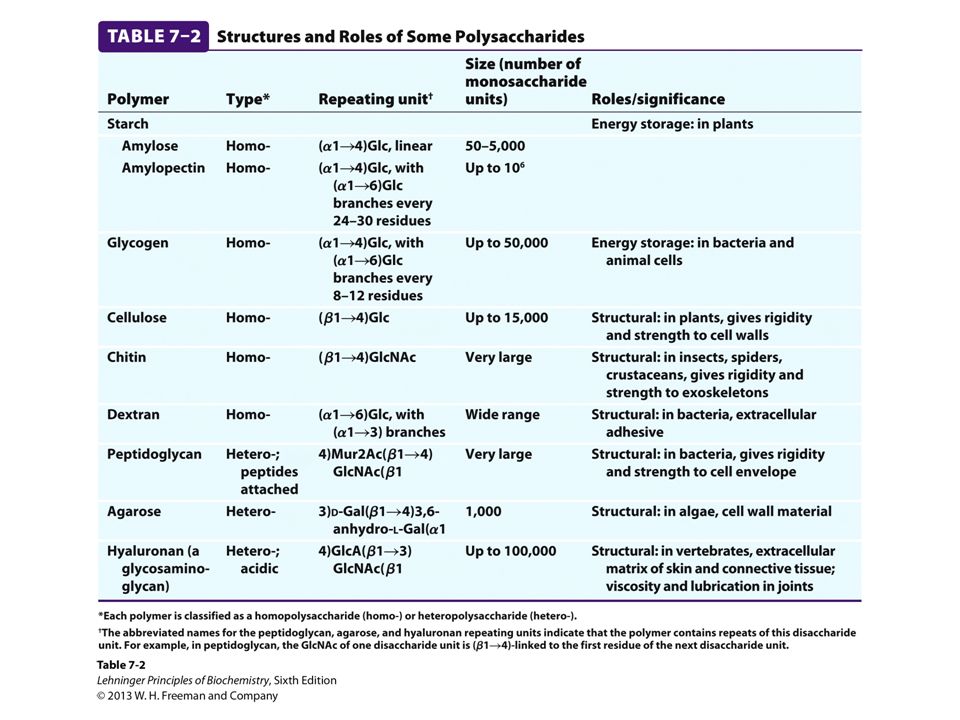
Polysaccharides Natural carbohydrates are usually found as polymers
These polysaccharides can be homopolysaccharides heteropolysaccharides linear branched Polysaccharides do not have a defined molecular weight. This is in contrast to proteins because unlike proteins, no template is used to make polysaccharides

29
FIGURE 7-12 Homo- and heteropolysaccharides
FIGURE 7-12 Homo- and heteropolysaccharides. Polysaccharides may be composed of one, two, or several different monosaccharides, in straight or branched chains of varying length.

30
Glycogen Glycogen is a branched homopolysaccharide of glucose
Glucose monomers form (1 4) linked chains Branch-points with (1 6) linkers every 8–12 residues Molecular weight reaches several millions Functions as the main storage polysaccharide in animals
 linked chains. Branch-points with (1 6) linkers every 8–12 residues. Molecular weight reaches several millions. Functions as the main storage polysaccharide in animals.")
31
Starch Starch is a mixture of two homopolysaccharides of glucose
Amylose is an unbranched polymer of (1 4) linked residues Amylopectin is branched like glycogen but the branch-points with (1 6) linkers occur every 24–30 residues Molecular weight of amylopectin is up to 200 million Starch is the main storage polysaccharide in plants
 linked residues. Amylopectin is branched like glycogen but the branch-points with (1 6) linkers occur every 24–30 residues. Molecular weight of amylopectin is up to 200 million. Starch is the main storage polysaccharide in plants.")
32
Glycosidic Linkages in Glycogen and Starch
FIGURE 7-13a,b Glycogen and starch. (a) A short segment of amylose, a linear polymer of D-glucose residues in (α1→4) linkage. A single chain can contain several thousand glucose residues. Amylopectin has stretches of similarly linked residues between branch points. Glycogen has the same basic structure, but has more branching than amylopectin. (b) An (α1→6) branch point of glycogen or amylopectin.
 A short segment of amylose, a linear polymer of D-glucose residues in (α1→4) linkage. A single chain can contain several thousand glucose residues. Amylopectin has stretches of similarly linked residues between branch points. Glycogen has the same basic structure, but has more branching than amylopectin. (b) An (α1→6) branch point of glycogen or amylopectin.")
33
Mixture of Amylose and Amylopectin in Starch
FIGURE 7-13c Glycogen and starch. (c) A cluster of amylose and amylopectin like that believed to occur in starch granules. Strands of amylopectin (black) form double-helical structures with each other or with amylose strands (blue). Amylopectin has frequent (α16) branch points (red). Glucose residues at the nonreducing ends of the outer branches are removed enzymatically during the mobilization of starch for energy production. Glycogen has a similar structure but is more highly branched and more compact.
 A cluster of amylose and amylopectin like that believed to occur in starch granules. Strands of amylopectin (black) form double-helical structures with each other or with amylose strands (blue). Amylopectin has frequent (α16) branch points (red). Glucose residues at the nonreducing ends of the outer branches are removed enzymatically during the mobilization of starch for energy production. Glycogen has a similar structure but is more highly branched and more compact.")
34
Cellulose Cellulose is a branched homopolysaccharide of glucose
Glucose monomers form (1 4) linked chains Hydrogen bonds form between adjacent monomers Additional H-bonds between chains Structure is now tough and water-insoluble Most abundant polysaccharide in nature Cotton is nearly pure fibrous cellulose
 linked chains. Hydrogen bonds form between adjacent monomers. Additional H-bonds between chains. Structure is now tough and water-insoluble. Most abundant polysaccharide in nature. Cotton is nearly pure fibrous cellulose.")
35
Hydrogen Bonding in Cellulose
FIGURE 7-14 Cellulose. (a) Two units of a cellulose chain; the D-glucose residues are in (β1→4) linkage. The rigid chair structures can rotate relative to one another.
 Two units of a cellulose chain; the D-glucose residues are in (β1→4) linkage. The rigid chair structures can rotate relative to one another.")
36
Cellulose Metabolism The fibrous structure and water-insolubility make cellulose a difficult substrate to act on Fungi, bacteria, and protozoa secrete cellulase, which allows them to use wood as source of glucose Most animals cannot use cellulose as a fuel source because they lack the enzyme to hydrolyze (1 4) linkages Ruminants and termites live symbiotically with microorganisms that produces cellulase Cellulases hold promise in the fermentation of biomass into biofuels
 linkages. Ruminants and termites live symbiotically with microorganisms that produces cellulase. Cellulases hold promise in the fermentation of biomass into biofuels.")
37
Chitin Chitin is a linear homopolysaccharide of N-acetylglucosamine
N-acetylglucosamine monomers form (1 4)-linked chains Forms extended fibers that are similar to those of cellulose Hard, insoluble, cannot be digested by vertebrates Structure is tough but flexible, and water-insoluble Found in cell walls in mushrooms, and in exoskeletons of insects, spiders, crabs, and other arthropods
-linked chains. Forms extended fibers that are similar to those of cellulose. Hard, insoluble, cannot be digested by vertebrates. Structure is tough but flexible, and water-insoluble. Found in cell walls in mushrooms, and in exoskeletons of insects, spiders, crabs, and other arthropods.")
38
Chitin FIGURE 7-16a Chitin. (a) A short segment of chitin, a homopolymer of N-acetyl-D-glucosamine units in (β1→4) linkage.
 A short segment of chitin, a homopolymer of N-acetyl-D-glucosamine units in (β1→4) linkage.")
39
FIGURE 7-16b Chitin. (b) A spotted June beetle (Pelidnota punctata), showing its surface armor (exoskeleton) of chitin.
 A spotted June beetle (Pelidnota punctata), showing its surface armor (exoskeleton) of chitin..")
40
Agar and Agarose Agar is a complex mixture of hetereopolysaccharides containing modified galactose units Agar serves as a component of cell wall in some seaweeds Agarose is one component of agar Agar solutions form gels that are commonly used in the laboratory as a surface for growing bacteria Agarose solutions form gels that are commonly used in the laboratory for separation DNA by electrophoresis

41
Agar and Agarose FIGURE 7-21 Agarose. The repeating unit consists of D-galactose (β1→4)-linked to 3,6-anhydro-L-galactose (in which an ether bridge connects C-3 and C-6). These units are joined by (α1→3) glycosidic links to form a polymer 600 to 700 residues long. A small fraction of the 3,6-anhydrogalactose residues have a sulfate ester at C-2 (as shown here). The open parentheses in the systematic name indicate that the repeating unit extends from both ends.
-linked to 3,6-anhydro-L-galactose (in which an ether bridge connects C-3 and C-6). These units are joined by (α1→3) glycosidic links to form a polymer 600 to 700 residues long. A small fraction of the 3,6-anhydrogalactose residues have a sulfate ester at C-2 (as shown here). The open parentheses in the systematic name indicate that the repeating unit extends from both ends.")
42
Glycosaminoglycans Linear polymers of repeating disaccharide units
One monomer is either N-acetyl-glucosamine or N-acetyl-galactosamine Negatively charged Uronic acids (C6 oxidation) Sulfate esters Extended hydrated molecule Minimizes charge repulsion Forms meshwork with fibrous proteins to form extracellular matrix Connective tissue Lubrication of joints
 Sulfate esters. Extended hydrated molecule. Minimizes charge repulsion. Forms meshwork with fibrous proteins to form extracellular matrix. Connective tissue. Lubrication of joints.")
43
FIGURE 7–22 (part 1a) Repeating units of some common glycosaminoglycans of extracellular matrix. The molecules are copolymers of alternating uronic acid and amino sugar residues (keratan sulfate is the exception), with sulfate esters in any of several positions, except in hyaluronan. The ionized carboxylate and sulfate groups (red in the perspective formulas) give these polymers their characteristic high negative charge. Therapeutic heparin contains primarily iduronic acid (IdoA) and a smaller proportion of glucuronic acid (GlcA, not shown), and is generally highly sulfated and heterogeneous in length. The space-filling model shows a heparin segment as its solution structure, as determined by NMR spectroscopy (PDB ID 1HPN). The carbons in the iduronic acid sulfate are colored blue; those in glucosamine sulfate are green. Oxygen and sulfur atoms are shown in their standard colors of red and yellow, respectively. The hydrogen atoms are not shown (for clarity). Heparan sulfate (not shown) is similar to heparin but has a higher proportion of GlcA and fewer sulfate groups, arranged in a less regular pattern.
 and a smaller proportion of glucuronic acid (GlcA, not shown), and is generally highly sulfated and heterogeneous in length. The space-filling model shows a heparin segment as its solution structure, as determined by NMR spectroscopy (PDB ID 1HPN). The carbons in the iduronic acid sulfate are colored blue; those in glucosamine sulfate are green. Oxygen and sulfur atoms are shown in their standard colors of red and yellow, respectively. The hydrogen atoms are not shown (for clarity). Heparan sulfate (not shown) is similar to heparin but has a higher proportion of GlcA and fewer sulfate groups, arranged in a less regular pattern.")
44
Heparin and Heparan Sulfate
Heparin is linear polymer, 3–40 kDa Heparan sulfate is heparin-like polysaccharide but attached to proteins Highest negative charge density biomolecules Prevent blood clotting by activating protease inhibitor antithrombin Binding to various cells regulates development and formation of blood vessels Can also bind to viruses and bacteria and decrease their virulence

46
Glycoconjugates: Glycoprotein
A protein with small oligosaccharides attached Carbohydrate attached via its anomeric carbon About half of mammalian proteins are glycoproteins Carbohydrates play role in protein-protein recognition Only some bacteria glycosylate few of their proteins Viral proteins heavily glycosylated; helps evade the immune system

47
FIGURE 7-30 Oligosaccharide linkages in glycoproteins
FIGURE 7-30 Oligosaccharide linkages in glycoproteins. (a) O-linked oligosaccharides have a glycosidic bond to the hydroxyl group of Ser or Thr residues (light red), illustrated here with GalNAc as the sugar at the reducing end of the oligosaccharide. One simple chain and one complex chain are shown. (b) N-linked oligosaccharides have an N-glycosyl bond to the amide nitrogen of an Asn residue (green), illustrated here with GlcNAc as the terminal sugar. Three common types of oligosaccharide chains that are N-linked in glycoproteins are shown. A complete description of oligosaccharide structure requires specification of the position and stereochemistry (α or β) of each glycosidic linkage.
 O-linked oligosaccharides have a glycosidic bond to the hydroxyl group of Ser or Thr residues (light red), illustrated here with GalNAc as the sugar at the reducing end of the oligosaccharide. One simple chain and one complex chain are shown. (b) N-linked oligosaccharides have an N-glycosyl bond to the amide nitrogen of an Asn residue (green), illustrated here with GlcNAc as the terminal sugar. Three common types of oligosaccharide chains that are N-linked in glycoproteins are shown. A complete description of oligosaccharide structure requires specification of the position and stereochemistry (α or β) of each glycosidic linkage.")
48
Glycoconjugates: Glycolipids
A lipid with covalently bound oligosaccharide Parts of plant and animal cell membranes In vertebrates, ganglioside carbohydrate composition determines blood groups In gram-negative bacteria, lipopolysaccharides cover the peptidoglycan layer

49
FIGURE 7-31 Bacterial lipopolysaccharides
FIGURE 7-31 Bacterial lipopolysaccharides. Schematic diagram of the lipopolysaccharide of the outer membrane of Salmonella typhimurium. Kdo is 3-deoxy-D-manno-octulosonic acid (previously called ketodeoxyoctonic acid); Hep is L-glycero-D-manno-heptose; AbeOAc is abequose (a 3,6-dideoxyhexose) acetylated on one of its hydroxyls. There are six fatty acid residues in the lipid A portion of the molecule. Different bacterial species have subtly different lipopolysaccharide structures, but they have in common a lipid region (lipid A), a core oligosaccharide also known as endotoxin, and an “O-specific” chain, which is the principal determinant of the serotype (immunological reactivity) of the bacterium. The outer membranes of the gram-negative bacteria S. typhimurium and E. coli contain so many lipopolysaccharide molecules that the cell surface is virtually covered with O-specific chains.
; Hep is L-glycero-D-manno-heptose; AbeOAc is abequose (a 3,6-dideoxyhexose) acetylated on one of its hydroxyls. There are six fatty acid residues in the lipid A portion of the molecule. Different bacterial species have subtly different lipopolysaccharide structures, but they have in common a lipid region (lipid A), a core oligosaccharide also known as endotoxin, and an O-specific chain, which is the principal determinant of the serotype (immunological reactivity) of the bacterium. The outer membranes of the gram-negative bacteria S. typhimurium and E. coli contain so many lipopolysaccharide molecules that the cell surface is virtually covered with O-specific chains.")
50
Glycoconjugates: Proteoglycans
Sulfated glucoseaminoglycans attached to a large rod-shaped protein in cell membrane Syndecans: protein has a single transmembrane domain Glypicans: protein is anchored to a lipid membrane Interact with a variety of receptors from neighboring cells and regulate cell growth

51
FIGURE 7-26a Two families of membrane proteoglycans
FIGURE 7-26a Two families of membrane proteoglycans. (a) Schematic diagrams of a syndecan and a glypican in the plasma membrane. Syndecans are held in the membrane by hydrophobic interactions between a sequence of nonpolar amino acid residues and plasma membrane lipids; they can be released by a single proteolytic cut near the membrane surface. In a typical syndecan, the extracellular amino-terminal domain is covalently attached (by tetrasaccharide linkers such as those in Fig. 7–25) to three heparan sulfate chains and two chondroitin sulfate chains. Glypicans are held in the membrane by a covalently attached membrane lipid (GPI anchor; see Fig. 11–15), but are shed if the bond between the lipid portion of the GPI anchor (phosphatidylinositol) and the oligosaccharide linked to the protein is cleaved by a phospholipase. All glypicans have 14 conserved Cys residues, which form disulfide bonds to stabilize the protein moiety, and either two or three glycosaminoglycan chains attached near the carboxyl terminus, close to the membrane surface.
 Schematic diagrams of a syndecan and a glypican in the plasma membrane. Syndecans are held in the membrane by hydrophobic interactions between a sequence of nonpolar amino acid residues and plasma membrane lipids; they can be released by a single proteolytic cut near the membrane surface. In a typical syndecan, the extracellular amino-terminal domain is covalently attached (by tetrasaccharide linkers such as those in Fig. 7–25) to three heparan sulfate chains and two chondroitin sulfate chains. Glypicans are held in the membrane by a covalently attached membrane lipid (GPI anchor; see Fig. 11–15), but are shed if the bond between the lipid portion of the GPI anchor (phosphatidylinositol) and the oligosaccharide linked to the protein is cleaved by a phospholipase. All glypicans have 14 conserved Cys residues, which form disulfide bonds to stabilize the protein moiety, and either two or three glycosaminoglycan chains attached near the carboxyl terminus, close to the membrane. surface.")
52
FIGURE 7-25 Proteoglycan structure, showing the tetrasaccharide bridge
FIGURE 7-25 Proteoglycan structure, showing the tetrasaccharide bridge. A typical tetrasaccharide linker (blue) connects a glycosaminoglycan— in this case chondroitin 4-sulfate (orange)—to a Ser residue in the core protein. The xylose residue at the reducing end of the linker is joined by its anomeric carbon to the hydroxyl of the Ser residue.
 connects a glycosaminoglycan— in this case chondroitin 4-sulfate (orange)—to a Ser residue in the core protein. The xylose residue at the reducing end of the linker is joined by its anomeric carbon to the hydroxyl of the Ser residue.")
53
Proteoglycans Different glycosaminoglycans are linked to the core protein Linkage from anomeric carbon of xylose to serine hydroxyl Our tissues have many different core proteins; aggrecan is the best studied

54
Extracellular Matrix (ECM)
Material outside the cell Strength, elasticity, and physical barrier in tissues Main components Proteoglycan aggregates Collagen fibers Elastin (a fibrous protein) ECM is a barrier for tumor cells seeking to invade new tissues Some tumor cells secrete heparinase that degrades ECM
 ECM is a barrier for tumor cells seeking to invade new tissues. Some tumor cells secrete heparinase that degrades ECM.")
55
Interaction of the Cells with ECM
Some integral membrane proteins are proteoglycans Syndecans Other integral membrane proteins are receptors for extracellular proteoglycans Integrins These proteins link cellular cytoskeleton to the ECM and transmit signals into the cell to regulate: cell growth cell mobility apoptosis wound healing

56
FIGURE 7-29 Interactions between cells and the extracellular matrix
FIGURE 7-29 Interactions between cells and the extracellular matrix. The association between cells and the proteoglycan of the extracellular matrix is mediated by a membrane protein (integrin) and by an extracellular protein (fibronectin in this example) with binding sites for both integrin and the proteoglycan. Note the close association of collagen fibers with the fibronectin and proteoglycan.
 and by an extracellular protein (fibronectin in this example) with binding sites for both integrin and the proteoglycan. Note the close association of collagen fibers with the fibronectin and proteoglycan.")
57
Oligosaccharides in Recognition
FIGURE 7-37 Role of oligosaccharides in recognition events at the cell surface and in the endomembrane system. (a) Oligosaccharides with unique structures (represented as strings of hexagons) are components of a variety of glycoproteins or glycolipids on the outer surface of plasma membranes. Their oligosaccharide moieties are bound by extracellular lectins with high specificity and affinity. (b) Viruses that infect animal cells, such as the influenza virus, bind to cell surface glycoproteins as the first step in infection. (c) Bacterial toxins, such as the cholera and pertussis toxins, bind to a surface glycolipid before entering a cell. (d) Some bacteria, such as H. pylori, adhere to and then colonize or infect animal cells. (e) Selectins (lectins) in the plasma membrane of certain cells mediate cell-cell interactions, such as those of leukocytes with the endothelial cells of the capillary wall at an infection site. (f) The mannose 6-phosphate receptor/lectin of the trans Golgi complex binds to the oligosaccharide of lysosomal enzymes, targeting them for transfer into the lysosome.
 Oligosaccharides with unique structures (represented as strings of hexagons) are components of a variety of glycoproteins or glycolipids on the outer surface of plasma membranes. Their oligosaccharide moieties are bound by extracellular lectins with high specificity and affinity. (b) Viruses that infect animal cells, such as the influenza virus, bind to cell surface glycoproteins as the first step in infection. (c) Bacterial toxins, such as the cholera and pertussis toxins, bind to a surface glycolipid before entering a cell. (d) Some bacteria, such as H. pylori, adhere to and then colonize or infect animal cells. (e) Selectins (lectins) in the plasma membrane of certain cells mediate cell-cell interactions, such as those of leukocytes with the endothelial cells of the capillary wall at an infection site. (f) The mannose 6-phosphate receptor/lectin of the trans Golgi complex binds to the oligosaccharide of lysosomal enzymes, targeting them for transfer into the lysosome.")
58
Glycoconjugates: Analysis
FIGURE 7-39 Separation and quantification of the oligosaccharides in a group of glycoproteins. In this experiment, the mixture of proteins extracted from kidney tissue was treated to release oligosaccharides from glycoproteins, and the oligosaccharides were analyzed by matrixassisted laser desorption/ionization mass spectrometry (MALDI MS). Each distinct oligosaccharide produces a peak at its molecular mass, and the area under the curve reflects the quantity of that oligosaccharide. The most prominent oligosaccharide here (mass u) is composed of 13 sugar residues; other oligosaccharides, containing as few as 7 and as many as 19 residues, were also resolved by this method.
. Each distinct oligosaccharide produces a peak at its molecular mass, and the area under the curve reflects the quantity of that oligosaccharide. The most prominent oligosaccharide here (mass u) is composed of 13 sugar residues; other oligosaccharides, containing as few as 7 and as many as 19 residues, were also resolved by this method.")
59
Chapter 7: Summary In this chapter, we learned about:
structures of some important monosaccharides structures and properties of disaccharides structures and biological roles of polysaccharides functions of glycosylaminoglycans as structural components of the extracellular matrix functions glycoconjugates in regulating a variety of biological functions

Similar presentations


 have empirical formulas of (CH 2 O) n, where n ≥ 3 Monosaccharides.>")


, D and L isomers of Glyceraldehyde.>")
 is the study of the chemical processes in living organisms.>")


 Objective: Understand classification and structure of carbohydrates Understand multistep.>")
