Download presentation
Presentation is loading. Please wait.

1
Tackling Injuries in Contact Athletes Joseph H. Guettler, M.D. Assistant Clinical Professor, OUWB Medical School Member, Board of Directors, AOSSM

2
Disclosure Educational and research support from Depuy Mitek, Arthrex, Biomet, and Smith & Nephew Consulting for Depuy Mitek and Smith and Nephew Speakers Bureau for Genzyme

3
Three Hot Topics in the Shoulder Clavicle Fractures – Pin vs. Plate? AC Separations – What to do with the Condroversial Grade III? Shoulder Dislocations – What do you tell the first time dislocator?

4
Three Hot Topics in the Knee ACL Tears – Who gets what graft? Patellar Dislocations – What do you do with the first-time dislocator? Meniscus Tears - When do you trim and how far do you push the envelop when it comes to repairing the meniscus?

5
Clavicle Fractures

6
Traditional Treatment of Clavicle Fractures “Put it in a sling and it should heal” Usually 4 to 6 weeks and then avoidance of contact activity for 2 to 3 months The Problems: –Not all fractures heal (fibrous union or nonunion) –Not all fractures heal right (malunion) –This can have a negative influence on the rest of the shoulder girdle
 –Not all fractures heal right (malunion) –This can have a negative influence on the rest of the shoulder girdle")
7
And Let’s Face it: Clavicle Fractures Can Land You in Jail

8
“Problem” Fractures Complete displacement with lack of bony apposition Shortening of greater than 1.5 to 2 cm High Energy and comminution Fractures in smokers Murray et al, JBJS 2013

9
The Trends Fix Fractures with significant displacement and/or shortening –Greater than 100% displacement –Shortening of greater than 1.5 to 2 cm 85% of fractures are midshaft and can be pinned or plated And hence the great debate…

10
Pin Versus Plate

11
Now I’m Not Afraid to Use a Plate

12
It’s OK to be a “Pinhead” When it Comes to Fixing Midshaft Clavicle Fractures

19
Post-op Pin Management Generally speaking, 4 weeks in a sling Pendulums and elbow ROM usually instituted at 2 weeks Motions to avoid early on: Elevation above 90 degrees and X-body adduction Formal rehab usually begins between 4 and 6 weeks (But not all need it) Pin removal 8 to 10 weeks Return to sport 3 to 4 weeks later
 Pin removal 8 to 10 weeks Return to sport 3 to 4 weeks later")
20
Pin vs. Plate – Meta-analysis After qualifying studies, four studies with 305 fractures included No significant differences pin vs. plate in regards to outcome scores, nonunion, infxn, fixation failure, and hardware removal More symptomatic hardware events occurred with plating –Duan et al, JSES 2011

21
Conclusions Excellent results can be obtained with either intramedullary or plate fixation Plate offers rigid fixation while pin acts as an internal splint Pin is less invasive, but most contact athletes really don’t care about the size of the incision Condition of the clavicle after hardware removal may have an impact on choice of fixation method

22
AC Separations

23
Anatomy and Biomechanics Posterior and Superior AC Ligaments –Important in Horizontal stability Conoid 45 mm away from distal clavicle Posterior ½ of the clavicle Trapezoid 30 mm away from distal clavicle Anterior ½ of the clavicle CC Ligaments important in Vertical Stability Image Courtesy of N Bontempo MD & A Mazzocca MD, Biomechanics and Treatment of AC and SC Joint Injuries, Br J Sports Med 2010

24
Mechanism of Injury Direct – Most Common –Usually a fall on the superolateral shoulder with the arm adducted Indirect –A fall on an outstretched arm or elbow with a superiorly directed force

25
Physical Exam Tenderness Deformity Pain with Cross-Body Adduction Pain with Obrien’s Type III can Usually be Reduced Type V Buttonholed through fascia

26
Radiographic Evaluation

27
Injury Classification Image Courtesy of N Bontempo MD & A Mazzocca MD, Biomechanics and Treatment of AC and SC Joint Injuries, Br J Sports Med 2010

28
The Controversial Grade 3 Another Meta-analysis of the Literature A meta-analysis of the current evidence base Of 724 citations, six retrospective case series studies met eligibility criteria Operative management results in better cosmetic outcome, but greater duration of sick leave compared to non-op No difference in strength, pain, throwing ability, and osteoarthritis One study showed a higher Constant score for operatively managed separation Smith et al, J Orthop Traumatol 2011

29
The Controversial Grade 3 What About the Aussies? 14 Australian Rules Football Players; 8 non-op, 6 operative, 2 converted to operative Quicker return to sport-specific training & competive games, as well as higher subjective scores at final follow-up for the operative group Cardone et al, J Sci Med Sport 2002

30
The Controversial Grade 3 A Common Sense Approach Recognized recent trend toward initial non-op management Offered a treatment algorithm with more aggressive management based on factors including type of sport or labor, timing of injury relative to athletic season, or throwing demands in the dominant arm Trainer, Arciero, Mazzocca, Clin J Sport Med 2008

31
Repair Acute Synthetic vs. Reconstruction Chronic Biologic Image Courtesy of P Brady MD, AC Repair Using Dog Bone Button Technology, Arthrex Inc, 2013 Image Courtesy of A Mazzocca MD, R Arciero MD, & A Romeo MD, Anatomic Coracoclavicular Reconstruction Surgical Technique, Arthrex Inc, 2012 In 2016

32
Repair of Acute Separations

35
Results of Button Fixation Twelve patients with Grade III and IV AC separations Treated with double-button fixation through mini-op incision Mean age 27.5; Follow-up 18.25 months Excellent Constant, Dash, and VAS scores CC distance maintained and comparable to opposite shoulder No radiographic AC arthritis Beris et al, Injury 2013

36
Post-Op Protocol Sling for 1 st 6 weeks Pendulums at 2 weeks Gentle PT with ROM at 5 weeks Gentle Strengthening with acute repair at 6 weeks (8 weeks for reconstruction) Nothing aggressive for 3 months either procedure Return to contact sport 4 months (repair) and 6 months (reconstruction)
 Nothing aggressive for 3 months either procedure Return to contact sport 4 months (repair) and 6 months (reconstruction)")
37
Traumatic 1 st Time Shoulder Dislocation Traumatic 1 st Time Shoulder Dislocation

38
Mechanism of Injury: Acute Anterior Dislocation

39
The Bankart Tear The Essential Lesion

40
The Trend: Stabilize High-Risk 1 st Time Dislocators

41
Contemporary Operative Considerations Arthroscopic Bankart repair using suture anchors Open Bankart repair using suture anchors Addition of: –Rotator Interval Closure –Posterior Plication Suture –SLAP Repair Treatment of Bony Deficiency of the Anterior/Inferior Labrum or Significant Engaging Hill Sachs Lesion

45
The Latest Debate

46
Arthroscopic vs. Open Repair Another Meta-Analysis Included 11 trials and 1022 patients Open Bankart repair produced a more stable shoulder but the poorer motion compared to arthroscopic repair Wang et al, Med Sci Monit, 2015

47
Ten Year Follow-up of Acute Arthroscopic Bankart Repair for Initial Shoulder Dislocation in Young Patients 21 Patients under age 25 underwent arthroscopic Bankart repair within 30 days of injury 35% failure rate with 5 recurrent dislocations and 2 subluxations Chapus et al, Orthop Traumatol Surg Res, 2015

48
Don’t Be Afraid to Go

49
Conclusions First-time dislocations in young contact athletes should be repaired Results are likely better when repaired early and when they are “ripe for the picking” Trend back toward open repair in high risk contact athletes

50
The Contact Athlete’s Knee ACL

51
The Problem ACL injuries do not heal The best we can hope for is that the ACL scars to the PCL and offers modest stability It’s truly a finicky little ligament

52
It needs to be Reconstructed, But… Only after out of the acute inflammatory phase Swelling down and motion restored Quads need to be “up and firing” An additional MCL injury, especially proximal, and even if low grade, might slow things down

53
Graft Options & Differences Patellar Tendon: Bone plugs grow into bone tunnels Hamstrings: Soft tissue graft must scar to bony walls of tunnels Patellar Tendon Allograft: Body takes longer to grow into foreign tissue Soft Tissue Allograft

54
The Traditional Way to Reconstruct an ACL

55
Is Still the Gold Standard It’s my “college football player graft” Most NFL and D1 Team Docs Agree Clinically very stable Higher incidence of anterior knee pain Flexion Contracture & OA

57
Hamstrings is the Contemporary Approach Smaller incision Very strong graft Avoids the anterior knee Persistent hamstrings weakness?

58
And You Have to be Creative with your fixation

60
Grafts in 2016 Younger contact athletes do better with autograft Hamstrings vs. Patallar Tendon? –It’s generally accepted that HS not quite as tight as BTB –But more stiffness and potential OA with BTB Trend back toward the gold BTB in higher risk patients

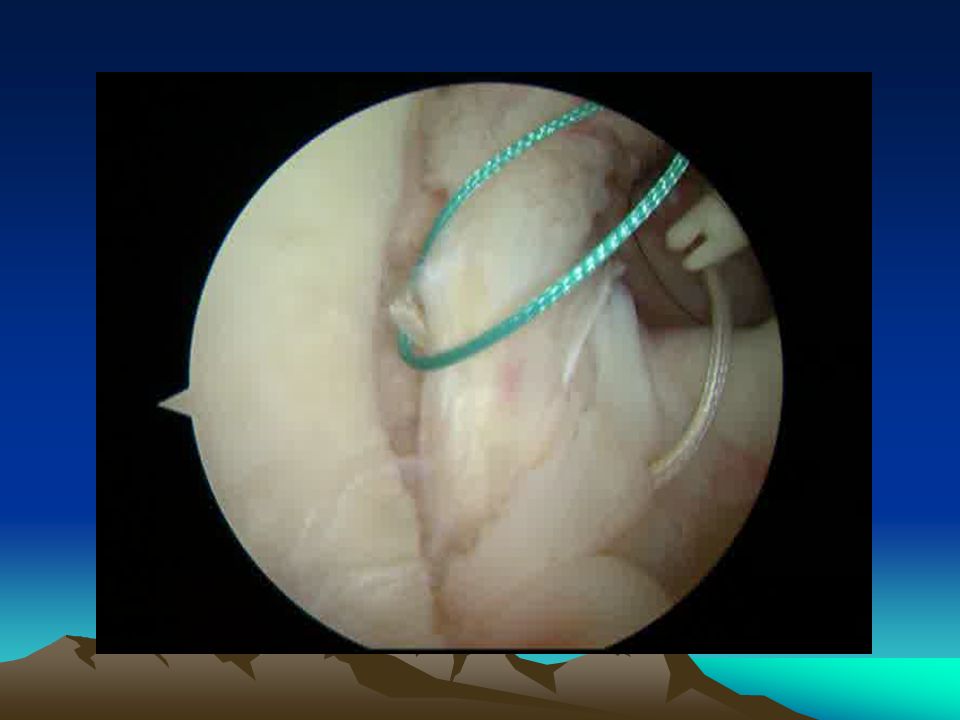
61
Regardless of Graft, Tunnels Need to be “Anatomic”

62
The Latest Craze Relates to the “ALL” A Review of the Anterolateral Ligament of the Knee Arthroscopy 2015 Pomajzl, Maerz, Shams, Guettler, Bicos

63
“Just Have a Cocktail” Zach Leonard MD, Joseph Guettler MD, Kevin Baker MS, Heather Aho MS, John Reddan, PhD, Kenneth Jurist MD: The Influence of Combined Growth Factors and Catabolic Inhibitors on ACL Healing Podium Presentation, AAOS Annual Meeting, New Orleans, 2010

64
Patellar Dislocation

65
MPFL and VMO are Major Restraints to Lateral Patellar Displacement MPFL provides 50 to 80% of the restraining force to lateral displacement VMO more important as dynamic stabilizer

66
The Essential Lesion Injury to the MPFL can be considered the essential lesion to acute patellar dislocation in the growing athlete The MPFL can tear off of the patella, medial epicondyle, or attenuate In higher energy injuries, retinacular rents, VMO avulsions, and proximal MCL injuries can accompany MPFL injury

67
Mechanism of Injury Usually a noncontact injury with foot planted while the knee incurs an IR force while in valgus position Similar mechanism to that of ACL injury

68
The Main Risk Factor in This Population Patella alta has been validated as the major risk factor in this young athletic population Atkin et al., Am J Sports Med 2000 Arendt et al., Clin Sports Med 2002 Hinton et al., Orthop Clin N Am 2003 Patella alta leads to later engagement of the patella on the trochlea Many high energy sporting maneuvers take place at lower degrees of flexion, so…

69
The Acute Patellar Dislocation Most patellar dislocations spontaneously reduce Reduction can be carried out by slowly extending knee with gentle antero-medial force on patella Most athletes seeking medical attention have experienced a subluxation event (< 20% have had true dislocation)
")
70
Imaging – X-ray AP, Lateral in 20 to 30 o flexion, Merchant View taken at 45 o flexion

71
The Million Dollar Question: Which of these is Going to Recur?

72
Example 1

73
Example 2

74
Risk of Recurrence Recurrent dislocation following an initial traumatic dislocation 15 to 44% Arendt et al., Clin Sports Med 2002 Cofield et al., J Trauma 1977

75
Operative vs. Non-Operative Treatment: What Does the Literature Tell Us? Buchner et al., Clin J Sports Med 2005 Retrospective clinical trial, 126 patients, no significant difference between conservative and operative groups in redislocation, re-operation, level of activity, functional/subjective outcomes –Authors surgical indications (Medial repair and lateral release): “Major patellar instability,” palpable defect in medial structures, persistent subluxation, chondral or osteochondral fragments –Authors concluded that conservative management is treatment of choice as long as “generally accepted indications for surgery are given due consideration”
: Major patellar instability, palpable defect in medial structures, persistent subluxation, chondral or osteochondral fragments –Authors concluded that conservative management is treatment of choice as long as generally accepted indications for surgery are given due consideration .")
76
Conservative Treatment Conservative treatment has a definite role in most first-time dislocators

77
Surgical Treatment First-time dislocators cannot simply be randomized Some patients are best-served with early surgical intervention The challenge is choosing the appropriate patient

78
Findings the “Tip the Scale” in First-Time Dislocator Chondral or osteochondral injury requiring arthroscopic intervention Persistent patellar subluxation on MRI with frank MPFL disruption and/or avulsion Medial retinacular disruption or VMO avulsion accompanying MPFL disruption Persistent and significant subluxation on the Merchant view following patellar reduction Subjective complaints of patellar instability during rehab

79
Results of Medial Repair in First- Time Dislocator Ahmed et al., Am J Sports Med 2000 Demonstrated no recurrent dislocations and a 96% satisfaction rate in patients managed with acute open repair of the medial patellar stabilizers in 1st-time dislocators

80
My Approach to MPFL Repair

81
Identify MPFL Injury Dissect reticulum off of underlying capsule Palpate capsular thickening - MPFL Identify zone of injury – medial patella vs. medial epicondyle

82
Repair MPFL Utilize one or two bioabsorbable anchors Use fluoroscopy to avoid injury to distal femoral physis

83
Retinacular Closure Close retinaculum in a pant’s-over-vest fashion Repair any retinacular rents Assess patellar mobility through arc of motion

84
The Repaired MPFL MPFL

85
The Finished Product

86
Post-op Protocol Crutches, TTWB, Hinged knee brace locked in extension for 2 weeks Add daily passive ROM increasing to 90 o weeks 2 – 4 Advance weightbearing and institute PT starting one month post-op Patellar stabilizing brace for 3 months post-op with ambulation then with activity for additional 3 months or 1 st season back

87
Meniscal Injuries

88
Vascularity of the Meniscus

89
Partial Meniscectomy Kirsten, Guettler et al, Presented AOSSM 2010

90
The Torn Meniscus There are Easy Decisions

91
The Torn Meniscus BucketedReduced There are Appropriate Decisions

92
The Torn “Normal” Meniscus Bucketed Reduced There are Hard Decisions

93
Indications for Repair Repair tears in the red vascular zone and extend indications in younger patients to tears propagating through the gray zone Repair if > 10-15mm in length, although lower threshold with newer implants All-inside technique for nonbucketed, moderately unstable tears Inside-out technique for grossly unstable bucket- handle tears Trephinate if partial thickness stable tears Reconstruction of ACL improves healing potential of repaired meniscus

94
Saucerized but Unstable

95
Repaired

96
Results of Repair Eggli et al, Am J Sports Med 1995 52 patients underwent meniscal repair, F/u 7.5 years, Over-all failure rate 27%, 64% of failures in first 6 months More failures when rim width greater than 3 mm and when tears repaired with resorbable sutures Tear length less than 2.5 cm, time from injury to surgery less than 8 weeks, tear in the lateral meniscus, and age less than 30 led to better clinical result MRI showed persistent signal and not reliable in assessing meniscal healing Tenuta and Arciero, Arthroscopy 1994 54 meniscus repairs evaluated with second-look arthroscopy, Average of 11 months post-repair 35/54 (65%) completely healed, 9 partially healed, over-all 81% showed healing No tear with a rim width > 4mm healed Meniscus repair in patients less than 30, combined with ACL reconstruction, and performed within 19 weeks of injury exhibited higher healing rate
 completely healed, 9 partially healed, over-all 81% showed healing No tear with a rim width > 4mm healed Meniscus repair in patients less than 30, combined with ACL reconstruction, and performed within 19 weeks of injury exhibited higher healing rate")
97
Peripheral Tear Lateral Meniscus

98
Peripheral Tear Medial Meniscus

99
Reduced Bucket-Handle Medial Menicus

100
Devices for All Inside Repair of meniscus tears

101
Biologic Enhancement of Meniscal Healing Mesenchymal and Synovial Stem Cells, Fibroblast Growth Factor, Platelet- Derived Growth Factor shown to positively influence meniscal healing Interleukin-1 and Tumor Necrosis Factor shown to inhibit healing Bioabsorbable Conduits shown to allow migration of cells and growth factors to avascular areas

102
PRP Enhancement of Meniscus Repair

103
Future Directions Cocktails of growth factors and catabolic inhibitors Genetically enhanced, stem cell-based tissue engineering Tissue Engineered Meniscus Scaffolds

104
Thank You

Similar presentations


 Is one of a pair of ligaments that are found in the middle.>")


, F.R.C.S.(C )>")


