Download presentation
Presentation is loading. Please wait.

1
PHARMACEUTICAL MICROBIOLOGY I PHT 226 Dr. Rasheeda Hamid Abdalla Assistant Professor E-mail rasheedahamed12@hotmail. com

2
Medically Important Fungi

3
OBJECTIVES Cutaneous Mycosis Subcutaneous mycoses Systemic Mycoses

4
Cutaneous Mycosis Also called dermatophytoses The dermatophytes fall into three genera,each with many species : Trichophyton Epidermatophyton Microsporum The causative organisms of the dermatophytoses are often distinguished according to their natural habitats:

5
Cutaneous Mycosis Anthrophilic-residing on human skin Zoophilic –residing on the skin of domestic and farm animals Geophilic-residing in the soil Most human infections are by anthrophilic and zoophilic organisms Transmission from human to human,or animal to human,is by infected skin scales

6
Pathology A defining characteristic of the dermatophytes is their ability to use keratin as a source of nutrition This ability allows them to infect keratinized tissues such as skin,hair and nails All three organisms attack the skin,Microsporum does not infect nails,and Epidermophyton does not infect hair They do not invade underlying, nonkeratiniz ed tissues

7
Clinical Significance Dermatophytoses are characterized by itching,scaling skin patches that can become inflamed and weeping. Specific diseases are usually identified according to affected tissues A given disease can be caused by any one of several organism, and some organisms can cause more than one disease depending on the site of infection, condition of the skin,etc

8
Dermatophytoses The most common encounter dermatophytoses 1- Tinea pedis (athlete’s foot) Oraganisms most often isolated from infected tissues are: Trichophyton rubrum, Trichophyton mentagrophytes Epidermatophyton floccosum The infected tissue is initially between the toes, but can spread to the nails, which become yellow and brittle Id reaction :is an allergic rash caused by an inflammatory fungal infection (tinea) at a distant site
 Oraganisms most often isolated from infected tissues are: Trichophyton rubrum, Trichophyton mentagrophytes Epidermatophyton floccosum The infected tissue is initially between the toes, but can spread to the nails, which become yellow and brittle Id reaction :is an allergic rash caused by an inflammatory fungal infection (tinea) at a distant site")
9
Dermatophytoses Tinea corporis (ring worm) Organisms most often isolated are : Epidermatophyton floccosum, and several species of Trichophyton and Microsporum Lesions appear as advancing annular rings with scaly centers The periphery of the ring,which is the site of active fungal growth, is usually inflamed and vesiculated Lesions most often occur on nonhairy areas of the trunck,but any site on the body can be affected.
 Organisms most often isolated are : Epidermatophyton floccosum, and several species of Trichophyton and Microsporum Lesions appear as advancing annular rings with scaly centers The periphery of the ring,which is the site of active fungal growth, is usually inflamed and vesiculated Lesions most often occur on nonhairy areas of the trunck,but any site on the body can be affected.")
10
Tinea corporis (ring worm)
")
11
Tinea capitis (scalp ringworm) A number of species of Trichophyton and Microsporum have been isolated from scalp ringworm lesions Disease manifestations range from small, scaling patches,to involvement of the entire scalp with the extensive hair loss The shafts can become invaded by Microsporum hyphae
 A number of species of Trichophyton and Microsporum have been isolated from scalp ringworm lesions Disease manifestations range from small, scaling patches,to involvement of the entire scalp with the extensive hair loss The shafts can become invaded by Microsporum hyphae")
12
Tinea capitis (scalp ringworm)
")
13
Tinea cruris (“jock itch”) Caused by Epidermatophyton floccosum and Trichophyton rubrum Disease manifestations are similar to ringworm except that lesions occur in the moist groin area, where can spread from the upper thighs to the genitals Tinea unguium (onychomycosis) Caused by Trichophyton rubrum Nails are thickened, discolored, and brittle Treatment must be continued for three to four months until all infected portions of the nail grow out and are trimmed off
 Caused by Epidermatophyton floccosum and Trichophyton rubrum Disease manifestations are similar to ringworm except that lesions occur in the moist groin area, where can spread from the upper thighs to the genitals Tinea unguium (onychomycosis) Caused by Trichophyton rubrum Nails are thickened, discolored, and brittle Treatment must be continued for three to four months until all infected portions of the nail grow out and are trimmed off")
14
Tinea unguium (onychomycosis)
")
15
Treament Removal of the skin Followed by topical application of antifungal antibiotics such as miconazole or clotrimazole Refractory infections usually respond well to oral griseofulvin and itraconazole Infection of the hair and nails usually require systemic (oral) therapy
 therapy")
16
Subcutaneous mycoses Subcutaneous mycoses are fungal infections of the dermis,subcutaneous tissue,and bone. The causative organisms reside in the soil and in decaying or live vegetation. Subcutaneous fungal infections are almost always acquired through traumatic lacerations or puncture wounds Sporotrichosis for example, is often acquired from the prick of a thorn The infections are more common in individuals who have frequent contact with soil and vegetation, and who wear little protective clothing. The subcutaneous mycoses are not transmissible from human to human under ordinary conditions

17
Sporotrichosis Caused by Sporothrix schenckii,is a dimorphic fungus that exhibits a yeast form in infected tissue and the mycelial form upon laboratory culture The infection is characterized by granulomatous ulcer at the puncture site, and may produce secondary lesions along the puncture site Oral itraconazole is the standard treatment.

18
Chromomycoses (chromoblastomycosis) The infection is characteraized by warty nodules that spread slowly along the lymphatics, and develop crusty abscesses caused by several species,Phialophora and Cladosporium Treatment by Surgical removal is effective Advanced stages treated with oral flucytosine combined with antihelminthic drug,thiabendazole
 The infection is characteraized by warty nodules that spread slowly along the lymphatics, and develop crusty abscesses caused by several species,Phialophora and Cladosporium Treatment by Surgical removal is effective Advanced stages treated with oral flucytosine combined with antihelminthic drug,thiabendazole")
20
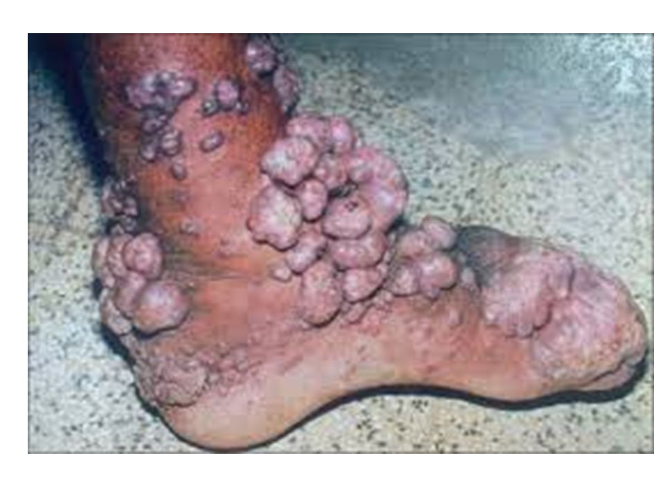
Mycetoma( Madura foot) Appears as a localized abscess,usually on the feet, that discharges pus,serum, and blood through sinuses (“abnoramal channel”) The infection can spread to the underlying bone and result in crippling deformities The most common causative pathogen Madurella grisea and Actinomadura madurae
 Appears as a localized abscess,usually on the feet, that discharges pus,serum, and blood through sinuses ( abnoramal channel ) The infection can spread to the underlying bone and result in crippling deformities The most common causative pathogen Madurella grisea and Actinomadura madurae")
21
Mycetoma( Madura foot) The defining characteristic of mycetoma is the presence of colored grains, composed of compacted hyphae, in the exudate. The color of the grains (black,white,red, or yellow) is characteristic of the causative organism, and thus useful in identifying the particular pathogen The treatment is usually surgical excision because there no effective chemotherapy.
 is characteristic of the causative organism, and thus useful in identifying the particular pathogen The treatment is usually surgical excision because there no effective chemotherapy..")
22
Madura foot

23
Systemic Mycoses The organisms responsible for systemic mycoses fall into two general categories True pathogens Opportunistic pathogens The most common systemic mycotic infection in the immunocompetent hosts in the united states caused by: Coccidiomycosis Histoplasmosis Blastomycosis

24
Systemic Mycoses The clinical manifestations closely resemble those seen in tuberculosis in that asymptomatic primary pulmonary infection is common, whereas chronic pulmonary or disseminated infection is rare Entry into the host is by inhalation of airborne spores, which germinate in the lungs, dissemination can occur to any organ of the body where the fungi can invade and destroy the tissue The fungi causing this diseases are dimorphic

25
Clinical Significance Most cases of coccidioidomycosis, histoplasmosis and paracoccidioidomycosis, present mild in healthy patients and self limiting. In immunosuppressed patients, the same infections can be life –threatening

26
Coccidioidomycosis Caused by Coccidioides immitis In the soil,the fungus generates spores by septation of hyphal filaments (arthrospores) These spores become readily airborne,and enter the lungs,where they germinate and develop into large spherules filled with many endospores. Rupture of the spherules releases the endospores,each of which can form new spherule Disseminated disease,lesion occur most often in the bone and CNS where they result in meningitis

27
Histoplasmosis Is caused by Histoplasma capsulatum In the soil, the fungus generates conidia Pulmonary infections may be: acute but relatively benign and self limiting or Chronic,progressive,and fatal AIDS patients who live in or travel through endemic areas are especially at risk The clinical manifestation of histoplasmosis often resembling tuberculosis

28
Blastomycosis Caused by Blastomyces dermatitidis Like Histoplasma, the fungus produces microconidia,most often in the soil, which become airborne and enter the lungs where they germinate into thick-walled yeast cells Initial pulmonary infections rarely disseminated to other sites When dissemination occurs, the secondary sites are skin (70%),bone (30%),and genitourinary tract (20%)
,bone (30%),and genitourinary tract (20%)")
29
Paracoccidioidomycosis Also called (South American blastomycosis) Caused by Paracoccidioides brasiliensis The clinical manifestations like that of histoplasmosis and blastomycosis except that,the common secondary site of the infection is the mucosa of the mouth and nose,where painful,destructive lesions may develop Over 90% of patients with symptomatic disease are mature males. It is speculated that female sex hormones may inhibit formation of the yeast form

30
Opportunistic pathogens Absidia corymbifera Aspergillus fumigatus Candida albicans Cryptococcus neoformans Pneumocystis carinii Rhizomucor pusillus Rhizopus oryzae (R.arrhizus)
")
31
The opportunistic mycoses Afflict debilitated and/or immunocompromised individuals,and which are rare in normal individuals The use of immunosuppressive drugs for organ transplantation, the use of chemotherapy in cancer treatment, and AIDS, resulted in expansion of immunocompromised population Fungal infections represent approximately 15% of all nosocomial infections in ICU in the U.S.A with candida species being the most commonly occurring fungal pathogen The opportunistic mycoses most commonly encounter today include the following:

32
Candidiasis(candidosis) Caused by yeast Candida albicans and other candida species which are normal body flora found in the skin,mouth,vagina, and intestines Although termed a yeast, Candida albicans is dimorphic Candida infections have various manifestations depending on the site of infection, for example,oral candidiasis (thrush) presents as raised, white plaques on the oral mucosa,tongue, or gums Vaginal candidiasis presents as itching and burning pain of the vulva and vagina accompanied by a thick or thin white discharge
 Caused by yeast Candida albicans and other candida species which are normal body flora found in the skin,mouth,vagina, and intestines Although termed a yeast, Candida albicans is dimorphic Candida infections have various manifestations depending on the site of infection, for example,oral candidiasis (thrush) presents as raised, white plaques on the oral mucosa,tongue, or gums Vaginal candidiasis presents as itching and burning pain of the vulva and vagina accompanied by a thick or thin white discharge")
33
Oral thrush

34
Cryptococcosis Caused by the yeast Cryptococcus neoformans The organism is especially abundant in soil containing bird (especially pigeon) droppings, although the birds themselves are not infected The organism has characteristic thick capsule that surrounds the budding yeast cell and observable on a background of Indian ink
 droppings, although the birds themselves are not infected The organism has characteristic thick capsule that surrounds the budding yeast cell and observable on a background of Indian ink")
35
Cryptococcosis The common form of cryptococcosis is a mild,subclinical lung infection In immunocompromised patients, the infection often disseminates to the brain and meninges, with fatal consequences In AIDS patients, cryptococcosis is the second most common fungal infection (after candidiasis) and is potentially the most serious
 and is potentially the most serious")
36
Aspergillosis Is caused by several species of the genus Aspergillus, but primarily by Aspergillus fumigatus Aspergillus is rarely pathogenic in the normal host, but can produce disease in immunosuppressed individuals, and patients treated with broad -spectrum antibiotics. The aspergilli are ubiquitous growing only as filamentous molds and producing prodigious numbers of conidiospores They reside in the soil, decomposing organic matter, and dust Aspergillosis manifested in several forms depend on the immunologic state of health of the patients

37
1-acute Aspergillosis Infections The most severe, and often fatal, form of aspergillosisis acute invasive infection of the lung, from which the infection can be disseminated to the brain,the GI,and other organs A less severe,noninvasive lung infection gives rise to fungus ball (aspergilloma) a mass of hyphal tissue that can form in lung cavities derived from prior diseases, such as tuberculosis Although the lung is the most common primary site of infections, the eye, ear,nasal sinuses, and skin can also be primary sites
 a mass of hyphal tissue that can form in lung cavities derived from prior diseases, such as tuberculosis Although the lung is the most common primary site of infections, the eye, ear,nasal sinuses, and skin can also be primary sites")
38
2- Allergic Reaction To Aspergillus A relatively rare condition, termed allergic aspergillosis Can rise from the mere inhalation of the spores The allergic reaction results in the formation of mucous that can block the bronchi

39
Mucormycosis Caused most often by Rhizopus oryazae (R.arrhizus) Less often caused by members of the order Mucorales, such as Absidia corymbifera, and Rhizomucor pusillus These organisms are ubiquitous in nature, and their spores are found in great abundance on rotting fruit and old bread
 Less often caused by members of the order Mucorales, such as Absidia corymbifera, and Rhizomucor pusillus These organisms are ubiquitous in nature, and their spores are found in great abundance on rotting fruit and old bread")
40
Mucormycosis Mucor infections occur worldwide, but are almost entirely restricted to individuals with some underlying predisposing condition such as burn,leukemias,and diabetes mellitus The most common fatal disease within a week is rhinocerebral mucormycosis, in which the infection begins in the nasal mucosa or sinuses,and progresses to the orbits, the palate, and brain

41
Pneumocystis carinii pneumonia (PCP) Caused by Pneumocystis carinii Previously considered a protozoan Lacks the ergosterol,which is essential component of most fungal membranes The disease was rare before the use of the immunosupressive drugs and onset of AIDS Currently is one of the most common opportunistic diseases of individuals infected with HIV-1 and 100% fatal if untreated
 Caused by Pneumocystis carinii Previously considered a protozoan Lacks the ergosterol,which is essential component of most fungal membranes The disease was rare before the use of the immunosupressive drugs and onset of AIDS Currently is one of the most common opportunistic diseases of individuals infected with HIV-1 and 100% fatal if untreated")
42
Laboratory identification Standard media –Sabouraud’s agar, potato dextrose agar, low P H 5.0,inhibits bacterial growth but allows fungal colonies to form cultures can be started from spores or hyphae fragments Specimens :blood, pus, CSF, sputum,tissue biopsies, skin scrapings, nail clippings Identification by morphology of conidia structure and carbohydrate assimilation tests

43
Laboratory Diagnosis of Fungal Infection Specimens Depend on the site of infection Systemic:- Blood culture antigen testing e.g. cryptococcal and histoplamosis Pneumonia:-Bronchoscopy washing or brushings for staining and fungal culture of bronchial biopsy

44
Laboratory Diagnosis of fungal infections Meningitis: Cerebral fluid for methylene blue staining and indian ink and cryptococcal antigen and fungal culture If skin infection require skin scrapings If nail infection require nail clippings Galactomannan antigen testing for aspergillus infection

45
Types of Test Carried Out Fungal staining-methylene blue staining or preparation wet using KOH to dissolve tissue material Fungal culture on media that encourages fungal growth e. g. PDA Antigen Testing i.e. to test for antigen present in the wall of fungus e.g cryptococcal antigens, galactomannan used in serum and CSF samples PCR not used on a routine basis on samples

46
Management of Fungal infections Some such as superficial skin infections require topical therapy only with cream e.g. pessaries for vaginal candidiasis Some require oral therapy for skin and nail infection up to 1 year e.g. terbinafine In the immunocompromised systemic therapy require e.g. voriconazol, fluconazole or amphotericin

47
Management of Fungal Infection Important to diagnose fungal infections early in the immunocompromised as there is a high mortality associated with infection Empirical therapy often started in advance of laboratory diagnosis in these patients

Similar presentations
>")


