Download presentation
Presentation is loading. Please wait.

1
Management of acute Pancreatitis By Ibrahim ALanbari Fahed Almutairi Abdullah Mubarki

3
Anatomy The pancreas is retroperitoneal organ. Divided into : head, neck, body and tail The head lies within the curve of the duodenum The tip of the pancreatic tail extends up to the splenic hilum Pancreas has exocrine cells – 98% and endocrine cells – 2% The head received arterial supply from coeliac and superior mesenteric arteries, and the body and the tail from splenic artery The body and neck of the pancreas drain into splenic vein; the head drains into the superior mesenteric and portal veins

4
Acinar cells produce digestive enzymes – amylase, lipase,Trypsinogen … Hormone producing cells are islets of langerhans, containing :B cells (producing insulin), A cells(producing glucagon); D cells (producing somatostatin) pancreatic polypeptide The main pancreatic duct joins the common bile duct to enter the duodenum as a single duct at the ampulla of vater
, A cells(producing glucagon); D cells (producing somatostatin) pancreatic polypeptide The main pancreatic duct joins the common bile duct to enter the duodenum as a single duct at the ampulla of vater")
5
Acute Pancreatitis It is acute inflammation of pancreas, which occurs in previously normal pancreas and returns to normal after resolution.

7
Symptoms Sudden Severe, constant, epigastric pain, with radiation to the back. It can be experienced in either hypochondrium. Associated with: nausia, vomiting and retching

8
Leaning forward

9
Signs 1- Tendernass and gaurding in the epigastric region which is much less than might expected from the history. Generalized peritonism warrant further investigation to exclude other intra-abdominal pathology. 2- Tachycardia, tachypnia and hypotension indicate severe pancreatitis 3- Jaundice : due to impacted stone in the ampulla or due to the pressure of edematous pancreas on the distal common bile duct. It should raise the possibility of co-existing cholangitis.

10
Specific clinical signs in severe necrotizing Pancreatitis Cullen signs – periumblical bruising Greyturner’s sign – flank bruising

11
Investigations serum amylase concentration. The diagnostic cut-off for serum amylase is three times the upper reference limit. Serum lipase is alternative and more specific. Other underlying causes of hyperamylasemia in patients with abdominal pain include : mesentric vascular ischemia, small bowel strangulation, perforated doudenal ulcer, rupture aortic aneurysm, ruptured ectopic pregnancy, acute cholecystitis. Others :CBC, urea and electrolytes, liver function test, LDH, lipid profile, coagulation profile, serum calcium, C-reactive protein, blood glucose.

12
Imaging Chest X-ray should be done to look for pleural effusion due to acute pancreatitis, and it may show air under the diaphragm in cases of perforated peptic ulcer. Ultrasound abdomen to look for the presence of gallstone as underlying cause of acute pancreatitis Abdominal computed tomography is indicated to clarify the diagnosis if the diagnosis of acute pancreatitis still in doubt, to look for the complications of acute pancreatitis or to assess for any evidence of necrotizing pancreatitis.

13
Assessment of severity of acute pancreatitis Ranson’s Criteria APACHE II score ≥ 8 APACHE II Organ failure Substantial pancreatic necrosis (at least 30% glandular necrosis according to contrast-enhanced CT)CT
CT")
15
Treatment Most of attacks of acute pancreatitis can be improve with conservative treatment including : 1. Pain relief : opiates administration 2. Fluid resuscitation : patients with severe pancreatitis require large volumes of fluid to maintain adequate urine output and blood pressure. Adequate early resuscitation in such cases is the most important consideration in early treatment. 3. Antibiotics prophylaxis: it is indicated in severe necrotizing pancreatitis and the recommended antibiotics is the imipenem or meropenem 4. Nutritional support : patients with severe pancreatitis who is unable to resume normal oral diets within 72 hours require nutritional support. This is best delivered by an enteral rather than parenteral route.

16
Endoscopic treatment: gallstone pancreatitis is due to the transient impaction of a stone at the papilla causing pancreatic duct obstruction. ERCP with sphincterotomy is indicated in patient with acute pancreatitis with persistent obstructive jaundice with or without cholangitis. Surgical treatment: patients with gallstone related acute pancreatitis should undergo cholecystectomy ( mild pancreatitis during the index admission while severe pancreatitis interval cholecystectomy in 8 – 12 weeks after resolution of the attach of severe acute pancreatitis). Note: Surgery is indicated in patient with infected necrotizing pancreatitis or in patient with sterile necrotizing pancreatitis who deteriorate and developed progressive multi-organ failure.
. Note: Surgery is indicated in patient with infected necrotizing pancreatitis or in patient with sterile necrotizing pancreatitis who deteriorate and developed progressive multi-organ failure..")
17
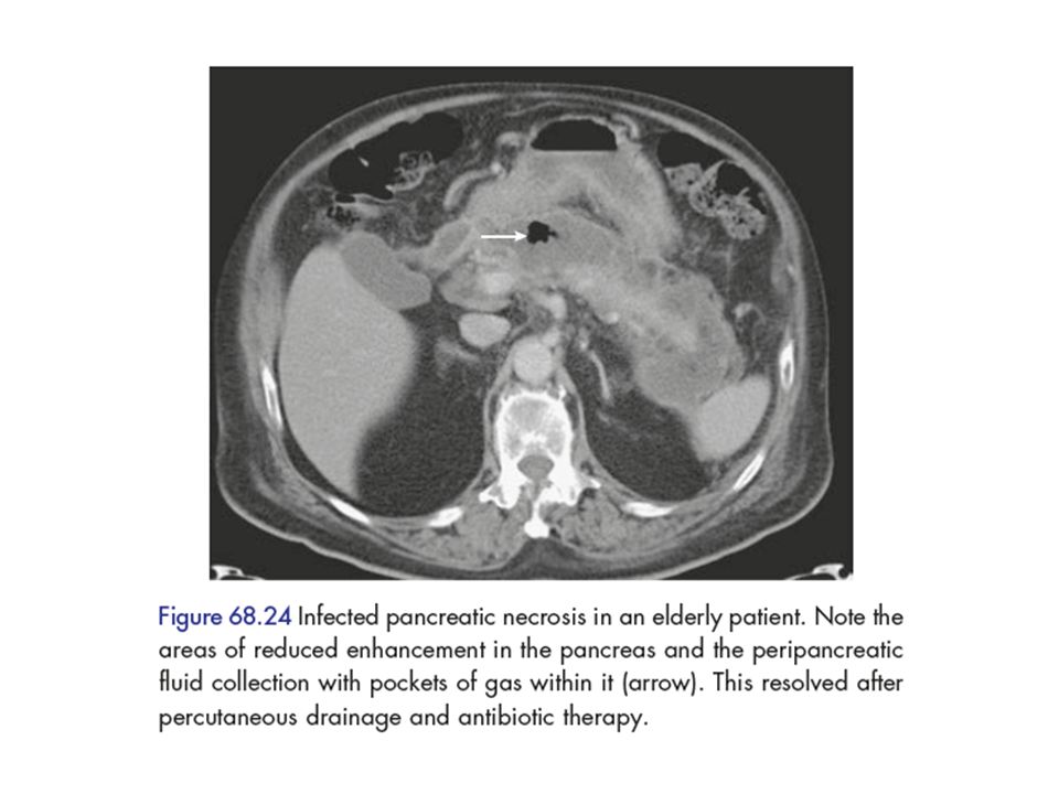
: Complications Infected pancreatic necrosis It is a serious complication which can develops in acute necrotizing pancreatitis with high mortality rate. It can be diagnosed by presence of gas bubbles in CT scan at the area of pancreas or by fine needle aspiration under imaging guidance. patients need intensive management with antibiotics coverage and maybe surgical intervention: Surgical debridement, Percutaneous drainage, debridement Endoscopic drainage

20
Pancreatic pseudocyst It is a collection of pancreatic secretions and inflammatory exudate enclosed in a wall of fibrous or granulomatous tissue. It is differs from a true cyst in that collection has no epithelial lining. It form commonly in the lesser sac near the pancreas and persist for 4 weeks or more from the onset of acute pancreatitis. It need drainage if : it persists more than 6 week after the attack of pancreatitis, the size > 6 cm and symptomatic

22
Pancreatic abscess It is a circumscribed intra-abdominal collection of pus, usually in proximity to the pancreas. This is a result of infection of a pseudocyst. It need urgent drainage.

23
Gastrointestinal bleeding Severe pancreatitis may be complicated by bleeding from gastritis, erosion or duodenal ulceration. Thrombosis of splenic vein can lead to splenomegaly, gastric fundal varices which can result in massive upper gastrointestinal bleeding ( sinistral, left sided, compartmental portal hypertension).
..")
24
Progressive jaundice Due to impacted stone in the papilla or compression of distal common bile duct by enlarged edematous pancreas.

Similar presentations


![GI Endoscopy ~ BASIC ~ ESOPHAGUS - EOSINOPHILIC ESOPHAGITIS ESOPHAGUS - EOSINOPHILIC ESOPHAGITIS EOSINOPHILIC ESOPHAGITIS IN CHILDREN [LECTURE] EOSINOPHILIC.](/20/5943603/big_thumb.jpg "GI Endoscopy ~ BASIC ~ ESOPHAGUS - EOSINOPHILIC ESOPHAGITIS ESOPHAGUS - EOSINOPHILIC ESOPHAGITIS EOSINOPHILIC ESOPHAGITIS IN CHILDREN [LECTURE] EOSINOPHILIC.>")


