Download presentation
Presentation is loading. Please wait.

1
The liver Surgical anatomy - Largest solid organ 1200-1800g Position: wedge shape from RT hypochondrium-epigastric- LT hypochondrium. surfaces (2 ) parietal & visceral surfaces (2 ) parietal & visceral Lesser omentum: fold of peritoneum connect lesser curvature of stomach with the visceral surface of liver. The free border contains :CBD- hepatic A- portal vein. Blood supply & venous drainage : Blood supply & venous drainage : - hepatic A from coeliac trunk - portal vein formed by union of SMV &splenic V. - Three hepatic V drains into IVC Lymphatic drainage: LN at porta-hepatis – coeliac LN – some--- thoracic duct. LN at porta-hepatis – coeliac LN – some--- thoracic duct.
 parietal & visceral surfaces (2 ) parietal & visceral Lesser omentum: fold of peritoneum connect lesser curvature of stomach with the visceral surface of liver. The free border contains :CBD- hepatic A- portal vein. Blood supply & venous drainage : Blood supply & venous drainage : - hepatic A from coeliac trunk - portal vein formed by union of SMV &splenic V. - Three hepatic V drains into IVC Lymphatic drainage: LN at porta-hepatis – coeliac LN – some--- thoracic duct. LN at porta-hepatis – coeliac LN – some--- thoracic duct..")
4
Surgical physiology - liver is a busy organ-1.5 L of blood/ min. 2/3 portal V – 1/3 hepatic A. Functions : 1- formation & secretion of bile. 2- CHO, protein & fat metabolism. 3- metabolism of many drugs & hormones. 4- removal of ammonia. 5- liver is the storage house of glycogen, vit.B12, vit.A, iron & copper. 6- reticuloendothelial cells clear the blood from bacteria that can escape from the intestine to the portal circulation.

5
INVESTIGATIONS A – Liver function tests: 1- total serum bilirubin. Direct & indirect. 2- AST serum aspartate aminotransferase. 3- ALT serum alanine aminotransferase. 4- alkaline phosphotase. 5- serum albumin. 6- prothrombin time. B – Imaging of liver: US- CT- MRI- arteriography C – Liver biopsy: per cutaneous – laparoscopy- laparotomy.

6
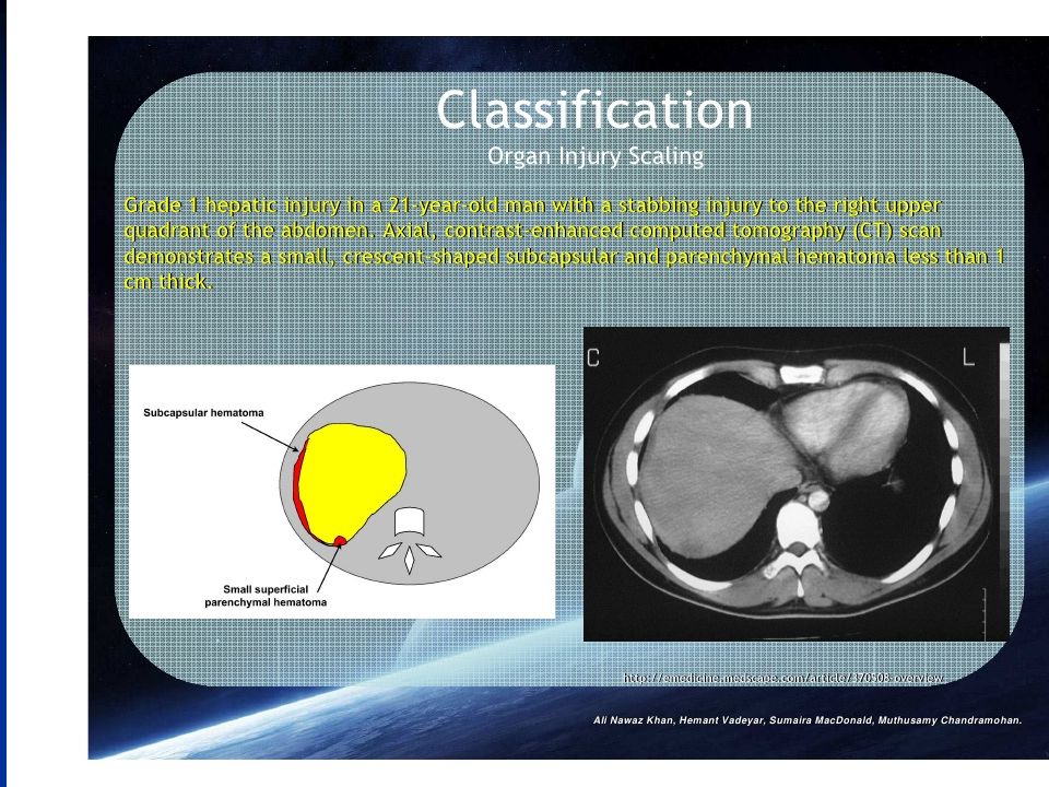
Liver trauma - 2 nd common solid organ after spleen. - associated injury: ribs, pleura, lung, colon & spleen. Aetiology: 1- accidental trauma: blunt (RTA) & penetrating (bullet, stab). 2- iatrogenic injury: percut biopsy, PTC. 3- spontaneous rupture (eclampsia, hepatic tumour). Types of injury: 1- small subcapsular hematoma. 2- small superficial tear or tears. 3- large subcapsular or intrahepatic hematoma. 4- large deep tear or tears. 5- shattered liver. 6- vascular injury : most difficult hepatic V Consequences: 1- bleeding. 2- hematobilia.
 & penetrating (bullet, stab). 2- iatrogenic injury: percut biopsy, PTC. 3- spontaneous rupture (eclampsia, hepatic tumour). Types of injury: 1- small subcapsular hematoma. 2- small superficial tear or tears. 3- large subcapsular or intrahepatic hematoma. 4- large deep tear or tears. 5- shattered liver. 6- vascular injury : most difficult hepatic V Consequences: 1- bleeding. 2- hematobilia..")
7
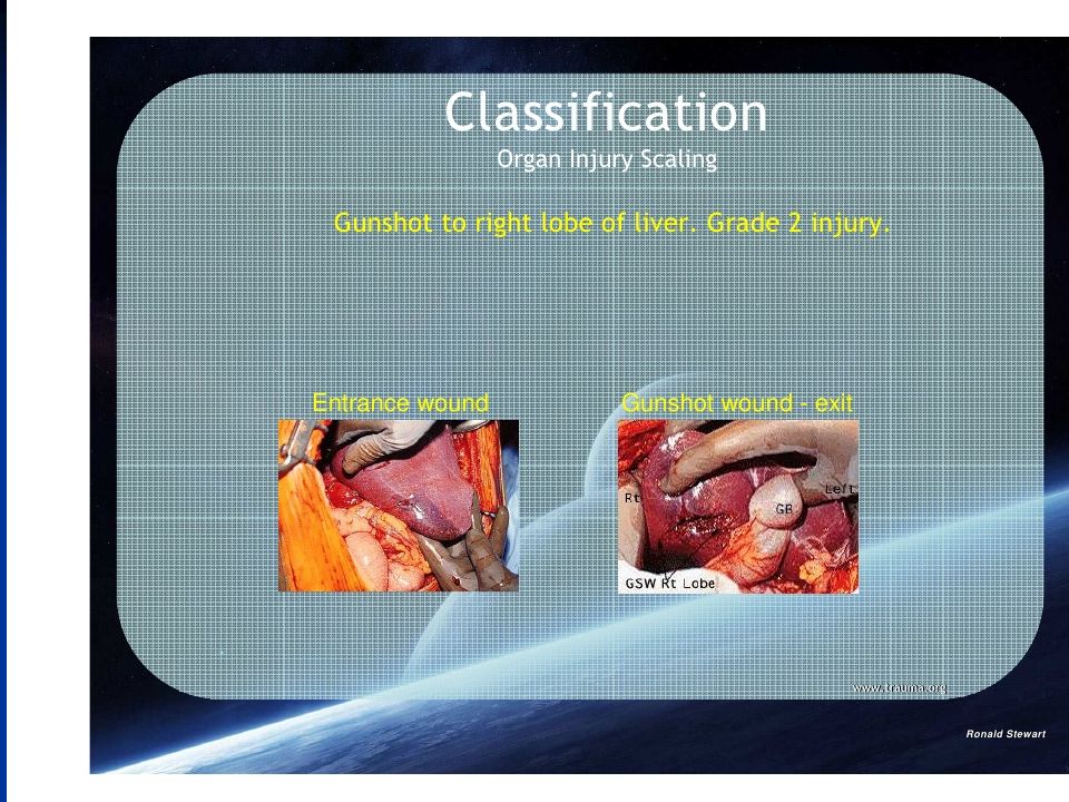
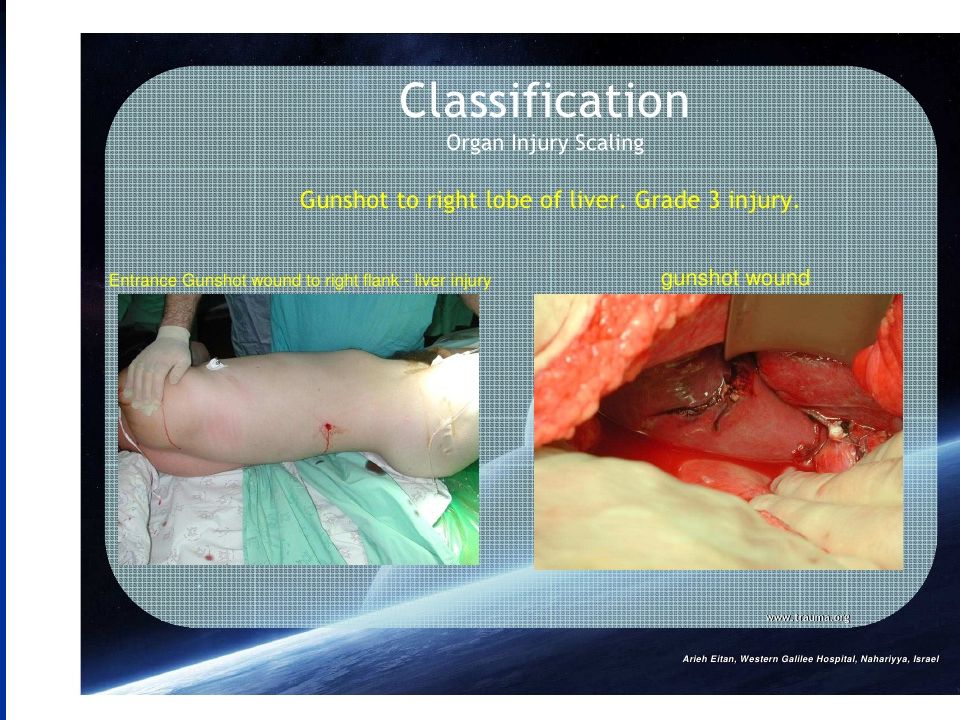
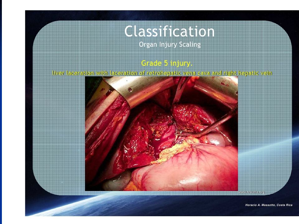
The AAST Liver injury grading system is as follows The AAST Liver injury grading system is as follows grade I : grade I : o haematoma : sub capsular, < 10% surface area o haematoma : sub capsular, < 10% surface area o laceration : capsular tear, < 1cm depth grade II : grade II : o haematoma: sub capsular, 10 - 50% surface area o haematoma: sub capsular, 10 - 50% surface area o haematoma : intraparenchymal < 10cm diameter o haematoma : intraparenchymal < 10cm diameter o laceration: capsular tear, 1 - 3cm depth, < 10cm length grade III : grade III : o haematoma : sub capsular, > 50% surface area, or ruptured with active bleeding o haematoma : intraparenchymal > 10 cm diameter o laceration : capsular tear, > 3 cm depth grade IV : grade IV : o haematoma : ruptured intraparenchymal with active bleeding o laceration : parenchymal distruption involving 25 - 75% hepatic lobes or o 1 - 3 Couinaud segments (within one lobe) Couinaud segments Couinaud segments grade V : grade V : o laceration : parenchymal distruption involving >75% helpatic lobe or o > 3 Couinaud segments (within one lobe) Couinaud segments Couinaud segments o vascular : juxtahepatic venous injuries (IVC, major hepatic vein) grade VI : vascular : hepatic avulsion grade VI : vascular : hepatic avulsion
 Couinaud segments Couinaud segments grade V : grade V : o laceration : parenchymal distruption involving >75% helpatic lobe or o > 3 Couinaud segments (within one lobe) Couinaud segments Couinaud segments o vascular : juxtahepatic venous injuries (IVC, major hepatic vein) grade VI : vascular : hepatic avulsion grade VI : vascular : hepatic avulsion")
13
Clinical features & diagnosis: 1- history of trauma. 2- abdominal pain. 3- abdominal tenderness & rigidity. 4- lower rib fractures. 5- massive bleeding--- hemorrhagic shock. 6- minor bleeding---DPL, US, CT. 7- during laparotomy. Treatment: 1- minor hemorrhage & small tear can be conservatively followed by regular CT. 2- continuous bleeding calls for surgical interference. 3- serious liver injuries require urgent laparotomy.

14
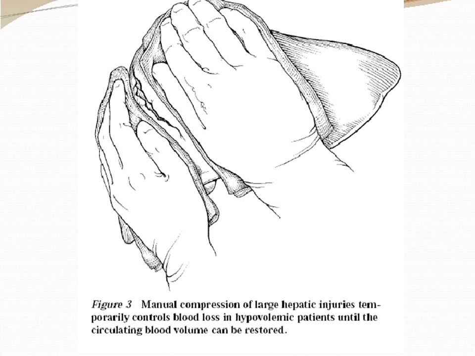
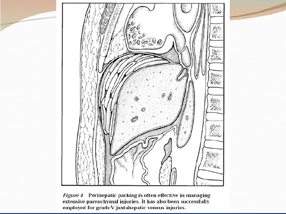
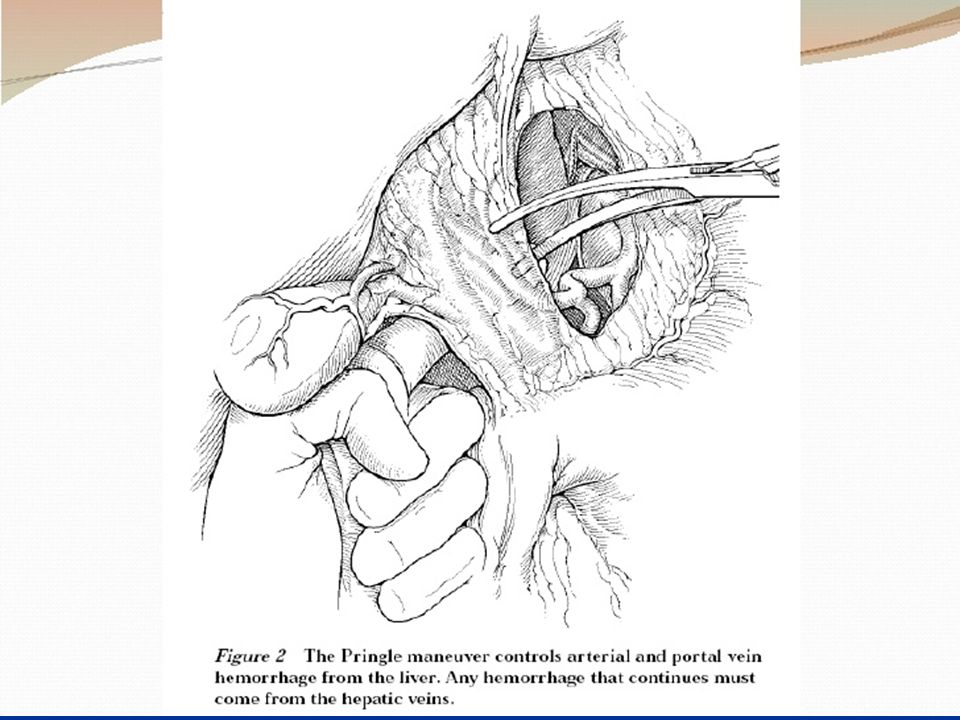
Principles of surgical management : 1- adequate exposure – longitudinal incision. 2- arrest bleeding. -small tear---spontaneous. -brisk liver hemorrhage --- packing--- Pringle`s maneuver (clamp, finger) for 20 min. -tying sutures over pedicled omentum, deep transverse mattress sutures using special liver needle. 3- hematoma – explore – ligation damaged vessels & ducts & excise dead tissue. 4- shattered lobe --- excise the lobe. 5- firm packing of inaccessible & difficult bleeding areas e.g hepatic veins. Mortality rate 15-20% & this percentage increase if there are associated injuries.
 for 20 min. -tying sutures over pedicled omentum, deep transverse mattress sutures using special liver needle. 3- hematoma – explore – ligation damaged vessels & ducts & excise dead tissue. 4- shattered lobe --- excise the lobe. 5- firm packing of inaccessible & difficult bleeding areas e.g hepatic veins. Mortality rate 15-20% & this percentage increase if there are associated injuries..")
Similar presentations