Download presentation
Presentation is loading. Please wait.

2
THE TISSUES Laboratory Manual for Anatomy and Physiology. Custom edition for Miami Dade College-Kendall Campus. BSC2085L by Michael G. Wood.

3
ALFONSO A. PINO MD.

4
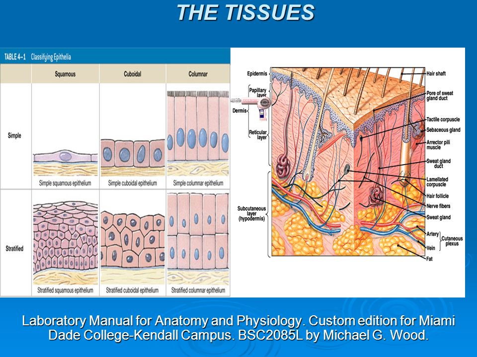
TISSUE CONCEPT- It is a collection of specialized cells & cell products that are organized to perform functions 4 TYPES : EPITHELIAL TISSUE CONNECTIVE TISSUE MUSCLE TISSUE NEURAL TISSUE

5
it covers exposed surfaces (skin), EPITHELIAL Tissue- it covers exposed surfaces (skin), lines internal passages & chambers in (Digestive, respiratory, reproductive and, urinary systems) it forms glands it fills internal spaces, CONNECTIVE Tissue- it fills internal spaces, provides structure support, transports material within the body & stores energy it contracts to perform movement. MUSCLE TISSUE- it contracts to perform movement. it generates heat that warms the body It carries information from one part of NEURAL TISSUE- It carries information from one part of the body to another by electrical impulses

6
OF THE EPITHELIAL TISSUES FUNCTIONS OF THE EPITHELIAL TISSUES from abrasion, dehydration, PHYSICAL PROTECTION- from abrasion, dehydration, chemical or biological agents. regulated by hormones, CONTROL Permeability- regulated by hormones, transport ions & nutrients. touch receptors, PROVIDES SENSATIONS- touch receptors, neuroepithelium conteins sensory cells that produce sensations of smell, taste, sigth, equillibrium or hearing. glands produces PROVIDES SECRETIONS- glands produces secretions for physical protection, chemical messangers in interstitial fluid & blood.

7
CLASSIFICATION OF THE EPITHELIA - SQUAMOUS-thin & flat SHAPE- SQUAMOUS-thin & flat CUBOIDAL- like little hexagonal boxes COLUMNAR-taller & more slender NUMBER OF LAYERS SIMPLE- single layer of cells STRATIFIED- several layers of cells COMBINATIONS OF SHAPE & NUMBER

8
Classification of Epithelia Classes of Epithelia Based on shape Squamous epithelia: thin and flat Cuboidal epithelia: square shaped Columnar epithelia: tall, slender rectangles Based on layers Simple epithelium: single layer of cells Stratified epithelium: several layers of cells ALFONSO A. PINO MD.

9
Martini pg 111

10
WITH EXAMPLES TYPES OF EPITHELIAL TISSUES WITH EXAMPLES Lung, serous membranes, lining heart SIMPLE SQUAMOUS- Lung, serous membranes, lining heart epidermis, mouth, throat, vagina STRATIFIED SQUAMOUS- epidermis, mouth, throat, vagina glands, ducts, kidney tubules SIMPLE CUBOIDAL- glands, ducts, kidney tubules linings some ducts STRATIFIED CUBOIDAL- linings some ducts - urinary bladder, ureters TRANSITIONAL- urinary bladder, ureters SIMPLE COLUMNAR- stomach, gallbladder, uterine tubes PSEUDOSTRATIFIED CILIATED COLUMNAR EPITHELIUM- respiratory tract salivary glands ducts STRATIFIED COLUMNAR EPITHELIUM- salivary glands ducts

11
ALFONSO A. PINO MD.

12
Squamous Epithelia Simple squamous epithelium Absorption and diffusion Mesothelium Lines body cavities Endothelium Lines heart and blood vessels

14
Transitional epithelium Appearance changes as stretching occurs, Tolerates repeated cycles of stretching and recoiling and returns to its previous shape without damage.

15
ALFONSO A. PINO MD. Simple columnar epithelium

16
ALFONSO A. PINO MD. Stratified columnar epithelium

17
ALFONSO A. PINO MD. Pseudostratified ciliated columnar epithelium

18
Connective Tissue Connect epithelium to the rest of the body (basal lamina) Provide structure (bone) Store energy (fat) Transport materials (blood) Have no contact with environment
 Provide structure (bone) Store energy (fat) Transport materials (blood) Have no contact with environment")
19
FUNCTIONS OF THE CONNECTIVE TISSUE It forms an structural framework It transports fluid & materials It protects delicates organs It supports, surrounds & interconnects tissues It storages energy It contains cells that defend the body from microorganisms Characteristics of Connective Tissues Specialized cells Solid extracellular protein fibers Fluid extracellular ground substance The extracellular components of connective tissues (fibers and ground substance) make up the matrix
 make up the matrix")
20
Connective Tissue Classification of Connective Tissues Connective tissue proper Connect and protect Fluid connective tissues Transport Supportive connective tissues Structural strength

21
Connective Tissue Categories of Connective Tissue Proper Loose connective tissue More ground substance, less fibers For example, fat (adipose tissue) Dense connective tissue More fibers, less ground substance For example, tendons
 Dense connective tissue More fibers, less ground substance For example, tendons")
22
Copyright © 2009 Pearson Education, Inc., publishing as Pearson Benjamin Cummings Connective Tissues Fibroblasts Fibrocytes Macrophages Adipocytes Mesenchymal cells Melanocytes Mast cells Lymphocytes Microphages Nine Cell Types of Connective Tissue Proper

23
ALFONSO A. PINO MD. CHARACTERISTICS OF THE CONNECTIVE TISSUE COLLAGEN FIBERS They are & unbranched They are long, straigth & unbranched They are a bundle of fibrous protein subunits wound together They have little stretch, but great tensile strengh They form tendons and ligaments RETICULAR FIBERS They have same subunits than collagen fibers But with a But with a different physical arrngement They form a network that resists forces apply From many directions and They stabilize relative position of cells, organs, blood vessels, nerves & other structures

24
ELASTIC FIBERS They contein protein elastin They are branched & wavy After streching, they can return to their original length They lack the tensile strength of collagen They dominate in elastic ligaments

25
TYPES OF CONNECTIVE TISSUES AREOLAR ADIPOSE RETICULAR DENSE REGULAR DENSE IRREGULAR ELASTIC CARTILAGE BONE BLOOD LYMPH

26
Areolar tissue Areolar Tissue Least specialized Open framework Viscous ground substance Elastic fibers Holds blood vessels and capillary beds For example, under skin (subcutaneous layer)
")
27
Adipose tissue

28
Reticular tissue

29
Dense regular connective tissue

30
Dense irregular connective tissue

31
Elastic tissue

32
Supportive Connective Tissues Support soft tissues and body weight Cartilage Gel-type ground substance For shock absorption and protection Bone Calcified (made rigid by calcium salts, minerals) For weight support
 For weight support")
33
Copyright © 2009 Pearson Education, Inc., publishing as Pearson Benjamin Cummings Supportive Connective Tissues Cartilage Matrix Proteoglycans derived from chondroitin sulfates Ground substance proteins Chondrocytes (cartilage cells) surrounded by lacunae (chambers)
 surrounded by lacunae (chambers)")
34
Supportive Connective Tissues Cartilage Structure No blood vessels: Chondrocytes produce antiangiogenesis factor Perichondrium: Outer, fibrous layer (for strength) Inner, cellular layer (for growth and maintenance)
 Inner, cellular layer (for growth and maintenance)")
35
Supportive Connective Tissues Figure 4–13 The Growth of Cartilage.

36
Supportive Connective Tissues Types of Cartilage Hyaline cartilage Stiff, flexible support Reduces friction between bones Found in synovial joints, rib tips, sternum, and trachea Elastic cartilage Supportive but bends easily Found in external ear and epiglottis Fibrous cartilage (fibrocartilage) Limits movement Prevents bone-to-bone contact Pads knee joints Found between pubic bones and intervertebral discs
 Limits movement Prevents bone-to-bone contact Pads knee joints Found between pubic bones and intervertebral discs")
37
Supportive Connective Tissues Figure 4–14 The Types of Cartilage.

38
Supportive Connective Tissues Figure 4–14 The Types of Cartilage.

39
Supportive Connective Tissues Figure 4–14 The Types of Cartilage.

40
Bone tissue

41
ALFONSO A. PINO MD. Blood

42
Muscle Tissue Specialized for contraction Produces all body movement Three types of muscle tissue Skeletal muscle Large body muscles responsible for movement Cardiac muscle Found only in the heart Smooth muscle Found in walls of hollow, contracting organs (blood vessels; urinary bladder; respiratory, digestive, and reproductive tracts)
")
43
Copyright © 2009 Pearson Education, Inc., publishing as Pearson Benjamin Cummings

44
Muscle Tissue Classification of Muscle Cells Striated (muscle cells with a banded appearance) Nonstriated (not banded; smooth) Muscle cells can have a single nucleus Muscle cells can be multinucleate Muscle cells can be controlled voluntarily (consciously) Muscle cells can be controlled involuntarily (automatically )
 Nonstriated (not banded; smooth) Muscle cells can have a single nucleus Muscle cells can be multinucleate Muscle cells can be controlled voluntarily (consciously) Muscle cells can be controlled involuntarily (automatically )")
45
Copyright © 2009 Pearson Education, Inc., publishing as Pearson Benjamin Cummings Muscle Tissue Skeletal Muscle Cells Are long and thin Are usually called muscle fibers Do not divide New fibers are produced by stem cells (myosatellite cells)
")
46
Muscle Tissue Cardiac muscle cells Are called cardiocytes Form branching networks connected at intercalated discs Are regulated by pacemaker cells

47
Copyright © 2009 Pearson Education, Inc., publishing as Pearson Benjamin Cummings Muscle Tissue Smooth muscle cells Are small and tapered Are small and tapered Can divide and regenerate Can divide and regenerate Figure 4–18 Muscle Tissue.

48
Neural Tissue Also called nervous nerve tissue Also called nervous or nerve tissue Specialized for conducting electrical impulses Specialized for conducting electrical impulses Rapidly senses internal or external environment Rapidly senses internal or external environment Processes information and controls responses Processes information and controls responses Neural tissue is concentrated in the Neural tissue is concentrated in the central nervous system Brain Spinal cord

49
Neural Tissue Two Kinds of Neural Cells Neurons Nerve cells Perform electrical communication Neuroglia Supporting cells neuronsRepair and supply nutrients to neurons

50
Neural Tissue Cell Parts of a Neuron Cell body Contains the nucleus and nucleolus Dendrites Short branches extending from the cell body Receive incoming signals Axon (nerve fiber) Long, thin extension of the cell body Carries outgoing electrical signals to their destination
 Long, thin extension of the cell body Carries outgoing electrical signals to their destination")
51
REMEMBER, GO TO THE TUTORING ROOM AND PRACTICE WITH MODELS! ROOM 3326

Similar presentations