Download presentation
Presentation is loading. Please wait.

1
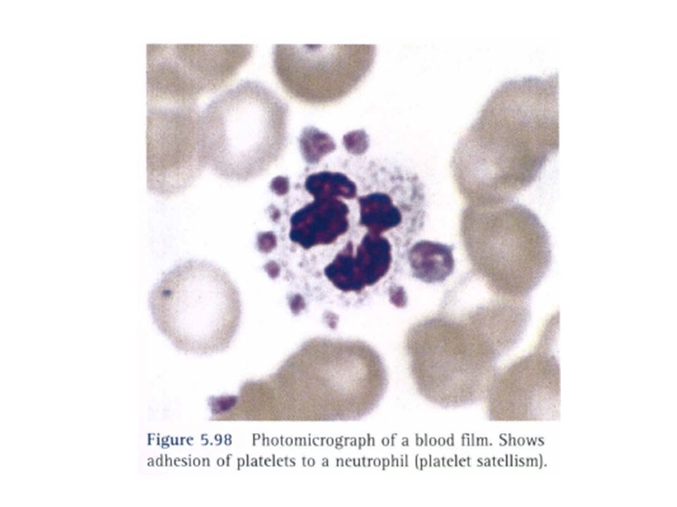
PLATELET MORPHOLOGY Normal platelets are I-3micron in diameter. They are irregular in outline with fine red granules that may be scattered or centralized. A small number of larger platelets, up to 5 micron in diameter, may be seen in normal films

2
Larger platelets are seen in the blood when platelet production is increased and in hyposplenism

7
Occasionally, platelets may be seen adhering to neutrophils This haspatients who have demonstrable antiplatelet autoantibodies, but it is more commonly seen in apparently healthy individuals. It is not seen in films made directly from blood that has not been anticoagulated.

11
Related&Erythrocyte Abnormalities

13
Mature Red Blood Precursor Cells: Polychromatic and Orthochromatic Erythroblasts (Normoblasts) and Reticulocytes
 and Reticulocytes")
44
Elliptocytes are often present in large numbers in hereditary elliptocytosis. In hereditary pyropoikilocytosis, elliptocytes are only one of the many types of poikilocyte present

45
Southeast Asian ovalocytosis is characterized by the presence of a variable number of elliptocytes, macro-ovalocytes, and stomatocytes

48
The terminology applied to spiculated cells has been confusing because the same terms have been used to designate different types of cells. For this reason the term "burr cell" should be discarded and the terms recommended by Bessis7 should be adopted

49
four typ schistocyte, keratocyte, acanthocyte, and echinocyte. The term echin0cyte is used for the crenated cell. It is differentiated from the acanthocyte on the basis of the number, shape, and disposition of the spicules of spiculated cell

50
Schistocytes or erythrocyte fragments are found in many blood diseases. They are smaller than normal red cells and of varying shape. Sometimes they have sharp angles or spines (spurs

51
and sometimes they are round in contour, usually staining deeply but occasionally palely as the result of loss of haemoglobin at the time of fragmentation.

55
Keratocytes have pairs of spicules, usually either one pair or two pairs. They may be formed either by removal of a Heinz body by the pitting action of the spleen or by mechanical damage The terms "helmet cell" and "bitecell" have sometimes been used to describe keratocytes

58
The term acanthocytosis was introduced to describe an abnormality of the red cell in which there are a small number of spicules of inconstant length, thickness, and shape, irregularly disposed over the surface of the cell

59
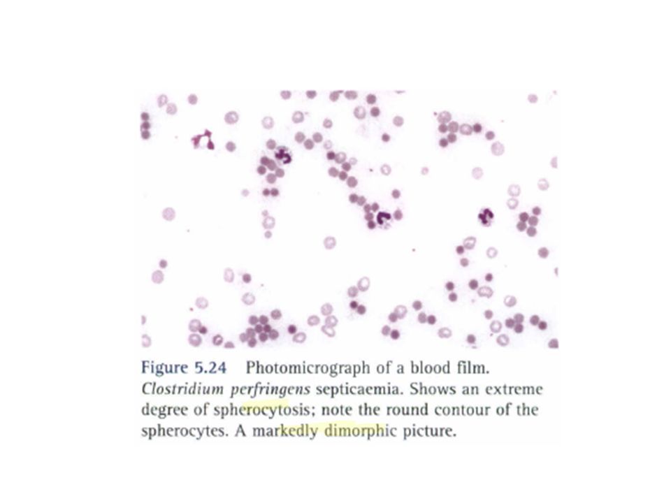
They are present in varying numbers following splenectomy and in hyposplenism. A similar cell occurs in severe liver disease ("spur cell" anaemia).
..")
61
Echinocytosis or crenation describes the process by which red cells develop many or numerous short, regular projections from their surface

62
Crenation regularly develops if blood is allowed to stand overnight at 20°C before films are made in freshly made blood films made from patients suffering from a variety of illnesses, especially uraemia

63
It is also seen in films from patients undergoing cardiopulmonary bypass. Marked echinocytosis has been reported in premature infants after exchange transfusion or transfusion of normal red cells.

64
Crenation also occurs as an artefact if red cells are washed free from plasma and suspended in 9 g/I NaCl between glass surfaces, particularly at a raised pH; it also occurs in the presence of traces of fatty substances on the slides on which films are made and in the presence of traces of chemicals that at higher concentrations cause lysis.

65
If echinocytosis is observed in a film, it usually represents a storage artefact caused by delay imaking the film. It is a warning that morphologic features in the blood film cannot be assessed reliably. If present in films made from fresh blood, it is a clinically significant observation.

66
The term leptocytosis has been used to describe unusually thin red cells, as in severe iron deficiency or thalassaemia in which the cells may stain as rings of membrane with a little attached haemoglobin with large, almost unstained, central areas

78
Cells containing them areregularly present after splenectomy and where there has been splenic atrophy. Usually only a few such cells are present, but they may be numerous in cases of coeliac disease in which there is splenic atrophy and coexisting folate deficiency.

80
Pappenheimer Bodies Pappenheimer bodies are small peripherally sited basophilic (almost black) erythrocyte inclusions They are composed of haemosiderin,and their presence is related to iron overload and hyposplenism
 erythrocyte inclusions They are composed of haemosiderin,and their presence is related to iron overload and hyposplenism")
84
Rouleaux and Autoagglutination This pseudoagglutination owing to massive rouleaux formation may be distinguished from true agglutination in two ways

85
1):By noting that the red cells, although forming parts of larger clumps, are mostly arranged side by side as in typical rouleaux.
:By noting that the red cells, although forming parts of larger clumps, are mostly arranged side by side as in typical rouleaux.")
86
2. By adding 3-4 volumes of 9 g/I NaCl to the preparation. Pseudoagglutination owing to massive rouleaux formation should either disperse completely or transform itself into typical rouleaux. The addition of saline to blood that has undergone true agglutination may cause the agglutinates to break up somewhat, but a major degree of it is likely to persist and typical rouleaux will not be seen

89
Polychromasia In practice, it means that some of the red cells stain shades of bluish grey

90
Polychromasia

91
In myelofibrosis and carcinomatosis, the number of erythroblasts is often disproportionately high for the degree of anaemia, and a few immature granulocytes are usually also present (so-called leucoerythroblastic anaemia)
")
97
Iron Deficiency and Blood Cell Analysis Focusing on the erythrocyte morphology is the quickest and most efficientway to investigate hypochromic anemia when the serum iron has dropped below normal values

98
In hypochromic anemiawith iron and hemoglobin deficiency (whether due to insufficient iron intake or an increased physiological iron requirement), erythrocyte size and shape does not usually vary much
, erythrocyte size and shape does not usually vary much")
99
Only in advanced anemias (from approx. 11 g/dl, equivalent to 6.27 mmol/l Hb) are relatively small erythrocytes (microcytes) with reduced MCV and MCH and grayish stained basophilic erythrocytes (polychromatic erythrocytes) seen, indicating inadequate hemoglobin content.
 are relatively small erythrocytes (microcytes) with reduced MCV and MCH and grayish stained basophilic erythrocytes (polychromatic erythrocytes) seen, indicating inadequate hemoglobin content..")
Similar presentations


. Introduction Hereditary spherocytosis is a class of hemolytic anemia. The disease occurs due to an intrinsic “membrane.>")


 أطياف بتضلها أطياف.>")


