Download presentation
Presentation is loading. Please wait.

1
Computed Tomography(CT, was CAT, Computerized Axial Tomography) rev 2016-02-20 this is now slide 1do not print it to pdf things to do (check off when complete): add revision date to cover page remove triangles create list for pages to print in the handout 2-10,12-17,20-22,29-31 add captions for photo slides incorporate notes taken during presentation add Key Points page 3 useful characters: ° degreesΩ ohmsμ micro ☑ checkbox ☐ slash-zero ØCO 2 O 2 SpO 2 N 2 O ® ™ trademarks à 224 E0 á 225 E1 â 226 E2 ã 227 E3 ä 228 E4 å 229 E5 æ 230 E6 ç 231 E7 Ð 208 D0 è 232 E8 é 233 E9 ê 234 EA ë 235 EB ì 236 EC í 237 ED î 238 EE ï 239 EF Ñ 209 D1 Ò 210 D2 Ó 211 D3 Ô 212 D4 Õ 213 D5 Ö 214 D6 Ø 216 D8 ß 223 DF Þ 222 DE Ù 217 D9 Ú 218 DA Û 219 DB Ü 220 DC Ý 221 DD Þ 222 DE ß 223 DF
 rev this is now slide 1do not print it to pdf things to do (check off when complete): add revision date to cover page remove triangles create list for pages to print in the handout 2-10,12-17,20-22,29-31 add captions for photo slides incorporate notes taken during presentation add Key Points page 3 useful characters: ° degreesΩ ohmsμ micro ☑ checkbox ☐ slash-zero ØCO 2 O 2 SpO 2 N 2 O ® ™ trademarks à 224 E0 á 225 E1 â 226 E2 ã 227 E3 ä 228 E4 å 229 E5 æ 230 E6 ç 231 E7 Ð 208 D0 è 232 E8 é 233 E9 ê 234 EA ë 235 EB ì 236 EC í 237 ED î 238 EE ï 239 EF Ñ 209 D1 Ò 210 D2 Ó 211 D3 Ô 212 D4 Õ 213 D5 Ö 214 D6 Ø 216 D8 ß 223 DF Þ 222 DE Ù 217 D9 Ú 218 DA Û 219 DB Ü 220 DC Ý 221 DD Þ 222 DE ß 223 DF")
2
Computed Tomography [was: Computerized Axial Tomography] CT scanners [or CAT scanners] © D. J. © D. J. McMahon 150504rev cewood 2016-02-20
![Computed Tomography [was: Computerized Axial Tomography] CT scanners [or CAT scanners] © D.](http://images.slideplayer.com/32/10024450/slides/slide_2.jpg "J. © D. J. McMahon rev cewood")
3
Key Points Computed Tomography (CT): Know the essential components of a CT scanner (handout image) Know the general idea of how CT scanners capture an image What value do more detectors offer? Understand the principle of Multislice Spiral Computed Tomography (MSCT)
.")
4
“Tomography” : an image from many slices (Greek: tomos = slice) Digital geometry processing is used to generate a three-dimensional image of the inside of the body from a large series of two-dimensional X-ray images taken around a single axis of rotation.
 Digital geometry processing is used to generate a three-dimensional image of the inside of the body from a large series of two-dimensional X-ray images taken around a single axis of rotation.")
5
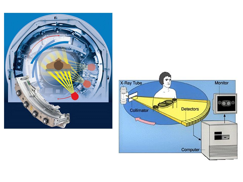
Individual slice data is generated using an X-ray source that rotates around the patient; X-ray sensors are positioned on the opposite side of the circle from the X-ray source and also move, staying 180° from the source. These sensors use scintillation systems based on photo diodes. Many data scans are progressively taken as the body is gradually passed through the gantry. The images are combined together by mathematical procedures known as tomographic reconstruction. Contrast between soft tissues can be increased by injection of iodine-based solutions.

6
Evolution of CT scanning - Increased beam size, and increased number of detectors. Most current installations are third and fourth generation.

7
https://www.youtube.com/watch?v=M-4o0DxBgZk

9
More detectors and smaller detectors give better resolution or faster image capture (e.g. to capture a beating heart) or both. Enhanced image data can be reconstructed thru algorithms Current CT machines can have 320 detectors yielding 256 slices.
 or both. Enhanced image data can be reconstructed thru algorithms Current CT machines can have 320 detectors yielding 256 slices..")
10
Multislice Spiral Computed Tomography (MSCT) Sampling Patterns of a 4-slice spiral scan at different pitch values. At pitch 1 and 2, each z-position is sampled 4 and 2 times respectively. The spacing between samples decreases from d to d/2 when going from pitch 1 to pitch 1.5, then increases again to d when increasing the pitch to 2. At a pitch of 4, each sample is acquired once and the sampling distance is d (d denotes the slice collimation)
.")
11
64-slice CT unit -

12
TX-ray tube DX-ray detectors XX-ray beam RGantry rotation

13
That gantry rotates at 180 - 240 r.p.m. Open CT runninghttp://www.youtube.com/watch?v=2CWpZKuy-NE

14
Toshiba “Aquilion” - 80 detectors, 160 slices http://medical.toshiba.com/products/ct/aquilion-prime/index.php

15
SpecificationsFirst CT Scanner (circa 1970) Recent CT Scanner Time to acquire one CT image:5 minutes0.5 seconds Pixel size:3 mm x 3 mm0.5 mm x 0.5 mm Number of pixels in an image:6,400256,000 Early scan, ~1975Scan, ~2003
 Recent CT Scanner Time to acquire one CT image:5 minutes0.5 seconds Pixel size:3 mm x 3 mm0.5 mm x 0.5 mm Number of pixels in an image:6,400256,000 Early scan, ~1975Scan, ~2003")
16
Siemens “Definition”scanner: Dual-source CT Two x-ray tubes are operated at different tube potentials: one at a low energy (e.g. 80 or 100 kV), and the other at a high energy (e.g. 140 kV). Different tube energies allow for differentiation between materials such as iodine, calcium and uric acid, and is used to distinguish bone or iodinated contrast material, as well as the visualization of blood perfusion. Dual-energy CT can also be used to identify the composition of materials in the body, and to identify renal stone type or to detect uric acid deposits in joints.
, and the other at a high energy (e.g. 140 kV). Different tube energies allow for differentiation between materials such as iodine, calcium and uric acid, and is used to distinguish bone or iodinated contrast material, as well as the visualization of blood perfusion. Dual-energy CT can also be used to identify the composition of materials in the body, and to identify renal stone type or to detect uric acid deposits in joints..")
17
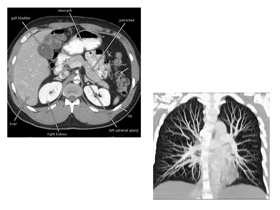
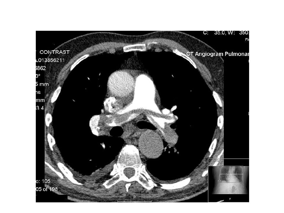
Typical applications for CT: Chest: Lungs, heart, esophagus, major blood vessels, the tissues in the center of the chest, lung cancer, pulmonary embolism. Abdomen: Infection, tumors, an aneurysm, foreign objects, bleeding. Urinary tract: Kidneys, urethra, and bladder (called a KUB). Liver: A CT scan can find liver tumors, liver diseases. Pancreas. Gallbladder and bile ducts. Adrenal glands. Spleen. Pelvis: Problems of organs in the pelvis. - Uterus, fallopian tubes, etc - Prostate
. Liver: A CT scan can find liver tumors, liver diseases. Pancreas. Gallbladder and bile ducts. Adrenal glands. Spleen. Pelvis: Problems of organs in the pelvis. - Uterus, fallopian tubes, etc - Prostate.")
20
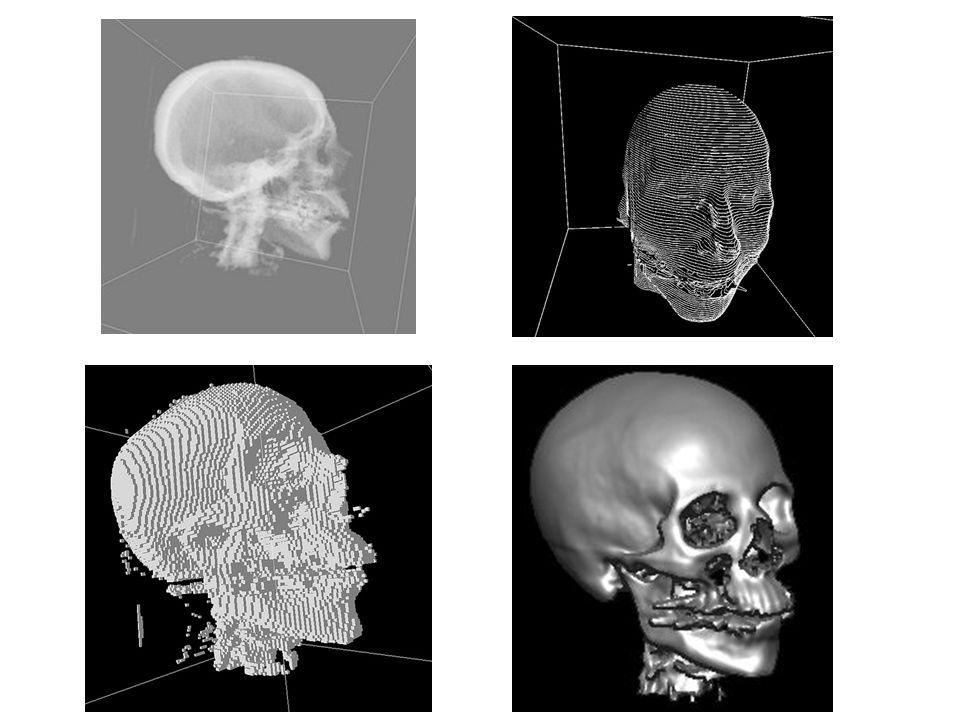
Typical CT study of a skull :

21
Three-dimensional CT scanning:

22
pixels voxels

25
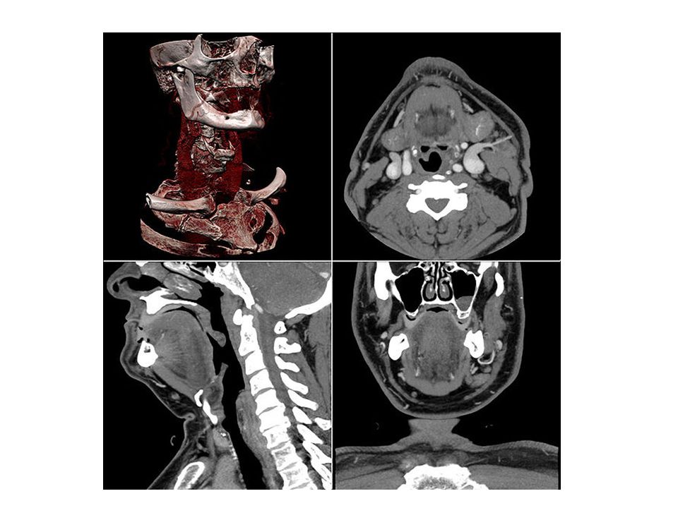
Revealing bone densities and vessels by manipulation of the data

26
http://medical.toshiba.com/resources/img/products/computed- tomography/aquilion-prime/Prime-Overview-Clincial-Example-01.gif

27
Two views of a femoral fracture -

28
Enhanced imaging --

29
How much ?? 4-slice scanner: $85,000 - $115,000 16-slice scanner:$145,000 - $225,000 64-slice scanner:$250,000 - $450,000 Service contracts:$100,000 - $135,000

30
CT tube replacement: $50,000 - $100,000

31
Major players in CT scanners: G.E. Medical ( GEMS) Philips Siemens Toshiba Major players in CT service and tubes:
 Philips Siemens Toshiba Major players in CT service and tubes:.")
Similar presentations


 theory was developed 1972: The CT scan was invented by Godfrey.>")
>")


 Dynamic scanning implies 15 or more scans in rapid sequence within one.>")


 CT scanning or (CAT scanning) is using X-rays to create a 3D image of the inside of an object. CT stands for computed tomography.>")
 Candidates should be able to show an understanding of the principles of CT scanning. (g) Candidates should be able to show an understanding.>")

 Topic:>")


 RAD 323. Reconstruction techniques dates back to (1917), when scientist (Radon) developed mathematical solutions to the problem of reconstructing.>")
