Download presentation
Presentation is loading. Please wait.

1
STRABISMUSBYDR. AMER ISMAIL ABU IMARA JORDANIAN BOARD OF OPHTHALMOLOGY I.C.O. PALESTINIAN BOARD IN OPHTHALMOLOGY

2
بسم الله الرحمن الرحيم - علم - فن - تخيل - قرار - متابعة ” لا يوجد علم دون تعب... ودراسة مستمرة... و متابعة حثيثة ” قال رسول الله صلى الله عليه و سلم : ( ان الله يحب اذا عمل احدكم عملا ان يتقنه ).
..")
3
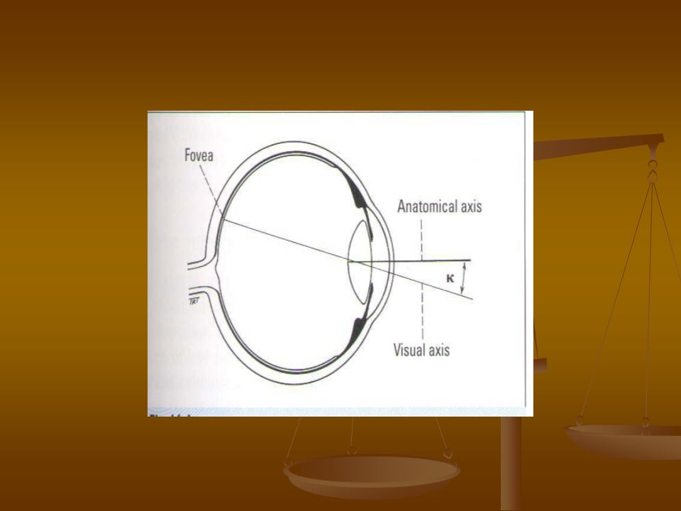
DEFENITIONS VISUAL AXIS (line of vision ) : VISUAL AXIS (line of vision ) : passes from the fovea through the nodal point of the eye to the point of fixation ( object of regard ). passes from the fovea through the nodal point of the eye to the point of fixation ( object of regard ). In normal binocular single vision (BSV) the two visual axes intersect at the point of fixation, with the images from the two eyes being aligned by the fusion reflex and combined by binocular responsive
. In normal binocular single vision (BSV) the two visual axes intersect at the point of fixation, with the images from the two eyes being aligned by the fusion reflex and combined by binocular responsive.")
4
cells in the visual cortex to give BSV. ORTHOPHORIA Implies perfect ocular alignment in the absence of any stimulus for fusion which is uncommon. HETEROPHORIA ( PHORIA ) Implies a tendency of the eyes to deviate when fusion is blocked (latent squint ). Slight phoria is present in most normal individuals and is overcome by the fusion reflex.
 Implies a tendency of the eyes to deviate when fusion is blocked (latent squint ). Slight phoria is present in most normal individuals and is overcome by the fusion reflex..")
5
It can be either esophoria or exophoria. When fusion is insufficient to control the imbalance the phoria is described as decompensating and is often associated with symptoms of binocular discomfort or double vision ( diplopia ). HETEROTROPIA It implies a manifest deviation in which the visual axes do not intersect at the the point of fixation.
. HETEROTROPIA It implies a manifest deviation in which the visual axes do not intersect at the the point of fixation..")
6
The images from the two eyes are misaligned so that either double vision is present or more commonly in children, the image from the deviating eye is suppressed at cortical level. Why squint in childhood ? 1- failure of the normal development of binocular fusion mechanisms. 2- oculomotor imbalance secondary to differences in refraction of the two eyes

7
Why squint in adult ? 1- failure of fusion, for example secondary to poor vision in one eye. 2- weakness of muscles. 3- restriction of muscles. 4- damage to nerve supply.

8
- Horizontal deviation ( latent or manifest ) is the most common form of strabismus. - A vertical deviation almost invariably reflects abnormal ocular motility. - Upward displacement of one eye relative to the other is termed hypertropia and a controlled upward imbalance a hyperphoria. - Downward displacement is termed a hypotropia and controlled imbalance a hypophoria.

9
ANATOMICAL AXIS Is a line passing from the posterior pole through the center of the cornea. Because the fovea is usually slightly temporal to the anatomical center of the posterior pole of the eye, the visual axis does not usually correspond to the anatomical axis of the eye.

10
ANGLE KAPPA Is the angle subtended by the visual and the anatomical axes and is usually about 5 degrees. The angle is positive when the fovea is temporal to the center of the posterior pole resulting in a nasal displacement of the corneal reflex and negative when the converse applies.

11
A large angle kappa may give the appearance of a squint when none is present ( pseudo- squint ) and is seen most commonly as a pseudo- exotropia following displacement of the macula in ROP where the angle may significantly exceed + 5 degrees.
 and is seen most commonly as a pseudo- exotropia following displacement of the macula in ROP where the angle may significantly exceed + 5 degrees.")
13
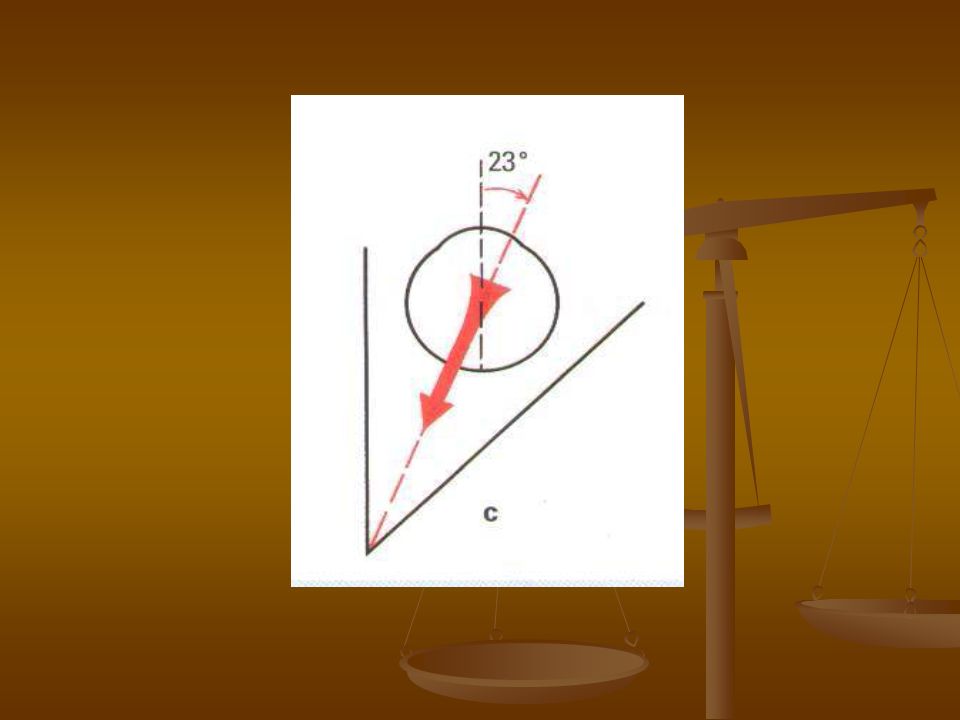
ANATOMY OF EXTRAOCULAR MUSCLES The lateral and medial orbital walls are at angle of 45 degrees with each other. The orbital axis therefore forms an angle of 22.5 degrees with both the lateral and medial walls. ~ 23 degrees

14
When the eye is looking straight a head at a fixed point on the horizon with the head erect ( primary position of gaze ), the visual axis forms an angle of 23 deg. With the orbital axis. The action of the extraocular muscles depend on the position of the globe at the time of muscle contraction.

16
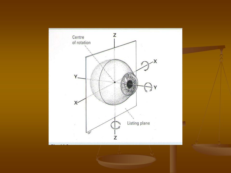
Primary action : of a muscle is it ’ s major effect when the eye is in the primary position. Subsidiary actions : are the additional effect which depend on the position of the eye. Listing plane : is an imaginary coronal plane passing through the center of rotation of the globe.the globe rotates on the X and Z axes of Fick, which intersect in Listing plane.

18
- The globe rotates left and right on the vertical Z axis. - The globe moves up and down on the horizontal X axis. - Torsional movements ( wheel rotation ) on the Y ( sagittal) axis which traverses the globe from front to back ( similar to the anatomical axis of the eye. - Intorsion occurs when the superior limbus rotates nasally and extorsion on temporal rotation.
 on the Y ( sagittal) axis which traverses the globe from front to back ( similar to the anatomical axis of the eye. - Intorsion occurs when the superior limbus rotates nasally and extorsion on temporal rotation..")
19
HORIZONTAL RECTUS MUSCLES When the eye is in the primary position, the horizontal recti are purely horizontal movers on the vertical Z axis and have only primary actions. MEDIAL RECTUS : originates at the annulus of Zinn at the orbital apex and inserts 5.5 mm behind the nasal limbus. it ’ s sole primary action is adduction. LATERAL RECTUS : originates at the annulus of Zinn and inserts 6.9 mm

20
Behind the temporal limbus.it ’ s sole primary action is abduction. VERTICAL RECTUS MUSCLES The vertical recti run in line with the orbital axis and are inserted in front of the equator. They therefore form an angle of 23 deg. With the visual axis.

22
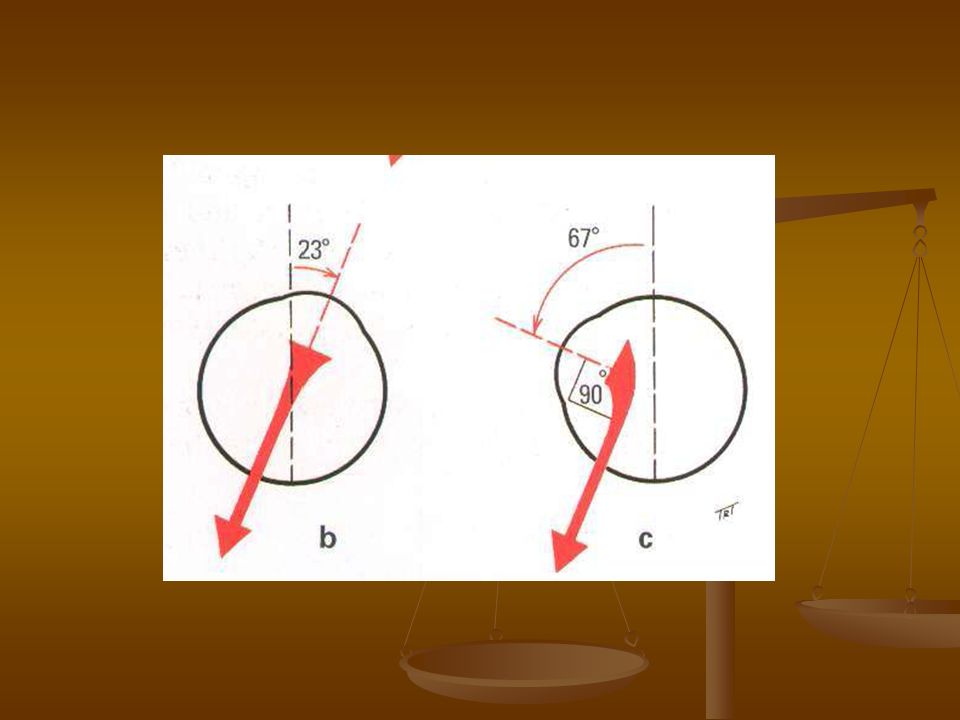
SUPERIOR RECTUS : originates from the upper part of the annulus of Zinn and inserts 7.7mm behind the superior limbus. The primary action is elevation, secondary actions are adduction and intorsion. when the globe is abducted 23 deg. The visual and orbital axes coincide. In this position it has no subsidiary actions and can only act as elevator.

23
This is therefore the optimal position of the globe for testing the function of the superior rectus muscle. If the globe was adducted 67 deg., the angle between the visual axis and orbital axis would be 90 deg. In this position the superior rectus could only act as intorter.

25
INFERIOR RECTUS : originates at the lower part of the annulus of Zinn and inserts 6.5 mm behind the inferior limbus. The primary action is depression, secondary actions are adduction and extorsion. When the globe is abducted 23 deg. The inferior rectus acts purely as a depressor. As for superior rectus, this is the optimal position of the globe for testing the function of the inferior rectus muscle.

26
If the globe was adducted 67 deg. The inferior rectus could only act as an extortor. SPIRAL OF TILLAUX The spiral is an imaginary line joining the insertions of the four recti and is an important anatomical land mark when performing surgery. The insertions get further away from the

27
Limbus and make a spiral pattern. Med.rectus 5.5 mm Inf.rectus 6.5 mm Lat. Rectus 6.9mm Superior rectus 7.7 mm

28
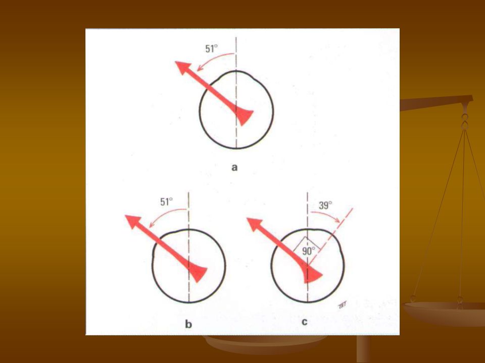
OBLIQUE MUSCLES The obliques are inserted behind the equator and form an angle of 51 deg with the visual axis.

29
SUPERIOR OBLIQUE Originates superomedial to the optic foramen. It passes forward through the trochlea at the angle between the superior and medial walls and is then reflected backwards and laterally to insert in the posterior upper temporal quadrant of the globe.

30
The primary action of the superior oblique is intorsion, secondary actions are depression and abduction. The anterior fibers of the superior oblique tendon are primarily responsible for intorsion and the posterior fibers for depression, allowing separate surgical manipulation of these two actions.

31
When the globe is adducted 51 deg. The visual axis coincide with the line of pull of the muscle. In this position it can only act as a depressor. This is therefore, the best position of the globe for testing the action of the superior oblique muscle. Thus, although the superior oblique has an abducting action in primary position, the main effect of the superior oblique weakness is seen as failure of depression in adduction.

32
When the eye is abducted 39 deg. The visual axis and the superior oblique make an angle of 90 deg. With each other. In this position the superior oblique can only cause intorsion.

34
INFERIOR OBLIQUE Originates from a small depression just behind the orbital rim lateral to the lacrimal sac. It passes backward and laterally to insert in the posterior lower temporal quadrant of the globe, close to the macula. The primary action is extorsion, secondary actions are elevation and abduction.

35
When the globe is adducted 51 deg., the inferior oblique acts only as an elevator. When the eye is abducted 39 deg., it ’ s main action is extorsion.

36
MUSCLE PULLEYS The four rectus muscles pass through condensations of connective tissue and smooth muscle just posterior to the equator. These condensations act as pulleys and minimize upward and downward movements of the of the bellies of the medial and lateral rectus muscles during up-gaze and down-gaze … and horizontal movements of the superior rectus and inferior rectus bellies in

37
Left and right gaze. Pulleys are the effective origins Of the rectus muscles and play an important role in the coordination of eye movements by reducing the effect of horizontal movements on vertical muscle actions and vice versa. Displacement of the pulleys can be one cause of abnormalities of eye movements such as V and A patterns.

38
NERVE SUPPLY LATERAL RECTUS :is supplied by the 6 th cranial nerve. ( abducent nerve – abducting muscle ). SUPERIOR OBLIQUE : is supplied by the fourth cranial nerve. ( trochlear nerve – muscle associated with the trochlea ). OTHER MUSCLES : together with the levator muscle of the upper lid and the ciliary and
. SUPERIOR OBLIQUE : is supplied by the fourth cranial nerve. ( trochlear nerve – muscle associated with the trochlea ). OTHER MUSCLES : together with the levator muscle of the upper lid and the ciliary and.")
39
Sphincter pupillae muscles are supplied by the 3 rd ( oculomotor ) nerve.
 nerve.")
40
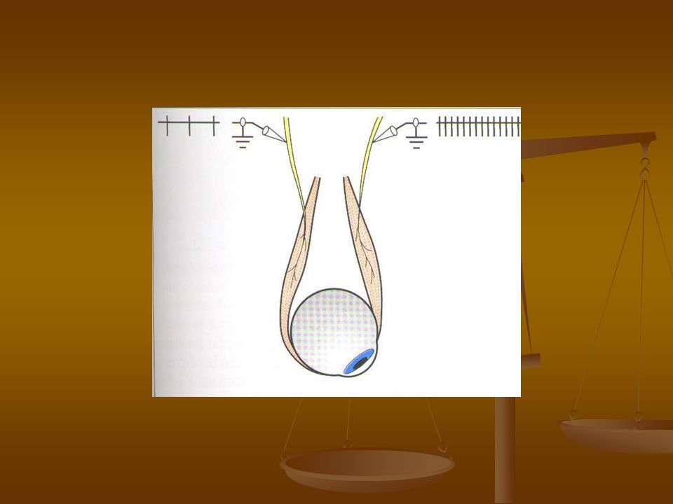
OCULAR MOVEMENTS DUCTIONS Ductions are monocular movements around the axes of Fick. They consist of adduction, abduction, elevation, depression, intorsion and extorsion. They are tested by occluding the fellow eye and asking the patient to follow a target in each direction of gaze. Torsional ductions are mainly observed in association with other abnormal eye movements.

41
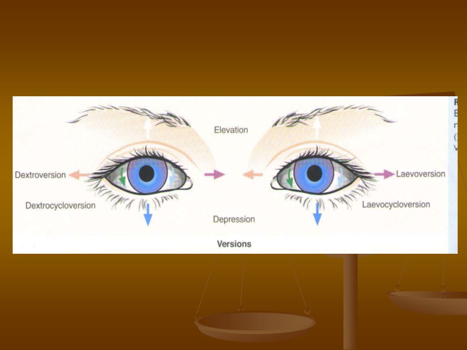
VERSIONS Versions are binocular, simultaneous, conjugate movements ( in the same direction ). Dextroversion and laevoversion ( gaze right, gaze left ), elevation ( up-gaze ) and depression ( down-gaze ). These four movements bring the globe into the secondary positions of gaze by rotation around either a vertical Z or a horizontal X axis of Fick.
, elevation ( up-gaze ) and depression ( down-gaze ). These four movements bring the globe into the secondary positions of gaze by rotation around either a vertical Z or a horizontal X axis of Fick..")
42
Dextroelevation and dextrodepression ( gaze up and right, gaze down and right ) and laevoelevation and laevodepression ( gaze up and left and gaze down and left ). These four oblique movements bring the eyes into the tertiary positions of gaze by rotation around oblique axes lying in Listing plane, equivalent to simultaneous movement about both horizontal and vertical axes.

44
Torsional movements to maintain upright images occur on tilting of the head. These are known as the righting reflexes. On head tilt to the right the superior limbi of the two eye rotate to the left, causing intorsion of the right globe and extorsion of the left.

45
VERGENCES Vergences are binocular, simultaneous, disjugate or disjunctive movements ( in opposite directions. Convergence is simultaneous adduction ( inward turning ), divergence is outwards movement from a convergent position. Convergence may be voluntary or reflex.
, divergence is outwards movement from a convergent position. Convergence may be voluntary or reflex..")
46
Reflex convergence has four components : - Tonic convergence : which implies inherent innervational tone to the medial recti, when the patient is awake. - Proximal convergence : is induced by psychological awareness of a near object. - Fusional convergence : is an optomotor reflex, which maintains BSV by insuring that similar images are projected onto corresponding retinal areas of each eye.

47
It is initiated by bitemporal retinal image disparity. - Accommodative convergence : is induced by the act of accommodation as part of the synkinetic - near reflex. Each dioptre of accommodation is accompanied by a constant increment in accommodative convergence, giving the accommodative convergence by accommodation ( AC/A ) ratio.
 ratio..")
48
This is the amount of convergence in prism dioptres per dioptre change in accommodation. The normal value is 3-5 prism dioptre. this means that one dioptre of accommodation is associated with 3-5 prism dioptres of accommodative convergence. this means that one dioptre of accommodation is associated with 3-5 prism dioptres of accommodative convergence. It will be shown later that abnormalities of the AC/A ratio play an important role in the aetiology of strabismus.

49
These changes in accommodation, convergence and pupil size, which occur in response to a change in the distance of viewing are known as the near triad and occur in response to both image blur and temporal image disparity.

50
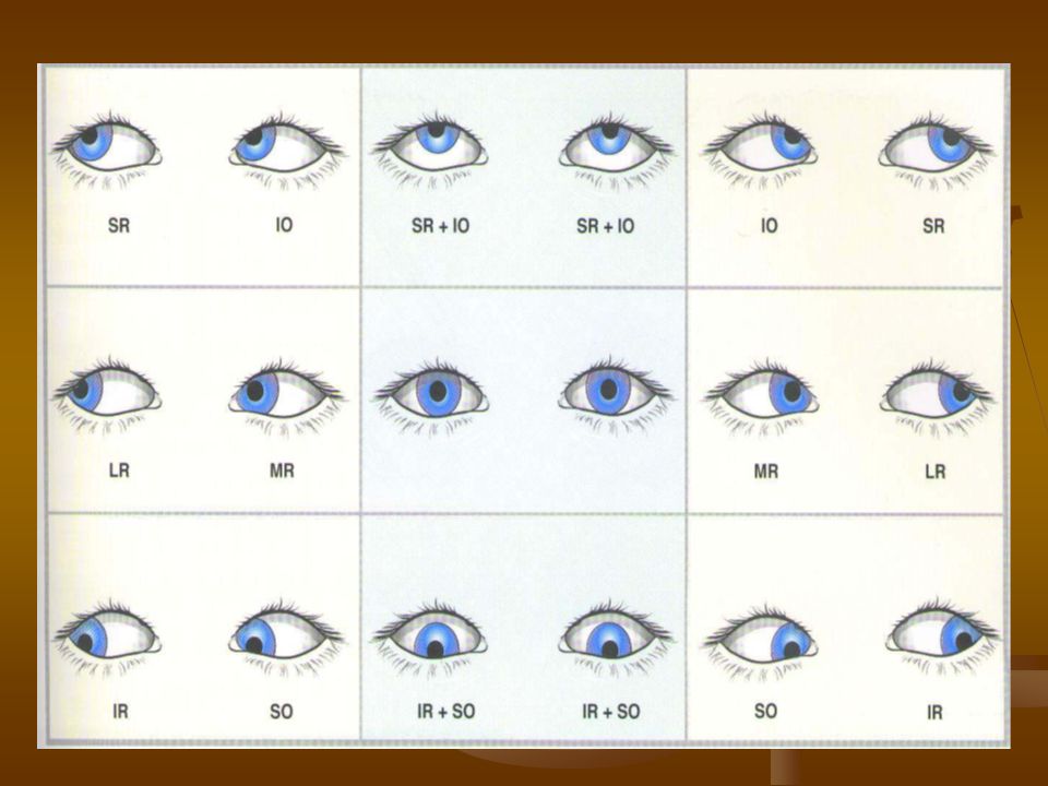
POSITIONS OF GAZE SIX CARDINAL :positions of gaze are those in which one muscle in each eye has moved the eye into that position as follows : - Dextroversion ( right lateral rectus and left medial rectus ). - Laevoversion ( left lateral rectus and right medial rectus ). - Dextroelevation ( right superior rectus and left inferior oblique ).
. - Dextroelevation ( right superior rectus and left inferior oblique )..")
51
- Laevoelevation ( left superior rectus and right inferior oblique ). - Dextrodepression ( right inferior rectus and left superior oblique ). - Laevodepression ( left inferior rectus and right superior oblique ).
. - Laevodepression ( left inferior rectus and right superior oblique )..")
52
NINE DIAGNOSTIC positions of gaze are those in which deviations are measured. They consist of the six cardinal positions, the primary position, elevation and depression ).
..")
54
LAWS OF OCULAR MOTILITY 1- AGONIST- ANTAGONIST : are muscles of the same eye that move the eye in opposite directions. The agonist is the primary muscle moving the eye in a given direction. The antagonist acts in the opposite direction to the agonist. Example : the right lateral rectus is the antagonist to the right medial rectus. 2- SYNERGISTS :are muscles of the same eye that move the eye in the same direction.

55
Example : the right superior rectus and the right inferior oblique act synergistically in elevation. 3- YOKE MUSCLES :( contralateral synergists ) are pairs of muscles, one in each eye, that produce conjugate ocular movements Example : left superior oblique – right inferior rectus.
 are pairs of muscles, one in each eye, that produce conjugate ocular movements Example : left superior oblique – right inferior rectus..")
56
SHERRINGTON LAW of reciprocal innervation ( inhibition ) states that increased innervation to an extraocular muscle ( e.g. right medial rectus ) is accompanied by a reciprocal decrease in innervation to its antagonist ( e.g. right lateral rectus ). This means that when the medial rectus contracts the lateral rectus automatically relaxes and vice versa. Sherrington law applies to both versions and vergences.
 is accompanied by a reciprocal decrease in innervation to its antagonist ( e.g. right lateral rectus ). This means that when the medial rectus contracts the lateral rectus automatically relaxes and vice versa. Sherrington law applies to both versions and vergences..")
58
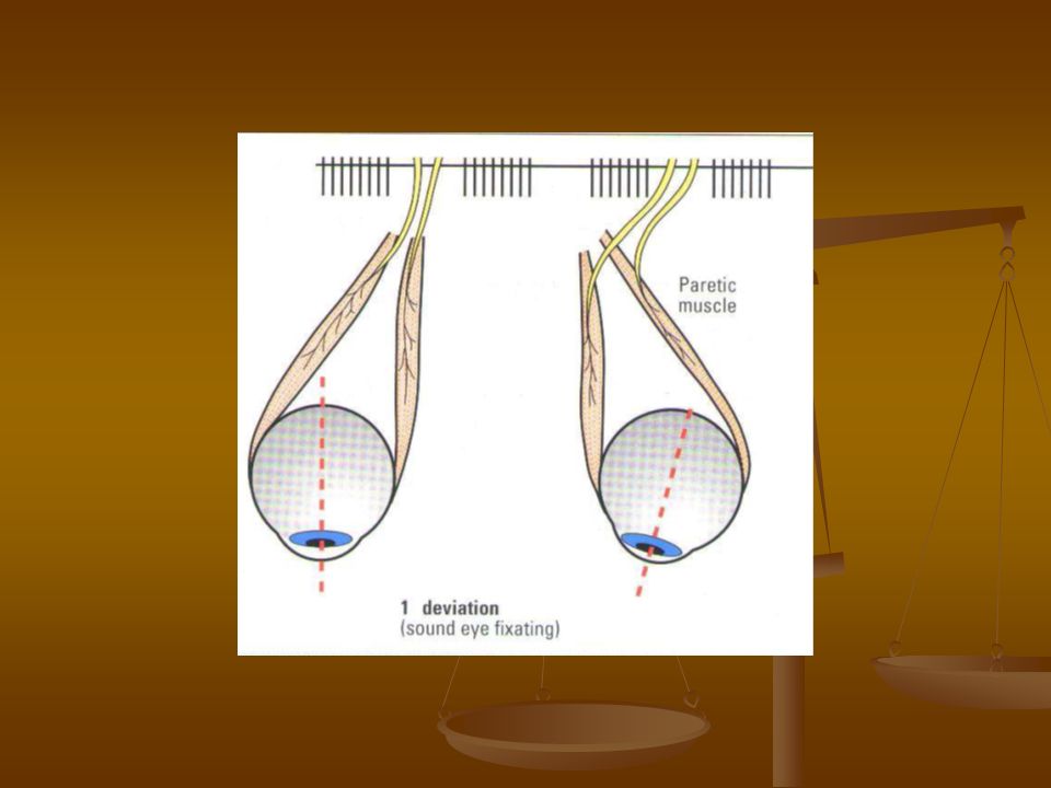
HERING LAW of equal innervation states that during any conjugate eye movement, equal and simultaneous innervation flows to the yoke muscles. In the case of a paretic squint, the amount of innervation to both eyes is symmetrical, and always determined by the fixating eye, so that the angle of deviation will vary according to which eye is used for fixation.

59
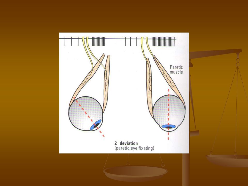
For example, if in the case of left lateral rectus palsy, the right normal eye is used for fixation, there will be an inward deviation of the left eye due to the unopposed action of the antagonist of the paretic left lateral rectus ( left medial rectus ). The amount of misalignment of the two eyes in this situation is called the primary deviation. If the paretic left eye is now used for fixation, additional innervation will flow

60
To the left lateral rectus, in order to establish this. However, according to Hering law, an equal amount of innervation will also flow to the right medial rectus ( yoke muscle ). This will result in an over action of the right medial rectus and an excessive amount of adduction of the right eye. The amount of misalignment between the two eyes in this situation is called the secondary deviation. In a paretic squint the secondary deviation exceeds the primary deviation.
. This will result in an over action of the right medial rectus and an excessive amount of adduction of the right eye. The amount of misalignment between the two eyes in this situation is called the secondary deviation. In a paretic squint the secondary deviation exceeds the primary deviation..")
63
MUSCLE SEQUELAE are the effects of the interactions described by these laws. They are of prime importance in diagnosing ocular motility disorders and in particular in distinguishing a recently acquired palsy from a longstanding one. The full pattern of changes takes time to develop and can be summarized as follows : - Primary under action ( e.g. Left superior oblique )
.")
64
- Secondary contracture of the unopposed direct antagonist ( left inferior oblique ). - Secondary contracture of the contralateral synergist or yoke muscle ( right inferior rectus, Hering law ). - Secondary inhibitional palsy ( right superior rectus, Sherrington law.
. - Secondary inhibitional palsy ( right superior rectus, Sherrington law..")
65
SECONDARY CONSIDERATIONS BASIC ASPECTS NORMAL BSV involves the simultaneous use of both eyes with bifoveal fixation, so that each eye contributes to a common single perception of the object of regard. This represents the highest form of binocular cooperation.

66
Conditions necessary for normal BSV : 1- normal routing of visual pathways with overlapping visual fields. 2- binocularly driven neurons in the visual cortex. 3- normal retinal ( retino - cortical ( correspondence ( NRC ) resulting in cyclopean viewing. 4- accurate neuromuscular development and coordination, so that the visual axes directed at, and maintain fixation on, the object of regard.
 resulting in cyclopean viewing. 4- accurate neuromuscular development and coordination, so that the visual axes directed at, and maintain fixation on, the object of regard..")
67
5- approximately equal image clarity and size for both eyes. BSV is based on NRC, which requires first an understanding of uniocular visual direction and projection.

68
VISUAL DIRECTION Is the projection of a given retinal element in a specific direction in the subjective space. PRINCIPAL visual direction is the direction in external space interpreted as the line of sight. This is normally the visual direction of fovea and is associated with a sense of direct viewing. SECONDARY visual directions are the projecting directions of extra- foveal points with respect to the principal direction of the fovea, associated with indirect ( eccentric ) viewing.
 viewing..")
69
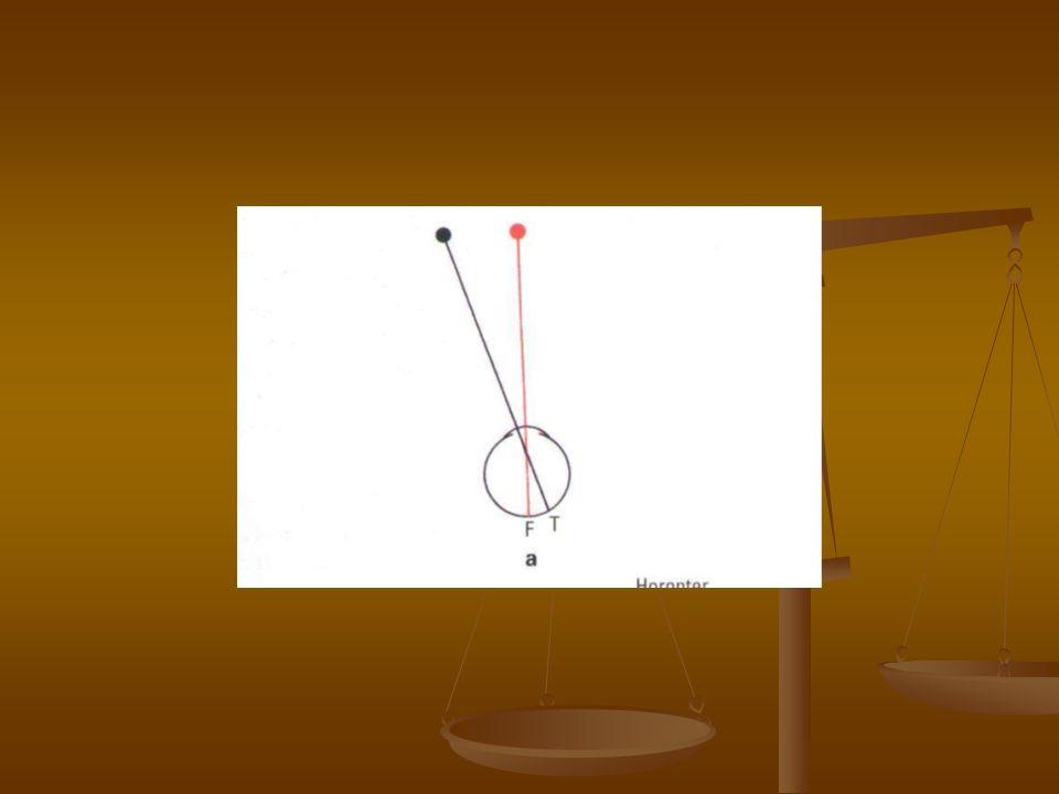
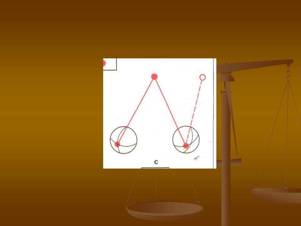
PROJECTION is the subjective interpretation of the position of an object in space on the basis of stimulated retinal elements. * If a red object stimulates the right fovea ( F ), and black object which lies in the nasal field stimulates a temporal retinal element (T), the red object will be interpreted by the brain as having originated from the straight a head position and the black object will be interpreted as having originated in the nasal field.
, and black object which lies in the nasal field stimulates a temporal retinal element (T), the red object will be interpreted by the brain as having originated from the straight a head position and the black object will be interpreted as having originated in the nasal field..")
71
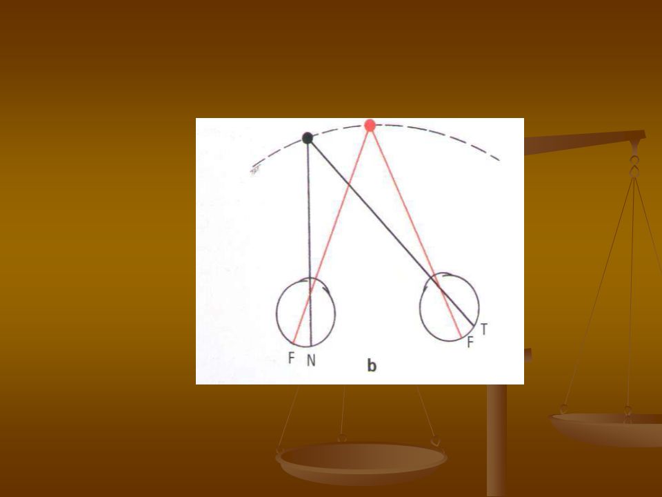
Similarly nasal retinal elements project into the temporal field, upper retinal elements into the lower field and vice versa. * With both eyes open, the red fixation object is now stimulating both foveae, which are corresponding retinal points. The black object is now not only stimulating the temporal retinal elements in the right eye but also the nasal elements of the left eye. The right eye therefore projects the object into its nasal field and the left eye projects the object into its temporal field.

72
Because both of these retinal elements are corresponding points, they will both project the object into the same position in space ( the left side ) and there will be no double vision.
 and there will be no double vision.")
74
RETINO – MOTOR VALUES The image of an object in the peripheral visual field falls on an extrafoveal element. To establish fixation on this object a saccadic version of accurate amplitude is required. Each extrafoveal retinal element therefore has a retino – motor value proportional to its distance from the fovea, which guides the amplitude of saccadic movements required to ( look at it ). Retino – motor value, zero at the fovea, increases progressively towards the retinal periphery.
. Retino – motor value, zero at the fovea, increases progressively towards the retinal periphery..")
75
CORRESPONDING POINTS Are areas on each retina that share the same visual direction ( for example : the foveae share the primary visual direction ). Points on the nasal retina of one eye have corresponding points on the temporal retina of the other eye and vice versa. For example, an object producing images on the right nasal retina and the left temporal retina will be projected into the right side of visual space. This is the basis of NRC. This retino – topic organization is reflected back a long the visual pathways, each eye maintaining separate images until the visual pathways converge onto binocularly responsive neurons in the primary visual cortex.

76
THE HOROPTER Is an imaginary plane in the external space, all points on which stimulate corresponding retinal elements and are therefore seen singly and in the same plane. This plane passes through the intersection of the visual axes and therefore includes the point of fixation in BSV.

77
PANUM FUSIONAL SPACE Is a zone in front of and behind the horopter in which objects stimulate slightly non corresponding retinal points ( retinal disparity ). Objects are seen singly and the disparity information is used to produce a perception of binocular depth ( stereopsis ). Objects in front of and behind Panum space appear double. This is the basis of physiological diplopia.
. Objects in front of and behind Panum space appear double. This is the basis of physiological diplopia..")
78
Panum space is shallow at fixation ( 6 sec. of arc ) and deeper towards the periphery ( 30 – 40 seconds of arc at 15 deg. From the fovea ).
 and deeper towards the periphery ( 30 – 40 seconds of arc at 15 deg. From the fovea )..")
79
Therefore objects on the horopter are seen singly and in one plane. Objects in Panum fusional area are seen singly and stereoscopically. Objects outside Panum fusional area appear double. Physiological diplopia is usually accompanied by physiological suppression and many subjects remain unaware of this phenomenon.

80
The retinal areas stimulated by images falling within Panum fusional space are termed Panum fusional areas.

81
BSV Is characterized by the ability to fuse the images from the two eyes and to perceive binocular depth. - SENSORY FUSION :involves the integration by the visual areas of the cerebral cortex of two similar images, one from each eye, into one image. It may be central, which integrate the image falling on the foveae, or peripheral, which integrates parts of the image falling outside the foveae.

82
It is possible to maintain fusion with a central visual deficit in one eye, but peripheral fusion is essential to BSV and may be affected in patients with field loss as in advanced glaucoma. - MOTOR FUSION : involves the maintenance of motor alignment of the eyes to sustain bifoveal fixation. It is driven by retinal image disparity,which stimulates fusional vergences.

83
FUSIONAL VERGENCE Involves disjugate eye movements to overcome retinal image disparity. Fusional vergence amplitudes can be measured with prisms or in the synoptophore. Normal values are : Convergence : about 15-20 ∆ for distance and 25 ∆ for near. Divergence : about 6-10 ∆ for distance and 12-14 ∆ for near.

84
Vertical : 2-3 ∆ Cyclovergence : about 2-3 ° Fusional convergence helps to control an exophoria whereas fusional divergence helps to control an esophoria. The fusional vergence mechanism may be decreased by fatigue or illness, converting a phoria to a tropia. The amplitude of fusional vergence mechanisms can be improved by orthoptic exercises, particularly in the case of near fusional convergence for the relief of convergence insufficiency.

85
STEREOPSIS Is the perception of depth ( the third dimension, the first two being the height and width ). It arises when objects behind and in front of the point of fixation ( but within Panum fusional space ) stimulate horizontally disparate retinal elements simultaneously. The fusion of such disparate images results in a single visual impression perceived in depth. A solid object is seen stereoscopically ( in 3D ) because each eye sees a slightly different aspect of the object.
 stimulate horizontally disparate retinal elements simultaneously. The fusion of such disparate images results in a single visual impression perceived in depth. A solid object is seen stereoscopically ( in 3D ) because each eye sees a slightly different aspect of the object..")
86
SENSORY PERCEPTIONS At the onset of a squint two sensory perceptions arise based on the normal projection of the retinal areas stimulated : Confusion and pathological diplopia may result. These require simultaneous ( visual ) perception i.e. the ability to perceive images from both eyes simultaneously.
 perception i.e. the ability to perceive images from both eyes simultaneously..")
87
CONFUSION Is the simultaneous appreciation of two superimposed but dissimilar images caused by stimulation of corresponding retinal points ( usually the foveae ) by images of different objects.
 by images of different objects.")
88
PATHOLOGICAL DIPLOPIA Is the simultaneous appreciation of two images of the same object in different positions and results from images of the same object falling on non- corresponding retinal points. - In esotropia the diplopia is homonymous ( uncrossed ). - In exotropia the diplopia is heteronymous ( crossed ).
. - In exotropia the diplopia is heteronymous ( crossed )..")
90
SENSORY ADAPTATION TO STRABISMUS The ocular sensory system in children has the ability to adapt to anomalous states ( confusion and diplopia ) by two mechanisms : A- suppression B- abnormal retinal correspondence. These occur because of the plasticity of the developing visual system in children under the age of 7-8 years. Occasional adults who develop sudden – onset strabismus are able to ignore the second image after a time and therefore do not complain of diplopia.

91
SUPPRESSION Involves active inhibition, in the visual cortex, of an image from one eye when both eyes are open. Stimuli for suppression include diplopia, confusion and a blurred image from one eye resulting from astigmatism / anisometropia. Clinically, suppression may be : 1- CENTRAL OR PERIPHERAL in central suppression the image from the fovea of the deviating eye is inhibited to avoid confusion. Diplopia on the other hand, is eradicated by the process of peripheral suppression, in which the image from the peripheral retina of the deviating eye is inhibited.

92
2-MONOCULAR OR ALTERNATING suppression is monocular when the image from the dominant eye always predominate over the image from the deviating eye ( or more ametropic ) eye, so that the image from the latter is constantly suppressed. This type of suppression leads to amblyopia. When suppression alternates ( switches from one eye to the other ) amblyopia does not develop.
 amblyopia does not develop..")
93
3- FACULTATIVE OR OBLIGATORY facultative suppression occurs only when the eyes are misaligned. Obligatory suppression is present at all times, irrespective of whether the eyes are deviated or straight. Examples are seen in intermittent exotropia and Duane syndrome.

94
ABNORMAL RETINAL CORRESPONDENCE Is a condition in which non-corresponding retinal elements acquire a common subjective visual direction, i.e. fusion occurs in the presence of a small angle manifest squint. The fovea of the fixating eye is thus paired with a non- foveal element of the deviated eye. ARC is a positive sensory adaptation to strabismus ( as opposed to suppression ), which allows some anomalous binocular vision in the presence of a heterotropia. Binocular responses in ARC are never as good as in normal bifoveal BSV. ARC is most frequently present in small angle esotropia ( microtropia ) associated with anisometropia.
, which allows some anomalous binocular vision in the presence of a heterotropia. Binocular responses in ARC are never as good as in normal bifoveal BSV. ARC is most frequently present in small angle esotropia ( microtropia ) associated with anisometropia..")
95
MICROTROPIA Is a small angle squint ( < 10 ∆ ) in which stereopsis is present but reduced and there is a relative amblyopia of the more ametropic eye. Microtropia has two forms : A- in microtropia with identity the point used for monocular fixation by the squinting eye also corresponds with the fovea of the straight eye under binocular viewing conditions. Therefore on cover test there is no movement of the squinting eye when it takes up monocular fixation.

96
B- in microtropia without identity the monocular fixation point of the squinting eye does not correspond with the fovea of the straight eye in binocular viewing. There is therefore a small movement of the deviating eye when it takes up monocular fixation on cover testing. ARC is less common in accommodative esotropia because of the variability of the angle of deviation, or in large angle deviations, because the separation of the images is too great.

97
CONSEQUENCES OF STRABISMUS - The fovea of the squinting eye is suppressed to avoid confusion. - Diplopia will occur, since non- corresponding retinal elements receive the same image. - To avoid diplopia, the patient will develop either peripheral suppression of the squinting eye or ARC. - If constant unilateral suppression occurs this will subsequently lead to strabismic amblyopia.

98
MOTOR ADAPTATION TO STRABISMUS Motor adaptation involves the adoption of an abnormal head posture (AHP ) and occurs primarily in children with congenitally abnormal eye movements who use the AHP to maintain BSV. In these children loss of an AHP may indicate loss of binocular function and the need for surgical intervention. These patients may present in adult life with symptoms of decompensation, often unaware of their AHP.

99
Acquired paretic strabismus in adults may be consciously controlled by an AHP provided the deviation is neither too large nor too variable with gaze ( incomitance ). The AHP eliminates diplopia and helps to centralize the binocular visual field. The patient will turn the head into the direction of the field of action of the weak muscle, so that the eyes are then automatically turned the opposite direction and as far as possible away from its field of action ( i.e. the head will turn where the eye cannot ).
..")
100
An AHP is analyzed in terms of the following three components : - Face turn to right or left. - Head tilt to right or left. - Chin elevation or depression. 1- A face turn will be adopted to control a purely horizontal deviation. For example, if the left lateral rectus is paralyzed, diplopia will occur in left gaze ; the face will be turned to the left which deviates the eyes to the right, away from the field of action of the weak muscle and area of diplopia. A face turn may also be adopted in a paresis of a vertically acting muscle to avoid the side where

101
The vertical deviation is greatest ( e.g. in a right superior oblique weakness the face is turned to the left ). 2- a head tilt is adopted to compensate for torsional and / or vertical diplopia. In left superior oblique weakness the left eye is relatively elevated and the head is tilted to the right, toward the hypotropic eye ; this reduces the vertical separation of the diplopic images and permits fusion to be regained.
. 2- a head tilt is adopted to compensate for torsional and / or vertical diplopia. In left superior oblique weakness the left eye is relatively elevated and the head is tilted to the right, toward the hypotropic eye ; this reduces the vertical separation of the diplopic images and permits fusion to be regained..")
102
If there is a significant torsional component preventing fusion, tilting the head in the same left direction will reduce this by invoking the righting reflexes ( placing the extorted right eye in a position which requires extorsion ). 3- chin elevation or depression may be used to compensate for weakness of an elevator or depressor muscle or to minimize the horizontal deviation when A or V pattern is present.

Similar presentations


 may turn in, out, up, or down can be present in one or both eyes cross-eyed, squint. Vergence Duction.>")


 Range of Movement and Ocular Alignment Establish.>")
 591-8860>")


>")


