Download presentation
Presentation is loading. Please wait.

1
Hypercalciuria Genetic and environmental basis Pascal Houillier
Paris-Descartes University

2
Hypercalciuria is a risk factor for calcium nephrolithiasis
Hypercalciuria is a well recognized risk factor for nephrolithiasis. This has been demonstrated in several studies. In one of them, reported here, we have studied members of families with idiopathic hypercalciuria. Some subjects had a quite normal urinary calcium excretion and others had a very high urinary calcium excretion, in excess of 0,15 mmol/kg/day. The first group served as the reference group with a relative risk of lithiasis equal to unity. In multivariate analysis, the risk of developing a calcium nephrolithiasis increased dose-dependently with the rate of urinary calcium excretion and became significantly higher than in the reference group for rates of urinary calcium excretion above 0,11 mmol/kg/day N Lerolle et al, Am J Med, 2002

3
Thiazide diuretics decreases the recurrence of stone
In a very nice study by Coe and colleagues, in 1988, it was also shown that drugs that decrease urinary calcium excretion are able to dramatically influence the recurrence of stones. In this study, patients with recurrent stones have been studied before and after the beginning of a medical treatment with thiazides. The horizontal lines represent the duration of follow-up and each dot represent the time of a stone episode. By comparison between the left and the right part of the figure, it is apparent that the number of stone episode is lower after the beginning of thiazide treatment than before FL Coe et al, Kidney Int, 1988

4
SHOK syndrome Stroke Hypertension Osteoporosis Kidney

5
Primary mechanisms resulting in hypercalciuria
Primary disorder Renal leak Primary disorder Bone resorption Primary disorder Intestine hyperabsorption ECF Ca ECF Ca ECF Ca Hypercalciuria may result from several primary disorders The primary disorder may be a renal leak of calcium that is a decrease in the ability of the renal tubule to reabsorb calcium. Under this condition, an adaptation of two other organs occur ; the intestinal absorption of calcium increases and the net bone release of calcium also increases. The primary disorder may involve the bones with a primary increase in net calcium release. Under this condition, intestinal calcium absorption does not change or decreases but urinary calcium excretion obviously increases Finally, the primary disorder may be an hyperabsorption of calcium from the gut ; the net bone calcium release is left unchanged but here again, the urinary calcium excretion increases Hypercalciuria Hypercalciuria Hypercalciuria

6
Idiopathic (genetic) hypercalciuria
Familial inheritance Heavy influence of environmental (dietary) factors Complex pathophysiology Idiopathic hypercalciuria is the most common condition in hypercalciuric patients. It has several characteristics that deserve to be mentionned First, idiopathic hypercalciuria is not a diagnosis but rather the translation of our lack of knowledge Second idiopathic hypercalciuria is frequently transmited within families Third the expression of idiopathic hypercalciuria is highly influenced by environmental factors Finally, idiopathic hypercalciuria has a complex pathophysiology. I shall comment on these points now
 factors. Complex pathophysiology. Idiopathic hypercalciuria is the most common condition in hypercalciuric patients. It has several characteristics that deserve to be mentionned. First, idiopathic hypercalciuria is not a diagnosis but rather the translation of our lack of knowledge. Second idiopathic hypercalciuria is frequently transmited within families. Third the expression of idiopathic hypercalciuria is highly influenced by environmental factors. Finally, idiopathic hypercalciuria has a complex pathophysiology. I shall comment on these points now.")
7
Gene1 Gene1 Gene2 Gene3 Gene4 Gene5 Low Ca excretion High Ca excretion
On the top of the figure, you can see a pedigree of a typical french family in which hypercalciuria is transmitted. Hypercalciuric patients are represented as black symbol. As you can see, hypercalciuric patients are present in each generation and the transmission does not match a recessive pattern and hypercalciuria can be transmitted from male to male. Therefore, some authors have proposed that idiopathic hypercalciuria can be an autosomal dominant trait. However, urinary calcium excretion is a continuous variable Low Ca excretion High Ca excretion

8
Influence of environmental factors
Low Ca excretion High Ca excretion

9
High Na intake increases urinary Ca excretion
Increased ECF volume Decreased proximal Na and Ca absorptions J Lemann, Jr, 1992

10
Thiazides reduce urinary calcium excretion through a decrease in ECF volume
T Nijenhuis et al, JCI, 2005

11
7 male patients with Dent syndrome (CLNC5 defect)
A. Blanchard, unpublished results.

12
Ncc inactivation is associated with an increased bone mineral density
Humans Mice L. Nicolet-Barousse et al, JBMR, 2005

13
High dietary protein intake increases urinary Ca excretion
Increased animal protein intake : Increased acid load Increased bone resorption ECF Ca Decreased tubular Ca reabsorption J Lemann, Jr, 1992 Hypercalciuria

14
Metabolic acidosis induces an increase in urinary calcium excretion
Acute Chronic P. Houillier et al, Kidney Int, 1996 J. Lemann Jr et al, N Engl J Med, 1979 µmol/min UCaV, Filtered load of Ca, Acid load

15
Metabolic acidosis induces a negative calcium balance
J Lemann et al, J Clin Invest, 1966 Sebastian et al, N Eng J Med, 1994

16
Carbohydrates induce an increase in urinary calcium excretion
J. Lemann Jr et al, N Engl J Med, 1969

17
Pathophysiology of human idiopathic hypercalciuria
ECF Ca Hypercalciuria Increased intestinal Ca absorption Increased Ca release (especially on a low Ca diet) Decreased renal tubular Ca reabsorption Primary or secondary disorders ?
 Decreased renal tubular. Ca reabsorption. Primary or secondary disorders")
18
Pathophysiology of rat idiopathic hypercalciuria
ECF Ca Hypercalciuria D. Bushinsky et al, Semin Nephrol, 1996 S. Tsuruoka et al, Kidney Int, 1997

19
Role of vitamin D receptor in intestinal epithelial cells
From Li, 1993

20
From Li, 1993

21
From Coe, 1991

22
Role of kidney in idiopathic hypercalciuria

24
Factors decreasing renal tubular calcium reabsorption
Reduced PTH NaCl intake (volume expansion) Protein intake (metabolic acidosis) Glucose, sucrose, ethanol Phosphate restriction Loop diuretics Calcium, magnesium
 Protein intake (metabolic acidosis) Glucose, sucrose, ethanol. Phosphate restriction. Loop diuretics. Calcium, magnesium.")
25
Acute response to hydrochlorothiazide
From Sutton, 1980 and Sakhaee, 1985

26
Calciuric response to an acute acid load
From Houillier, 1996

27
Calciuric response to furosemide
From Tsuruoka, 1997

28
Kidney as the primary defect : monogenic disease in humans and/or mice
CLC-5 OCRL ATP7B FAH G6PC NPT2 NHERF-1 AKr1b1 CAII TRPV5 VDR Calbindin-D28k WNK1-4 T Kallikrein NKCC2 ROMK CLC-Kb Barttin CaSR PCLN-1 ATP6V1B1 ATP6V0V4 SLC4A1 (AE1) SCNN1B and G (ENaC ß and g subunits) Kidney as the primary defect : monogenic disease in humans and/or mice
 SCNN1B and G. (ENaC ß and g. subunits) Kidney as the primary defect : monogenic disease in humans and/or mice.")
29
TRPV5 (ECaC 1)
")
30
TRPV5 (ECaC) is the gatekeeper for Ca absorption in the distal tubule
Copyright ©2000 American Physiological Society Hoenderop, J. G. J. et al. Am J Physiol Renal Physiol 278: F352-F Fig. 1.

31
Phenotype of Trpv5 -/- mice
J. Hoenderop et al, J Clin Invest, 2003

32
Phenotype of Trpv5 -/- mice
Decreased distal tubular Ca reabsorption Adaptive increase in intestinal Ca absorption

34
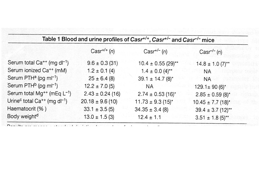
CaSR

35
CaSR and model of ion transport in the TAL
Na 2Cl K Ca, Mg - + Ca CaSR Lumen Cell Interstitium Cl PCLN-1 PTH

36
CaSR +/+ CaSR +/- CaSR -/-

38
Croisement de souris mutées pour CaSR et de souris hypoparathyroïdiennes (Gcm2-/-)
I : normales ; II : CaSR -/-, Gcm2+/+; III : CaSR+/+, Gcm2-/- ; IV : CaSR-/-, Gcm2 -/- ; V: CaSR+/-, Gcm2 +/+ ; VI : CaSR +/-,Gcm2-/-

39
Tu, Q. et al. J. Clin. Invest. 2003;111:1029-1037
Copyright ©2003 American Society for Clinical Investigation

40
Tu, Q. et al. J. Clin. Invest. 2003;111:1029-1037
Copyright ©2003 American Society for Clinical Investigation

41
CaSR +/+ CaSR -/- CaSR +/+ CaSR -/- CaSR +/- CaSR +/- Gcm2+/+ Gcm2+/+
Tu, Q. et al. J. Clin. Invest. 2003;111: Copyright ©2003 American Society for Clinical Investigation

42
Tu, Q. et al. J. Clin. Invest. 2003;111:1029-1037
Copyright ©2003 American Society for Clinical Investigation

43
Gain-of-function mutations in CASR gene induce a renal leak of calcium
Yamamoto et al, JCEM, 2000

46
Expression hétérologue du CaSR
Vargas-Poussou, JASN, 2002

47
CaSR in idiopathic hypercalciuria
Petrucci et al, 2000:No significant linkage between CaSR variants and idiopathic hypercalciuria Lerolle et al, 2002: No point mutation in CASR gene in families with idiopathic hypercalciuria Vezzoli et al, 2002: higher urinary Ca excretion in patients bearing the R990G polymorphism (ARQ/AGQ or AGQ/AGQ) Yao et al, 2005: GHS rats have a higher renal content in CaSR protein and mRNA
 Yao et al, 2005: GHS rats have a higher renal content in CaSR protein and mRNA.")
48
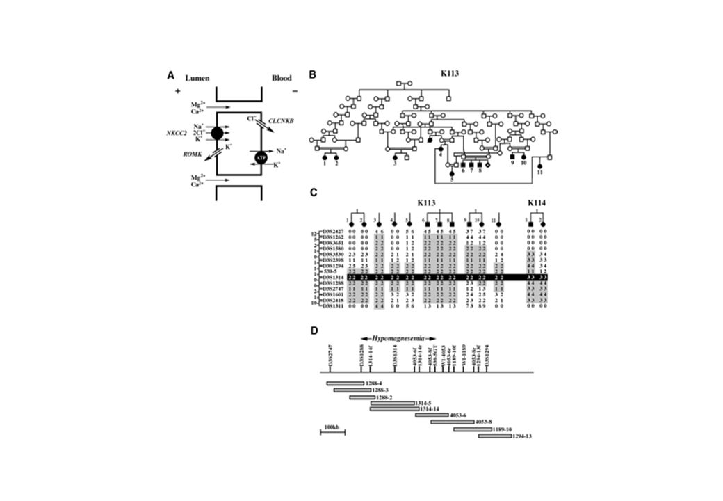
Paracellin-1 (Claudin 16) and hypercalciuria
 and hypercalciuria")
52
Tubular phenotype of patients with loss-of-function in PCLN-1 gene
HHF Controls 5 10 15 20 UV/GFR mmol/l GF Na + 0,1 Ca ++ baseline furo 0,2 Mg Cl - Na+ K+ 2 Cl- 3 Na+ 2 K+ Cl- Ca++ Mg++ ? Paracellin-1 A. Blanchard et al, Kidney Int, 2002

53
CLC5 and hypercalciuria

54
Canaux chlore : 3 familles distinctes
"Cystic fibrosis transmembrane conductance regulator (CFTR)" Cl- channel Extracellular-ligand gated (ELG), post synaptic Cl- channels CLC family : voltage-gated Cl- channels CLC-1 à CLC-7, CLC-Ka, CLC-Kb
 Cl- channel. Extracellular-ligand gated (ELG), post synaptic Cl- channels. CLC family : voltage-gated Cl- channels. CLC-1 à CLC-7, CLC-Ka, CLC-Kb.")
55
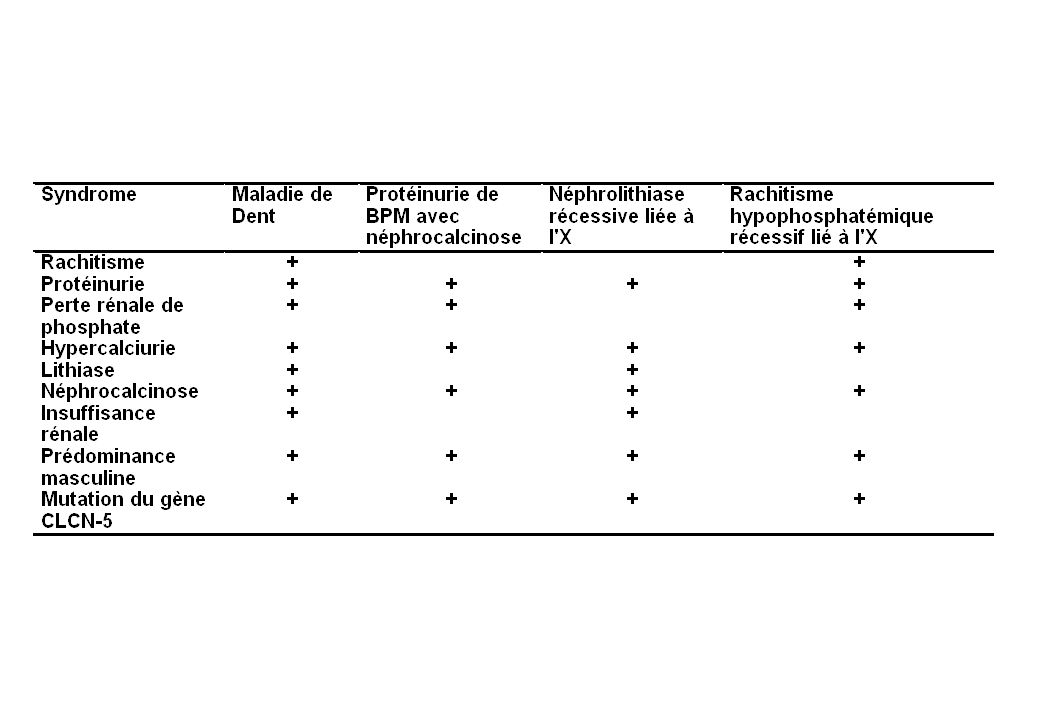
Néphrolithiase hypercalciurique liée à l'X
Maladie de Dent (Grande-Bretagne) Protéinurie de bas poids moléculaire avec hypercalciurie et néphrocalcinose (Japon) Néphrolithiase récessive liée à l'X (Etats-Unis, Canada) Rachitisme hypophosphatémique récessif lié à l'X (Italie, France)
 Protéinurie de bas poids moléculaire avec hypercalciurie et néphrocalcinose (Japon) Néphrolithiase récessive liée à l X (Etats-Unis, Canada) Rachitisme hypophosphatémique récessif lié à l X (Italie, France)")
58
Mutations du gène CLCN5 : perte de fonction du canal
Faux-sens Non sens Mutation d'un site d'épissage Insertion Délétion Absence de parallélisme entre le phénotype et le génotype

59
Colocalisation avec Rab4 et H+-ATPase

60
A-ClC-5 B-H+ATPase C=A+B D-CLC-5 E-2microglob. F=D+E G-CLC-5 H- 2microglob I=G+H 13min. ME CLC-5 Localisation de CLC-5 dans le tubule proximal

61
ß2 microglob Lactoglobuline lactoglobuline CLC-5 horseradish peroxydase FITC-dextran CLC-5

62
Mégaline CLC-5 CLC-5+mégaline Expression de la mégaline à la surface des cellules du tubule proximal en l’absence ou en présence de CLC-5

63
Physiopathologie diabète phosphaté
Diminution de l’expression apicale de NaPi-2 chez la souris CLC-5-/- Pas de modification de l’expression de NaPi-2 à la surface des cellules CLC-5- chez la souris CLC-5+/-. mégaline ClC-5 mégaline + CLC-5 NaPi 2 S1 +/+ -/- -/- +/- +/-

64
Fonction de CLC-5

65
Rôle de ClC5 dans le diabète phosphaté « hyperparathyroidisme »
PHYSIOPATHOLOGIE Rôle de ClC5 dans le diabète phosphaté Piwon Nils, Nature, 2000, vol 408, Hypothèse : la diminution de l ’expression basale de NaPi-2 est liée à une augmentation de PTH. Elévation luminale et non basolatérale ([PTH] systémique Nle). Diminution de l’endocytose [PTH] nle PTH filtrée [PTH]> Nle « hyperparathyroidisme » luminal (S3)
. Diminution de l’endocytose. [PTH] nle. PTH. filtrée. [PTH]> Nle. « hyperparathyroidisme » luminal (S3)")
66
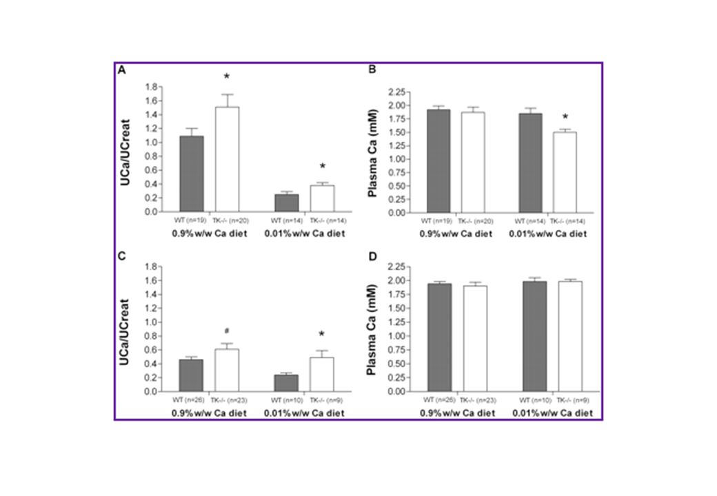
Tissue kallikrein and hypercalciuria

67
Tissue kallikrein and TRPV5 are coexpressed in the renal tubule

68
Meneton, Pierre et al. (2001) Proc. Natl. Acad. Sci. USA 98, 2634-2639
Copyright ©2001 by the National Academy of Sciences

71
Femelles C57Bl6/J

72
Males C57Bl6/J

73
Femelles 129Sv

74
Tissue kallikrein gene expression is controlled by calcium intake

75
Expression des transcrits des transporteurs

76
Mécanisme d’action de la TK
D. Gkika et al, EMBO J, 2006

77
L’effet de la TK est dépendant de la PKC

79
Effet des mutations des sites consensus de la PKC

80
La TK stabilise TRPV5 à la membrane

82
Monogenic hypercalciuria : clues to the genetics of idiopathic hypercalciuria ?
Intestine as the primary defect : > 5 genes, no gene encoding a Ca transporter Bone as the primary defect : Kidney as the primary defect : > 18 genes, only one gene encoding a Ca transporter (PCLN-1)
")
83
Selective genotyping of F2 (GHS female x normocalciuric male WKY rats)
Genetics of idiopathic hypercalciuria : lessons from genetic hypercalciuric rats Selective genotyping of F2 (GHS female x normocalciuric male WKY rats) Linkage between hypercalciuria and chromosomal regions Significant at D1Rat169 Suggestive to regions of Chr. 4, 7, 10, 14 No linkage with CaSR or VDR gene regions R. Hoopes et al, J.A.S.N., 2003
 Linkage between hypercalciuria and chromosomal regions. Significant at D1Rat169. Suggestive to regions of Chr. 4, 7, 10, 14. No linkage with CaSR or VDR gene regions. R. Hoopes et al, J.A.S.N.,")
84
Conclusion Hypercalciuria is a complex trait, and its expression depends on both Environmental factors Genetic factors Modification of dietary factors is efficient but not specific Continuing efforts are warranted - detailed proximal phenotype definition - study of monogenic causes of hypercalciuria - identification of loci linked to idiopathic hypercalciuria

87
Georges Pompidou Hospital Tenon Hospital
Pascal Houillier Anne Blanchard Marie Briet Marc Froissart Gérard Maruani Laurence Nicolet Tenon Hospital Eric Rondeau Pierre Ronco Brigitte Lantz Françoise Paillard INSERM Unit Nicolas Picard Nijmegen University Joost Hoenderop Rene Bindels

88
Pathophysiology of human idiopathic hypercalciuria
ECF Ca Hypercalciuria Adapted from Lemann, 1992 Adapted from Houillier, 1996

90
Monogenic renal hypercalciuria : clues to the genetics of idiopathic hypercalciuria ?
Trpv5 (ECaC1) CaSR Paracellin-1 (Claudin 16)
 CaSR. Paracellin-1 (Claudin 16)")
Similar presentations


 (by kidneys, liver, lungs, skin) BODY LOAD Metabolic production Metabolism to a new substance.>")
![H + Homeostasis by the Kidney. H + Homeostasis Goal: To maintain a plasma (ECF) pH of approximately 7.4 (equivalent to [H + ] = 40 nmol/L Action needed:](/15/4651439/big_thumb.jpg "H + Homeostasis by the Kidney. H + Homeostasis Goal: To maintain a plasma (ECF) pH of approximately 7.4 (equivalent to [H + ] = 40 nmol/L Action needed:>")


