Download presentation
Presentation is loading. Please wait.

2
Why Intubate and Ventilate ?
Improve Oxygenation (PaO2, SaO2). Improve ventilation (PaCO2). Relieve work of breathing. Unload Respiratory Muscle.
. Improve ventilation (PaCO2). Relieve work of breathing. Unload Respiratory Muscle.")
3
Evaluate before you sedate
How soon do you allow your patient to breath spontaneously? The sooner the better !!!

4
Does your ventilated patient appear to be struggling or uncomfortable?
If a patient is experiencing ineffective triggering the ventilator will not include the missed effort in the respiratory rate displayed on the ventilator. For example, the ventilator may display a breath rate of 25 while the multi-parameter is displaying a breath rate of 35. This is an indication the patient may be experiencing patient-ventilator asynchrony. Does your ventilated patient appear to be struggling or uncomfortable?

5
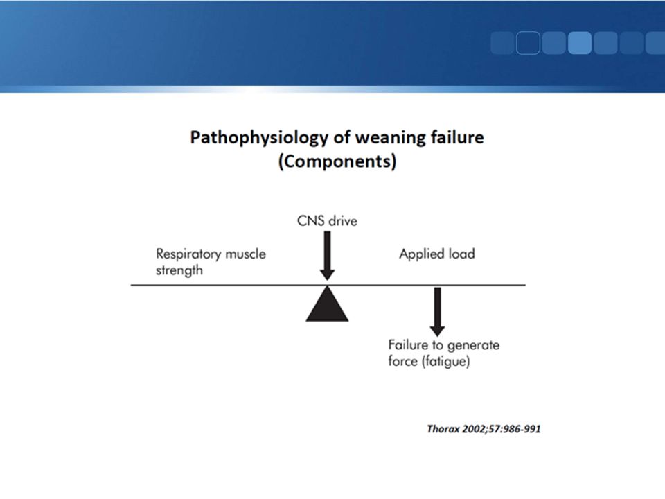
Lesson Learned !! Conventional modes of ventilation can not cope well with the ever-changing patients Ventilatory needs

6
Prevent Asynchrony with Ventilator
The Next Generation of Synchronization Ventilatory Support PAV+ Prevent Asynchrony with Ventilator

7
Ventilator Support is proportional to:
Instantaneous Effort (Pmus) Pulmonary Mechanics (Resistance, Compliance)
 Pulmonary Mechanics (Resistance, Compliance)")
8
How Does PAV+ Work?

9
Pmus + Paw = (Flow x R) + (Volume x E)
PAV+ measures patient demand by monitoring flow and volume every 5 milliseconds and measures compliance and resistance every 4 to 10 breathes Equation Of Motion= Pmus + Paw = (Flow x R) + (Volume x E)
 + (Volume x E)")
10
{ Pappl Ventilator Patient effort throughout inspiration .
S (ViL EL) S (ViY * RiSYS) ViY . ò ViY dt = ViL Patient effort throughout inspiration { Ventilator RLUNG CLUNG RET PiLUNG-THORAX PiET + PATIENT Inspiratory Effort ViI . VLUNG Pappl Trigger and Execute PAV PLUNG Sum: PiFLOW + PiVOLUME = PiY PiY=S (ViL * RiETT)+ S (ViL RL) + S (ViL Ers) .
 S (ViY * RiSYS) ViY. . ò ViY dt = ViL. Patient effort throughout inspiration. { Ventilator. RLUNG. CLUNG. RET. PiLUNG-THORAX. PiET + PATIENT. Inspiratory Effort. ViI. . VLUNG. Pappl. Trigger and Execute PAV. PLUNG. Sum: PiFLOW + PiVOLUME = PiY. PiY=S (ViL * RiETT)+ S (ViL RL) + S (ViL Ers) .")
11
Response to patient’s inspiratory effort “Mirror Image Breath
How does PS work ? The pt has to make a sufficient respiratory effort to trigger the ventilator , at this point the ventilator increases the pressure in the circuit but the amount of pressure support that the clinician has set , the ventilator then decrease or increase the flow going to the patient to maintain the preset pressure , will terminate the breath with the flow termination criteria by adjusting the E sensitivity level The actual termination of inspiration does not necessarily coincide with the pt pt breathing effort The flow pattern in PS does not look like the effort made by the pt , from pt perspective , the pressure in solid green represent what the pt is asking for whether the flow pattern generated by ventilator is what ventilator is giving in response

12
Response to patient’s inspiratory effort “Mirror Image Breath

13
Muscle Effort Amplifier

14
Results of pressure strategy with greater patient effort (PSV vs. PAV+)
")
15
PAV+ and Patient Control
Inspiration How deeply to breath? When the breath ends? PAV+ and Patient Control (A) Patient determines when inspiration begins when Flow senses at the Wye (B) The ventilator will continue to provide the gas as long as the patient inspiratory effort is continuing (C) Because pressure is proportional to pt inspiratory effort , when effort ends pressure rapidly declines and flow stops
 Patient determines when inspiration begins when Flow senses at the Wye. (B) The ventilator will continue to provide the gas as long as the patient inspiratory effort is continuing. (C) Because pressure is proportional to pt inspiratory effort , when effort ends pressure rapidly declines and flow stops.")
16
So How Does It Work ? Patient drives the ventilator

17
Q. How does the clinician know where to set the %Support?
Sound Clinical Assessment. 2. Work of Breathing (WOB) bar.
 bar.")
18
Clinical Assessment Signs of Respiratory Distress: Vital Signs ABG
Respiratory rate > 40 breaths/minute PLUS… Marked use of accessory muscles Diaphoresis Abdominal paradox Marked complaint of dyspnea Etc…

19
PAV+, Understanding the WOB Bar

21
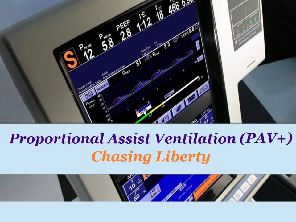
Proportional Assist Ventilation (PAV+), Chasing Liberty
, Chasing Liberty")
22
Literature Review

23
VCV VC Ventilator Pressure Patient Effort

24
Pressure Limited Ventilation (PCV, PS)
Pressurewye Time b c c Active Effort Flowwye b Trigger Only VolumeLUNG c Active Effort b Trigger Only

25
Physiological Mode VC More physiological PAV+ PCV/PS Ventilator
Pressure Patient Effort Proportional assist Ventialtion and Neurally Adjusted Ventilator Assist Robert Kacmarek PhD Resp Care Feb Vol. 56 No. 2

26
Now the patient’s respiratory center is in charge.
Rate , rhythm and depth

28
Complete controlled ventilation beyond 24 hours may cause Respiratory muscle Injury and can result in Ventilator Induced Diaphragmatic dysfunction (VIDD).[8] Approximately 40% of patients in medical ICUs require mechanical ventilation (MV). Approximately 20% to 25% of these patients will encounter difficulties in discontinuing MV. Multiple studies have suggested that MV has an unloading effect on the respiratory muscles that leads to diaphragmatic atrophy and dysfunction, a process called ventilator-induced diaphragmatic dysfunction (VIDD). VIDD may be an important factor affecting when and if MV can be discontinued. A sensitive and specific diagnostic test for VIDD could provide the physician with valuable information that might influence decisions regarding extubation or tracheostomy. Levine et al., Rapid Disuse Atrophy of Diaphragm Fibers in Mechanically Ventilated Humans, NEJM :
![Complete controlled ventilation beyond 24 hours may cause Respiratory muscle Injury and can result in Ventilator Induced Diaphragmatic dysfunction (VIDD).[8]](http://slideplayer.com/slide/4156970/13/images/28/Complete+controlled+ventilation+beyond+24+hours+may+cause+Respiratory+muscle+Injury+and+can+result+in+Ventilator+Induced+Diaphragmatic+dysfunction+%28VIDD%29.%5B8%5D.jpg "Approximately 40% of patients in medical ICUs require mechanical ventilation (MV). Approximately 20% to 25% of these patients will encounter difficulties in discontinuing MV. Multiple studies have suggested that MV has an unloading effect on the respiratory muscles that leads to diaphragmatic atrophy and dysfunction, a process called ventilator-induced diaphragmatic dysfunction (VIDD). VIDD may be an important factor affecting when and if MV can be discontinued. A sensitive and specific diagnostic test for VIDD could provide the physician with valuable information that might influence decisions regarding extubation or tracheostomy. Levine et al., Rapid Disuse Atrophy of Diaphragm Fibers in Mechanically Ventilated Humans, NEJM :")
29
Ventilator Induced Diaphragmatic Dysfunction
MV and Diaphragm Complete controlled ventilation beyond 24 hours may cause Respiratory Muscle Injury and can result in: Ventilator Induced Diaphragmatic Dysfunction Sassoon et al (2002). Altered Diaphragm Contractile Properties with Controlled Mechanical Ventilation. J Appl Physiol 92(6):
. Altered Diaphragm Contractile Properties with Controlled Mechanical Ventilation. J Appl Physiol 92(6):")
30
Frequent setting changes and increase sedation requirements
Length of ICU Stay Frequent setting changes and increase sedation requirements Increase Ventilation time , possible Muscle Atrophy and Increase Length of stay Asynchrony

31
PVA is common in Conventional Mode of Ventilation
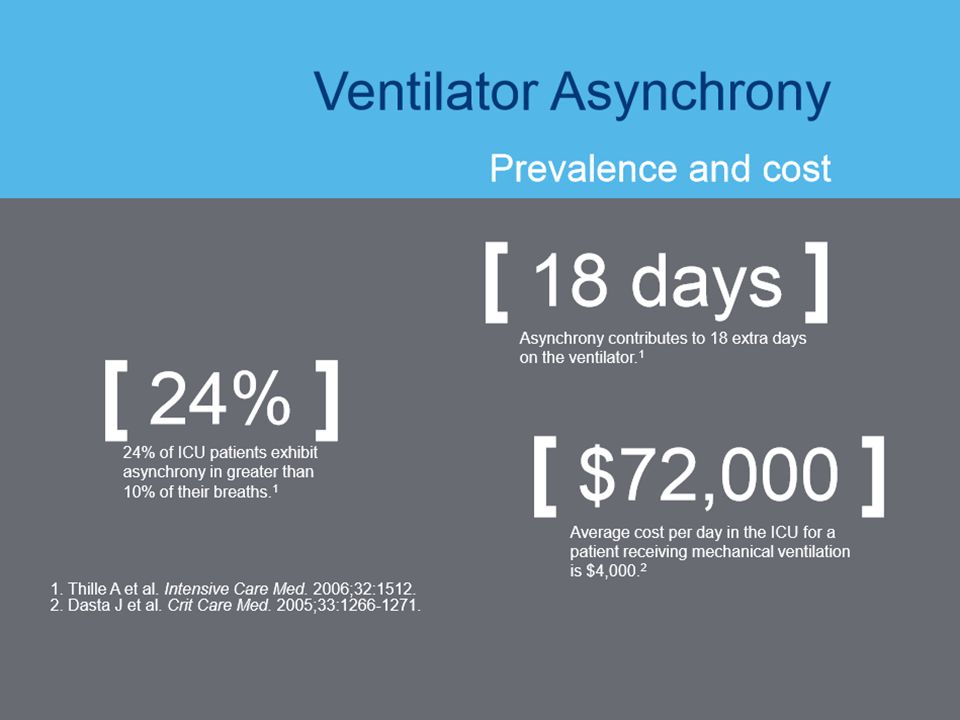
> 25% of patients on VC and PSV had higher incidence of Asynchrony. Asynchrony Index (AI) of > 10% of respiratory efforts. Higher incidence of Asynchrony is associated with prolonged duration of MV (25 vs. 7 days, p=.005). More likely to receive Tracheostomy (33% vs 4%) Thille et al .(2006) Intensive Care Med 32;
 of > 10% of respiratory efforts. Higher incidence of Asynchrony is associated with prolonged duration of MV (25 vs. 7 days, p=.005). More likely to receive Tracheostomy (33% vs 4%) Thille et al .(2006) Intensive Care Med 32;")
32
Failure to Synchronize
Deleterious Effects of PVA: Patient Fights Ventilator Higher Work of Breathing More Sedation Required Muscle Fatigue Dynamic Hyperinflation Delayed or prolonged weaning Longer ICU Stay Higher Costs. Respir Care (2005).50(2);
.50(2);")
35
Potential Clinical Advantages
• The patient ‘drives’ the ventilator • Avoids ventilator over-assistance • Less patient-ventilator asynchrony – Asynchrony associated with • More sedation (deWit, M. Journal of Critical Care 2009:24;74-80) • Longer duration of mechanical ventilation (Thille, A. Intensive Care Medicine 2006; deWit, M. Crit Care Med 2009) • Disrupted sleep (Bosma, K, Crit Care Med 2007, Fanfulla, F AJRCCM 2005)
 • Longer duration of mechanical ventilation. (Thille, A. Intensive Care Medicine 2006; deWit, M. Crit Care Med 2009) • Disrupted sleep. (Bosma, K, Crit Care Med 2007, Fanfulla, F AJRCCM 2005)")
36
PAV+ Increases patient safety by decreasing the risk of over-assisted ventilation
less likelihood of overventilation — With PAV+ software, there is no minimal delivered tidal volume like with other modes. If the % Support dialed in is more than is necessary, patients will down regulate their efforts. Because with PAV+, the pressure delivered is a function of effort. When effort decreases the pressure demand will also decrease. Accordingly, this feedback mitigates the tendency for overventilation. By contrast, with other modes the ventilator will continue to give the same pressure or volume regardless of what happens to effort, so long as the effort is enough to trigger. Also, in the case of airway artifacts like hiccups or heartbeats, no standard volume or pressure would be accidentally delivered because with PAV+, the ventilator would stop delivering gas as soon as the artifact was over. With other modes, cardiac artifacts may continue to cause frequent triggering and delivery of large volumes even when efforts cease completely. Imanaka H. Crit Care Med. 2000;28(2):
:")
37
PAV+ Increases patient safety by decreasing the risk of over-assisted ventilation
Preservation and enhancement of the patient’s control mechanisms — With PAV+ the breath is being driven by the patient’s own control center and reflexes. One such reflex is the Hering-Breuer reflex, which causes the inhibition of inspiratory efforts when tidal volume reaches a physiologically determined threshold, thus preventing over distension of the lung. Because the ventilator ceases its pressure when inspiratory effort is terminated, stimulation of this reflex would cause the breath to cycle off. Improved hemodynamics — Research has shown that when patients were switched from Volume Control to manual PAV+, cardiac output increased by 22% in septic patients. Patrick W, et al.. Am J Respir Crit Care Med. 1993;147:A61

38
PAV+ Increases patient safety by decreasing the risk of over-assisted ventilation
Weaning — The greater the reliability of ventilator rate as a measure of distress, the better the decision-making on tolerance. Because with PAV+ there are little or no ineffective efforts, ventilator rate and patient rate are the same. So, when ventilator rate increases as the assist level is reduced (e.g., during a weaning trial), it means that the patient’s rate has also increased, a sign that suggests this new level is not tolerated (distress). With other modes in which ineffective efforts may exist (e.g., PSV, volume - cycled), ventilator rate can be considerably less than the patient’s rate. Giannouli E. Am J Respir Crit Care Med. 1999;159(6): Thille AW,. Intensive Care Med. 2006;32(10): Leung P. Am J Respir Crit Care Med. 1997;155(6):
, it means that the patient’s rate has also increased, a sign that suggests this new level is not tolerated (distress). With other modes in which ineffective efforts may exist (e.g., PSV, volume - cycled), ventilator rate can be considerably less than the patient’s rate. Giannouli E. Am J Respir Crit Care Med. 1999;159(6): Thille AW,. Intensive Care Med. 2006;32(10): Leung P. Am J Respir Crit Care Med. 1997;155(6):")
39
PAV+ Increases patient safety by decreasing the risk of over-assisted ventilation
Because the number of ineffective efforts decreases, often dramatically, as assist level is decreased, the ventilator rate frequently jumps during a weaning trial despite the fact that patient’s rate has not changed. This can lead to the false diagnosis of weaning failure. The PAV+ may emerge as a useful tool in weaning because with PAV+ there is less likelihood of misinterpretation of actual respiratory rate. In addition, this type of ineffective muscle contraction has been associated with muscle injury. With PAV+, this is less likely to occur. Giannouli E. Am J Respir Crit Care Med. 1999;159(6): Van Der Meulen JH,. J Appl Physiol. 1997;83(3): Devor ST. J Appl Physiol. 1999;87(2):
: Van Der Meulen JH,. J Appl Physiol. 1997;83(3): Devor ST. J Appl Physiol. 1999;87(2):")
40
PAV+ Increases patient safety by decreasing the risk of over-assisted ventilation
Less-invasive technology. Unlike other approaches to measuring patient demand (e.g., esophageal manometry or diaphragm EMG), there is no need for an additional invasive procedure, which could lead to complication from incorrect placement. Cost and time are other obvious advantages to a less invasive approach. The PAV+ takes random measurements of compliance and resistance along with rapid samples of pressure and flow to determine the support pressure via a cuffed artificial airway.
, there is no need for an additional invasive procedure, which could lead to complication from incorrect placement. Cost and time are other obvious advantages to a less invasive approach. The PAV+ takes random measurements of compliance and resistance along with rapid samples of pressure and flow to determine the support pressure via a cuffed artificial airway.")
41
What Kind of Patient Would Benefit more from PAV+?
Comfort. Lower peak airway pressure. Less need for paralysis and/or sedation. Less likelihood for over ventilation. Preservation and enhancement of patient’s own control mechanisms such as metabolic ABG control and Hering-Breuer reflex. Improved efficiency of negative pressure ventilation. M Younes. Am Rev Respir Dis 1992;145:

42
Comfort 12 COPD patients Using VAS Breathing comfort was sig. with PAV
(38 vs. 11) Wysocki et al .(2002) Crit Care Med. 30(2);
 Wysocki et al .(2002) Crit Care Med. 30(2);")
43
Vt Variability 13 Ventilated COPD patients PAV vs. PSV
Wrigge et al .(1999) Intensive Care Med. 25;
 Intensive Care Med. 25;")
44
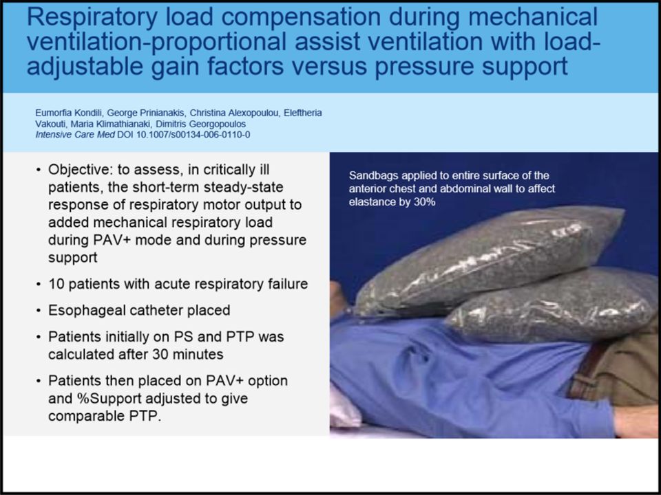
WOB Kondili et al .(2006) Intensive Care Med. 32;
 Intensive Care Med. 32;")
45
PVA Bosma et al .(2007) Crit Care Med. 35(4);
 Crit Care Med. 35(4);")
46
Settings Manipulation and Need for Sedation
Compared with PSV, PAV was associated with: Fewer manipulations of ventilator settings Fewer changes in sedative dosing.

47
Evaluate before Sedate
Careful evaluation of the patient prior to sedation may help reduce the use of unnecessary sedation. Siegel MD. Clin Chest Med 2003; 24 (4):
:")
48
Probability to remain on Spontaneous Mode
RCT of 208 critically ill patients on a controlled mode randomized to either PAV or PSV.

49
Probability to remain on Spontaneous Mode
Failure to transition: PAV 11%, PSV 22%. Proportion of patients who developed to asynchrony: PAV 5.6%, PSV 29%.

50
Probability to remain on Spontaneous Mode

51
Proportional assist Ventialtion and Neurally Adjusted Ventilator Assist Robert Kacmarek PhD Resp Care Feb Vol. 56 No. 2 As stated at the onset of this paper, PAV and NAVA are designed to achieve the same overall outcome, that is patient- driven ventilatory assist applied with improved patient- ventilator synchrony. Both modes are capable of achieving the goal, but there are some differences (Table2). They manage ventilatory assist using different variables.PAV does not require additional hardware; NAVA requires the placement of a special nasogastric tube. NAVA can be used for invasive and noninvasive ventilation; PAV also can be used invasively and noninvasively, but is not available for both applications on all ventilators. NAVA can be applied to patients of any age group: premature neonates to adults, PAV is reserved for patients 20 kg.NAVA is not affected by leaks or intrinsic PEEP, since its function is based on diaphragm EMG signals. PAV is unable to adjust to intrinsic PEEP. In the presence of intrinsic PEEP, PAV functions the same as any other mode. Both PAV and NAVA improve patient-ventilator synchrony, but further study of both is needed to determine if either improves patient outcomes.
. They manage ventilatory assist using different variables.PAV does not require additional hardware; NAVA requires the placement of a special nasogastric tube. NAVA can be used for invasive and noninvasive ventilation; PAV also can be used invasively and noninvasively, but is not available for both applications on all ventilators. NAVA can be applied to patients of any age group: premature neonates to adults, PAV is reserved for patients 20 kg.NAVA is not affected by leaks or intrinsic PEEP, since its function is based on diaphragm EMG signals. PAV is unable to adjust to intrinsic PEEP. In the presence of intrinsic PEEP, PAV functions the same as any other mode. Both PAV and NAVA improve patient-ventilator synchrony, but further study of both is needed to determine if either improves patient outcomes.")
59
Background 6% of ventilated patients are prolonged mechanically ventilated (PMV) 20% to 30% are difficult-to-wean. Weaning tends to be delayed -Exposing the patient to unnecessary discomfort -Increased risk of complications -Increasing the cost of care and mortality 12% vs 27% . Time spent in the weaning process → 40–50% of the total duration of mechanical ventilation.

60
Reasons contributing to weaning failure in anesthetized and critically ill patients
Intensive Care Med (2013) 39:1885–1895 DOI /s
 39:1885–1895. DOI /s")
61
Objective: This study was designed to determine the effect of PAV+ on adult difficult-to-wean PMV patients. Gulf Thoracic Congress March 13, 2014

62
Results: 13 adult Pts were included in this study.
9 of the Pts with Mean duration of MV was 53.2 days prior to PAV+ trial. On PAV+, NIF and P 0.1 measurements improved by 87% and 79% respectively from the baseline. They were successfully weaned off MV with an average weaning time of 5.8 days. 4 of the Pts were unsuccessfully weaned off MV and retained back to SIMV mode and went to be prolonged ventilator dependents. Gulf Thoracic Congress March 13, 2014

63
Gulf Thoracic Congress March 13, 2014
NIF and P 0.1 measurements throughout PAV+ trails Time Spent on Conventional Weaning and PAV+ Gulf Thoracic Congress March 13, 2014

64
Conclusion: PAV+ can be used safely and efficiently to wean adult difficult-to-wean PMV patients who failed multiple trails of conventional weaning. PAV+ provides opportunity for a respiratory muscle to recover and strengthen, increasing the likelihood of weaning success. Gulf Thoracic Congress March 13, 2014

67
PAV vs PSV in the weaning of Pt with AECOPD
60 Pts: 30 on PAV, 30 on PSV Weaning Success PAV vs PSV 90% vs 66.7% In PAV, less PVA 1.5 days reduction in mean days of MV 2 days reduction in mean days of ICU stay 1.8 days reduction in mean days of hospital stay

68
New Developments in PAV+
Recently, a new technology has been introduced that aims to monitor and improve patient– ventilator interaction. With PVI monitor, a signal representing an estimate of the patient’s total respiratory muscle pressure (Pmus,PVI) is calculated via the equation of motion, utilizing estimated values of resistance and elastance of the respiratory system, obtained noninvasively. The waveform of Pmus.PVI is continuously displayed online on a breath-by-breath basis and can be used to trigger the ventilator. It has been shown that this triggering method may substantially shorten the triggering delay (by approximately 70%), even in patients with dynamic hyperinflation. Theoretically, this system should increase the efficiency of PAV+ to support critically ill patients with dynamic hyperinflation. Younes M, et al. Intensive Care Med 2007; 33: 1337–1346 Kondili E, et al. Intensive Care Med 2010; 36: 648–655.
 is calculated via the equation of motion, utilizing estimated values of resistance and elastance of the respiratory system, obtained noninvasively. The waveform of Pmus.PVI is continuously displayed online on a breath-by-breath basis and can be used to trigger the ventilator. It has been shown that this triggering method may substantially shorten the triggering delay (by approximately 70%), even in patients with dynamic hyperinflation. Theoretically, this system should increase the efficiency of PAV+ to support critically ill patients with dynamic hyperinflation. Younes M, et al. Intensive Care Med 2007; 33: 1337–1346. Kondili E, et al. Intensive Care Med 2010; 36: 648–655.")
69
Why PAV+ is not Commonly Used?
PAV (1992) PAV+ (2005) PAV+ (2005) is more accurate, safe & effective. Failure to Knowledge Transfer??? * Application sometimes regarded as difficult** PAV+ had not been investigated thoroughly in weaning trails** . ** Boles, al et, Eur Respir J 2007; 29: 1033–1056
 PAV+ (2005) PAV+ (2005) is more accurate, safe & effective. Failure to Knowledge Transfer * Application sometimes regarded as difficult** PAV+ had not been investigated thoroughly in weaning trails** . ** Boles, al et, Eur Respir J 2007; 29: 1033–1056.")
70
Why PAV+ is not Commonly Used?

71
Conclusion PAV+ – Provides assistance in PROPORTION to patient effort
– Provides patient greater control of modulating VE – Reduces patient-ventilator asynchrony – Improves sleep quality for patients asynchronous on PSV PAV+ may help: – Preserve Respiratory Muscle Strength – Facilitate Weaning – Decrease Need for Sedation Does all this mean extra coffee breaks for clinicians?

72
Conclusion PAV is safe and effective mode of ventilation.
There is strong evidence that PAV provides some advantages related to patient comfort and better synchrony. PAV may be helpful with difficult to wean patients (muscle fatigues, changing lung mechanics) Understanding PAV physiology and operation is essential for save use of the mode.
 Understanding PAV physiology and operation is essential for save use of the mode.")
73
Questions Need to be Answered?
Non-invasive PAV+? PAV+ as Initial Setting Mode for Non-Fully Sedated or Paralyzed Patient? PAV+ in The Specialized Weaning Units? PAV+ in The Home Ventilators?

Similar presentations