Download presentation
Presentation is loading. Please wait.

1
Disorders of the Pleura, Mediastinum, Diaphragm, Chest Wall and Lung Cancer
Maximino G. Bello III, MD, FPCP, FPSMO Executive Secretary, Cancer Institute

2
Take note! All exams are Harrison based
Rapid advances in oncology, new findings may supersede Harrison Take note if I stressed a particular fact or statement Topics not discussed in today's lecture does NOT mean it would not be included in exams

3
Pleural diseases Pleural effusion Pneumothorax
Pleural space from the capillaries in the parietal pleura removed via lymphatics Interstitial spaces from the lung via visceral pleura Peritoneal cavity via diaphragm Pneumothorax Air in the pleural space

5
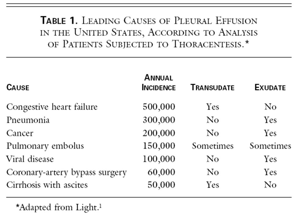
Diagnostic approach Transudate: systemic factors
Exudate: local factors Light’s Criteria Pleural fluid CHON /serum CHON >0.5 Pleural fluid LDH/serum LDH >0.6 Pleural fluid LDH more than 2/3 normal upper limit for serum

6
Light’s Criteria misidentifies ≈25% of transudates as exudates

7
Light’s Criteria Transudate CHF Cirrhosis PE Nephrotic syndrome
Peritoneal dialysis SVC Myxedema Exudate Infectious Neoplastic GI disease Collagen vascular dse P CABG etc

10
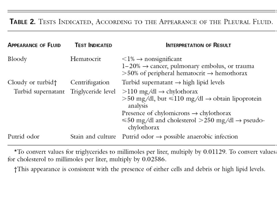
Parapneumonic effusions
Associated with bacterial infection, lung abscess or bronchiectasis Empyema: grossly purulent effusion Condensed milk “significant effusion” Lateral decubitus view shows 10mm layering of fluid drainage of effusion

11
Drainage of effusion Need for a more invasive procedure (other than thoracentesis) Loculated pleural effusion Pleural fluid pH <7.20 Pleural fluid glucose < 3.3mmol/L + gram stain or culture of the pleural fluid empyema

12
Effusion secondary to malignancy
Lung and breast carcinoma and lymphoma 75% of malignant effusion Dyspnea is NOT proportionate to the amount of effusion Lung metastasis Treatment Drainage of the fluid sclerosing agent treatment of the malignancy

13
Effusion secondary to mesothelioma
Primary tumor of the mesothelial cells Line the pleural cavity Significant asbestos exposure Imaging: Effusion, thick pleura, collapse hemithorax Treatment: Surgery pretexemed

16
Pneumothorax Primary spontaneous pneumothorax Treatment: aspiration
Rupture of apical bleb It typically occurs in tall, thin boys and men between the ages of 10 and 30 years rarely occurs in persons over the age of 40. Appears almost exclusively in smokers ½ will have recurrences Treatment: aspiration

17
Pneumothorax Secondary spontaneous pneumothorax Treatment COPD
More fatal lesser physiologic reserve Treatment Tube thoracostomy

19
Pneumothorax Traumatic pneumothorax Tension pneumothorax Penetrating
Non penetrating injuries Tension pneumothorax Medical emergency During resuscitation Cyanosis, hypotension

21
Most Diaphragmatic Hernia’s are detected in childhood.
Rare in adults!

22
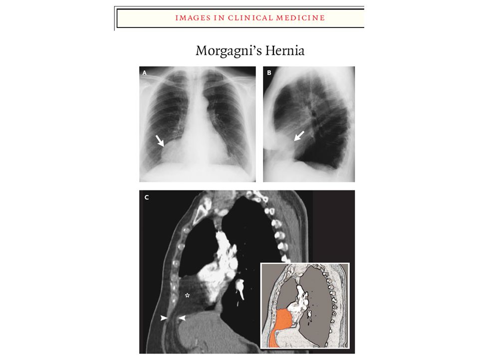
Diaphragmatic Hernia Congenital diaphragmatic hernia Bochdalek:
More common postero-lateral diaphragmatic hernia majority of Bochdalek hernias (80-85%) occur on the left side Morgagni Less common Anterior, right
 occur on the left side. Morgagni. Less common. Anterior, right.")
24
Mediastinum Occupies the central portion of the thoracic cavity
Boundaries: Lateral- pleural cavity Superior- thoracic inlet Inferior- diaphragm Anterior- sternum Posterior- chest wall anterior posterior middle De Vita, et al .Principles & Practice of Oncology 8th ed 24

25
Mediastinal tumors: Feature Thymoma Lymphoma Germ cell tumor
Mesenchymal Incidence - most common anterior Mediastinal neoplasm % of Mediastinal tumors - equal in male and female - ages % of primary Mediastinal masses - 2nd most common mass - Most Mediastinal lymphomas are seen in the anterosuperior mediastinum. -15% of anterior Mediastinal tumors in adults. (24% in children) - Rarely, they are found in the posterior mediastinum - 6% of Mediastinal tumors. - More than 50% are malignant
 - Rarely, they are. found in the posterior. mediastinum. - 6% of Mediastinal. tumors. - More than 50% are malignant.")
26
Mediastinal tumors: Feature Thymoma Lymphoma Germ cell tumor X-ray
Mesenchymal Radiographic findings: X-ray smooth mass in the upper half of the chest. Overlying the superior portion of the cardiac shadow. -The mass projects predominantly into one of the hemithoraces. Lobulated with enlargement of hilar and media- stinal lymph nodes. - well defined mass occasionally containing calcifications. - Mediastinal widening on CXR

27
Mediastinal tumors: Feature Thymoma Lymphoma Germ cell tumor
Mesenchymal Radiographic findings: CT scan - demonstrates uniform enhancement conglomerate of lymph nodes discrete enlarged LN with cystic degeneration lobulated, asymmetrical, homogenous tumors with/ without cystic components can determine components of tumor (fat, soft tissue) defines the relation of tumor to adjacent tissues.
 defines the relation of tumor to adjacent tissues.")
28
Mediastinal tumors: Feature Thymoma Lymphoma Germ cell tumor
Mesenchymal Signs and symptoms - 50% asymptomatic - symptoms due to myasthenia in 35% of patients - others with substernal pains, dyspnea, cough - Invasive thymoma cause local compression /svc syndrome -Majority of are symptomatic at diagnosis. - Common: fever, weight loss, night sweats - Compression symptoms: pain, dyspnea, stridor, or superior vena cava syndrome - Associated pleural effusions are common malignant tumors are symptomatic in 85% of patients: chest pain, hemoptysis, cough, fever, weight loss. - Superior vena caval syndrome is occasionally seen - Compressive sign and symptoms based on adjacent tissues involved.

29
Made Ridiculously simple!
Lung Cancer Made Ridiculously simple!

30
MORTALITY: TEN LEADING (10) LEADING CAUSES Number and rate/100,000 Population Philippines 5-Year Average ( ) & 2005
 LEADING CAUSES Number and rate/100,000 Population Philippines 5-Year Average ( ) & 2005")
32
US Mortality, 2005 No. of deaths % of all deaths Rank Cause of Death
1. Heart Diseases 652, 2. Cancer , 3. Cerebrovascular diseases 143, 4. Chronic lower respiratory diseases 130, 5. Accidents (unintentional injuries) 117, 6. Diabetes mellitus 75, 7. Alzheimer disease , 8. Influenza & pneumonia 63, Nephritis* , 10. Septicemia , Cancer accounts for nearly one-quarter of deaths in the United States, exceeded only by heart diseases. In 2005, there were 559,312 cancer deaths in the US. *Includes nephrotic syndrome and nephrosis. Source: US Mortality Data 2005, National Center for Health Statistics, Centers for Disease Control and Prevention, 2008.
 117, Diabetes mellitus 75, Alzheimer disease 71, Influenza & pneumonia 63, Nephritis* 43, Septicemia 34, Cancer accounts for nearly one-quarter of deaths in the United States, exceeded only by heart diseases. In 2005, there were 559,312 cancer deaths in the US. *Includes nephrotic syndrome and nephrosis. Source: US Mortality Data 2005, National Center for Health Statistics, Centers for Disease Control and Prevention,")
33
2008 Estimated US Cancer Deaths*
Men 294,120 Women 271,530 Lung & bronchus 31% Prostate 10% Colon & rectum 8% Pancreas 6% Liver & intrahepatic 4% bile duct Leukemia 4% Esophagus 4% Urinary bladder 3% Non-Hodgkin % lymphoma Kidney & renal pelvis 3% All other sites % 26% Lung & bronchus 15% Breast 9% Colon & rectum 6% Pancreas 6% Ovary 3% Non-Hodgkin lymphoma 3% Leukemia 3% Uterine corpus 2% Liver & intrahepatic bile duct 2% Brain/ONS 25% All other sites Lung cancer is, by far, the most common fatal cancer in men (31%), followed by prostate (10%), and colon & rectum (8%). In women, lung (26%), breast (15%), and colon & rectum (9%) are the leading sites of cancer death. ONS=Other nervous system. Source: American Cancer Society, 2008.
, followed by prostate (10%), and colon & rectum (8%). In women, lung (26%), breast (15%), and colon & rectum (9%) are the leading sites of cancer death. ONS=Other nervous system. Source: American Cancer Society,")
34
HOSPITAL TUMOR REGISTRY REPORT 2000-2007
TOP TEN CANCER SITES (both sexes) SITE MALE FEMALE TOTAL 1. BREAST Lungs Colon/rectum cervix uterii Nasopharynx Uterus/ endometrium 7. prostate thyroid gland blood/ bone marrow 10. lymphomas total
 SITE MALE FEMALE TOTAL 1. BREAST Lungs Colon/rectum cervix uterii Nasopharynx Uterus/ endometrium 7. prostate thyroid gland blood/ bone marrow 10. lymphomas total")
35
Change in the US Death Rates* by Cause, 1950 & 2005
Rate Per 100,000 1950 2005 ? Compared to the rate in 1950, the cancer death rate decreased slightly in 2005, while rates for other major chronic diseases decreased substantially during this period. Heart Diseases Cerebrovascular Diseases Influenza & Pneumonia Cancer * Age-adjusted to 2000 US standard population. Sources: 1950 Mortality Data - CDC/NCHS, NVSS, Mortality Revised. 2005 Mortality Data: US Mortality Data 2005, NCHS, Centers for Disease Control and Prevention, 2008.

36
Lung Cancer : 13% : 13% : 16%

37
Worldwide Incidence and Mortality for Lung Cancer
Lung cancer is the most common cancer in the world Smoking is the most important risk factor World Incidence1 World Mortality1 Lung Cancer 1.5 Mio 90% Worldwide Incidence and Mortality for Lung Cancer Lung cancer is the most common cancer in the world, with 1.3 million new cases diagnosed every year, and it is the leading cause of cancer-related mortality Asia accounts for 49% of the worldwide incidence and mortality of cancer, Europe accounts for 28% of the incidence and 29% of the mortality, and North America accounts for 17% of the incidence and 15% of the mortality Central and South America account for 4% of the worldwide incidence and mortality, while Africa and Oceania* each account for 1% of the incidence, with Africa accounting for 2% and Oceania accounting for 1% of worldwide cancer mortality * Includes Australia and New Zealand. Lung Cancer: Kamangar et al. J Clin Oncol. 2006;24:

38
Inherited cancer syndrome
Host Susecptibility Family Hx Inherited cancer syndrome P53 mutation EGFR mutation Retinoblastoma SNP variation at 15q24–15q25.1 susceptibility and risk also increase with reduced DNA repair capacity ERCC1 Molecular Evolution of Lung Cancer. Environmental factors, such as tobacco smoke, and genetic susceptibility interact to influence carcinogenesis. Factors that are unrelated to smoking — including genetic, hormonal, and viral (e.g., human papillomavirus) factors — have been suggested.2 Tissue injury (e.g., from tobacco smoke, reflected in the discolored smoking-related lungs) initially occurs in the form of genetic and epigenetic changes (e.g., mutations, loss of heterozygosity, and promoter methylation) and global transcriptome changes (e.g., inflammation and apoptosis pathways). These changes can persist long term3,4 and eventually lead to aberrant pathway activation and cellular function (e.g., dysregulated proliferation and apoptosis) to produce premalignant changes, including dysplasia and clonal patches. Additional changes can result in angiogenesis, invasion and early-stage cancer, and advanced cancer and metastasis.5 Many molecular changes in earliest-stage cancer also occur in advanced disease.6,7 Premalignant patches contain clones and subclones (inset), which can involve loss of heterozygosity, microsatellite instability, and mutations (e.g., in p53 and epidermal growth factor receptor [EGFR]). Lung cancers unrelated and related to smoking have strikingly different molecular profiles, including those of mutations in p53, KRAS, EGFR, and HER2. Smoking-related patches and primary cancers (usually squamous-cell carcinoma and small-cell lung cancer) most often develop in the central airway.4,8 Most tumors that are not related to smoking are adenocarcinomas and develop in the peripheral airways. Molecular markers can signify risk (in people without cancer), prognosis (outcome independent of treatment), and sensitivity to treatment through predictive markers. Such stage-specific markers can span the course of disease from its early stages through its late stages. They also can help define mechanisms of resistance to therapy.
 factors — have been suggested.2 Tissue injury (e.g., from tobacco smoke, reflected in the discolored smoking-related lungs) initially occurs in the form of genetic and epigenetic changes (e.g., mutations, loss of heterozygosity, and promoter methylation) and global transcriptome changes (e.g., inflammation and apoptosis pathways). These changes can persist long term3,4 and eventually lead to aberrant pathway activation and cellular function (e.g., dysregulated. proliferation and apoptosis) to produce premalignant changes, including dysplasia and clonal patches. Additional changes can result in. angiogenesis, invasion and early-stage cancer, and advanced cancer and metastasis.5 Many molecular changes in earliest-stage cancer. also occur in advanced disease.6,7 Premalignant patches contain clones and subclones (inset), which can involve loss of heterozygosity, microsatellite instability, and mutations (e.g., in p53 and epidermal growth factor receptor [EGFR]). Lung cancers unrelated and related to. smoking have strikingly different molecular profiles, including those of mutations in p53, KRAS, EGFR, and HER2. Smoking-related patches. and primary cancers (usually squamous-cell carcinoma and small-cell lung cancer) most often develop in the central airway.4,8 Most tumors. that are not related to smoking are adenocarcinomas and develop in the peripheral airways. Molecular markers can signify risk (in people without. cancer), prognosis (outcome independent of treatment), and sensitivity to treatment through predictive markers. Such stage-specific markers. can span the course of disease from its early stages through its late stages. They also can help define mechanisms of resistance to therapy.")
39
Clonal Evolution Changes in certain genes occur in nonmalignant lung tissue of smokers and patients with lung cancer Early events in the development of NSCLCA include loss of heterozygosity at chromosomal region 3p21.3 , 3p14.2, 9p21 (p16), and 17p13 (p53) Molecular Evolution of Lung Cancer. Environmental factors, such as tobacco smoke, and genetic susceptibility interact to influence carcinogenesis. Factors that are unrelated to smoking — including genetic, hormonal, and viral (e.g., human papillomavirus) factors — have been suggested.2 Tissue injury (e.g., from tobacco smoke, reflected in the discolored smoking-related lungs) initially occurs in the form of genetic and epigenetic changes (e.g., mutations, loss of heterozygosity, and promoter methylation) and global transcriptome changes (e.g., inflammation and apoptosis pathways). These changes can persist long term3,4 and eventually lead to aberrant pathway activation and cellular function (e.g., dysregulated proliferation and apoptosis) to produce premalignant changes, including dysplasia and clonal patches. Additional changes can result in angiogenesis, invasion and early-stage cancer, and advanced cancer and metastasis.5 Many molecular changes in earliest-stage cancer also occur in advanced disease.6,7 Premalignant patches contain clones and subclones (inset), which can involve loss of heterozygosity, microsatellite instability, and mutations (e.g., in p53 and epidermal growth factor receptor [EGFR]). Lung cancers unrelated and related to smoking have strikingly different molecular profiles, including those of mutations in p53, KRAS, EGFR, and HER2. Smoking-related patches and primary cancers (usually squamous-cell carcinoma and small-cell lung cancer) most often develop in the central airway.4,8 Most tumors that are not related to smoking are adenocarcinomas and develop in the peripheral airways. Molecular markers can signify risk (in people without cancer), prognosis (outcome independent of treatment), and sensitivity to treatment through predictive markers. Such stage-specific markers can span the course of disease from its early stages through its late stages. They also can help define mechanisms of resistance to therapy.
, and 17p13 (p53) Molecular Evolution of Lung Cancer. Environmental factors, such as tobacco smoke, and genetic susceptibility interact to influence carcinogenesis. Factors that are unrelated. to smoking — including genetic, hormonal, and viral (e.g., human papillomavirus) factors — have been suggested.2 Tissue injury (e.g., from tobacco smoke, reflected in the discolored smoking-related lungs) initially occurs in the form of genetic and epigenetic changes (e.g., mutations, loss of heterozygosity, and promoter methylation) and global transcriptome changes (e.g., inflammation and apoptosis pathways). These changes can persist long term3,4 and eventually lead to aberrant pathway activation and cellular function (e.g., dysregulated. proliferation and apoptosis) to produce premalignant changes, including dysplasia and clonal patches. Additional changes can result in. angiogenesis, invasion and early-stage cancer, and advanced cancer and metastasis.5 Many molecular changes in earliest-stage cancer. also occur in advanced disease.6,7 Premalignant patches contain clones and subclones (inset), which can involve loss of heterozygosity, microsatellite instability, and mutations (e.g., in p53 and epidermal growth factor receptor [EGFR]). Lung cancers unrelated and related to. smoking have strikingly different molecular profiles, including those of mutations in p53, KRAS, EGFR, and HER2. Smoking-related patches. and primary cancers (usually squamous-cell carcinoma and small-cell lung cancer) most often develop in the central airway.4,8 Most tumors. that are not related to smoking are adenocarcinomas and develop in the peripheral airways. Molecular markers can signify risk (in people without. cancer), prognosis (outcome independent of treatment), and sensitivity to treatment through predictive markers. Such stage-specific markers. can span the course of disease from its early stages through its late stages. They also can help define mechanisms of resistance to therapy.")
40
Lung Cancer: Histology The Clinical Importance
Histological types NSCLC 80% Non Small Cell Lung Cancer SCLC 20 % Small Cell Lung Cancer 10 % Large Cell Ca. 50 % Adeno-ca. 40 % Squamous-ca. NSCLC Histological Subtype non-squamous: 60% squamous: 40% Grouping bet. Squamous vs non squamous is an oncologic/clinical classification Clinical Classification are always clinically useful

41
Lung Cancer NSCLCA AJCC staging (I to IV) Less chemo sensitive
Less radio sensitive Established role of surgery Small Cell Lung Cancer Veterans Affairs Staging (limited vs. extensive) More chemo sensitive More radio sensitive No role for surgery
 More chemo sensitive. More radio sensitive. No role for surgery.")
43
The difference Squamous Harder to treat Not susceptible to TKI
Stronger smoking association Males TX: gemcitabine Non squamous More “easier to treat” Sensitive to TKI Lesser smoking association: adenocarcinoma Females: adenocarcinoma TX: TKI’s bevasizumab & pretexemed

45
Staging

46
Surgery + adjuvant chemo platinum + vinorelbine platinum + gemcitabine
NSCLC treatment Stage IIIB/IV Stage I Stage II Stage IIIA The majority Chemotherapy Surgery + chemotherapy Or chemoRT Surgery + adjuvant chemo Surgery Chemoradio therapy Chemotherapy 1st line 2nd line pemetrexed platinum + docetaxel platinum + vinorelbine docetaxel 3rd line erlotinib platinum + paclitaxel platinum + gemcitabine gefitinib (Asia) Platinum = Cisplatin or Carboplatin
 Platinum = Cisplatin or Carboplatin.")
48
NSCLC Tumor Stages: IIIB and IV
Stage IIIB Stage IV ERBITUX

49
Clinical Manifestations
Tumors in the large airways - cough, wheezing, hemoptysis With atelectasis and with pleural space involvement - pleuritic chest pain Tumors invading the chest wall - stabbing or burning radicular pain

50
Methods to Establish Tissue Diagnosis
Sputum Cytology -sensitivity is 65% (22%- 98%) -molecular techniques (p53, A2/B1 expression,k-ras) Percutaneous Fine-Needle Aspiration -fluoroscopic or CT-guided techniques -The positive yield exceeds 95% (even if lesions are less than 1 cm in diameter)
 -molecular techniques (p53, A2/B1. expression,k-ras) Percutaneous Fine-Needle Aspiration. -fluoroscopic or CT-guided techniques. -The positive yield exceeds 95% (even if. lesions are less than 1 cm in diameter)")
51
Methods to Establish Tissue Diagnosis
Bronchoscopy minimal morbidity,safe visualization of the tracheobronchial tree to the 2nd or 3rd segmental divisions cytologic or histologic specimens can be obtained -diagnostic yield of FOB with cytologic brushings or biopsy of visible lesions exceeds 90%

52
Methods to Establish Tissue Diagnosis
Mediastinoscopy, Mediastinotomy, and Endoscopic Ultrasound-Fine-Needle Aspiration most accurate technique to assess paratracheal, proximal peribronchial, and subcarinal lymph nodes in lung cancer patients indicated in any patient suspected of having locally advanced disease mediastinoscopy before surgical intervention for lung cancer has evolved during recent years

53
Methods to Establish Tissue Diagnosis
Thoracentesis identify inoperable, pleural disease (T4) unless malignant cells are identified, a bloody pleural effusion should be considered traumatic diagnosis of cancer in can be established in 70% of malignant effusions by thoracentesis Thoracoscopy Video-assisted thoracoscopy is frequently used for the diagnosis, staging, and resection of lung cancer valuable for evaluation and palliation of suspected pleural disease, particularly when thoracentesis has been nondiagnostic
 unless malignant cells are identified, a bloody pleural effusion should be considered traumatic. diagnosis of cancer in can be established in 70% of malignant effusions by thoracentesis. Thoracoscopy. Video-assisted thoracoscopy is frequently used for the diagnosis, staging, and resection of lung cancer. valuable for evaluation and palliation of suspected pleural disease, particularly when thoracentesis has been nondiagnostic.")
54
Methods to Establish Tissue Diagnosis
Thoracotomy diagnosis often can be obtained via multiple FNAs with immediate cytologic analysis, or incisional (or preferably excisional) biopsy with frozen section intraoperative biopsies of hilar and mediastinal lymph nodes resection of the primary lesion and complete mediastinal lymph node dissection
 biopsy with frozen section. intraoperative biopsies of hilar and mediastinal lymph nodes. resection of the primary lesion and complete mediastinal lymph node dissection.")
55
Comparison of First-Line Doublet Trials: Treatments
ECOG 1594 (n = 1,207) Paclitaxel 135 mg/m2 over 24 hrs day 1 Cisplatin 75 mg/m2 day 2 q 3 wks Paclitaxel 225 mg/m2 over 3 hrs day 1 Carboplatin AUC 6 day 1q 3 wks Gemcitabine 1,000 mg/m2 days 1, 8, 15 Cisplatin 100 mg/m2 day 1 q 4 wks Taxotere 75 mg/m2 over 1 hr day 1 Cisplatin 75 mg/m2 day 1q 3 wks SWOG 9509 (n = 408) Vinorelbine 25 mg/m2 /wk Cisplatin 100 mg/m2 day 1q 4 wks Paclitaxel 225 mg/m2 over 3 hrs day 1 Carboplatin AUC 6 day 1q 3 wks
 Paclitaxel 135 mg/m2 over 24 hrs day 1. Cisplatin 75 mg/m2 day 2 q 3 wks. Paclitaxel 225 mg/m2 over 3 hrs day 1. Carboplatin AUC 6 day 1q 3 wks. Gemcitabine 1,000 mg/m2 days 1, 8, 15. Cisplatin 100 mg/m2 day 1 q 4 wks. Taxotere 75 mg/m2 over 1 hr day 1. Cisplatin 75 mg/m2 day 1q 3 wks. SWOG 9509 (n = 408) Vinorelbine 25 mg/m2 /wk. Cisplatin 100 mg/m2 day 1q 4 wks. Paclitaxel 225 mg/m2 over 3 hrs day 1. Carboplatin AUC 6 day 1q 3 wks.")
56
Comparison of First-Line Doublet Trials: Treatments
TAX 326 (n = 1,218) Vinorelbine mg/m2 days 1, 8, 15, 22 Cisplatin 100 mg/m2 day 1q 4 wks Taxotere 75 mg/m2 over 1 hr day 1 Cisplatin 75 mg/m2 day 1q 3 wks Taxotere 75 mg/m2 over 1 hr day 1 Carboplatin AUC 6 day 1q 3 wks ILCP (n = 612) Vinorelbine 25 mg/m2 /wk 12 wks, then every other wk Cisplatin 100 mg/m2 day 1q 4 wks Paclitaxel 225 mg/m2 over 3 hrs day 1 Carboplatin AUC 6 day 1q 3 wks Gemcitabine 1,250 mg/m2 days 1, 8 Cisplatin 75 mg/m2 day 2q 3 wks
 Vinorelbine 25 mg/m2 days 1, 8, 15, 22. Cisplatin 100 mg/m2 day 1q 4 wks. Taxotere 75 mg/m2 over 1 hr day 1. Cisplatin 75 mg/m2 day 1q 3 wks. Taxotere 75 mg/m2 over 1 hr day 1. Carboplatin AUC 6 day 1q 3 wks. ILCP (n = 612) Vinorelbine 25 mg/m2 /wk 12 wks, then every other wk. Cisplatin 100 mg/m2 day 1q 4 wks. Paclitaxel 225 mg/m2 over 3 hrs day 1. Carboplatin AUC 6 day 1q 3 wks. Gemcitabine 1,250 mg/m2 days 1, 8. Cisplatin 75 mg/m2 day 2q 3 wks.")
57
Comparison of First-Line Doublet Trials: Median Survival Time
Median Survival (months) Vin + Cis Pac + Carbo Pac + Cis Gem + Cis Tax + Cis 10.1 11.3 9.9 9.5 10.0 9.8 7.8 8.1 7.4 8.6 9.4 P = 0.044 Tax + Carbo
 Vin + Cis. Pac + Carbo. Pac + Cis. Gem + Cis. Tax + Cis P = Tax + Carbo.")
58
for patients across all histologies
Achievements in NSCLC for patients across all histologies Median OS Months 1950‘s 1970‘s 1990‘s 1998 - 1995 2007 2008 3rd Generation Chemotherapy 11,12 5, 10 9, 8 9, 3, 6, 7 2, 8, 4, 7 4, 5, 6, 7 3, 4 2 1 30 years: step by step increase in median OS ranged from 1-2 months 1. Pirker et al, JCO 2008; 18S Abstract 3; 2. Scagliotti et al. JTO 2007; 2, 8 (Suppl 4), 308 (Abstr. PRS-03); 3. Fosella et al. JCO 2003; 21: ; 4. Schiller et al., NEJM 2002; 346: 92–98; 5. Bonomi et al. JCO 2000; 18: ; 6. Kelly et al. JCO 2001; 19:3210–3218; 7. Scagliotti et al. JCO 2002; 21: ; 8. Alberola et al. JCO 2003; 9. Wozniak et al. JCO 1998; 16: ; 10. Cardenal et al. JCO 1999; 17: 12-18; 11. Roszkowiski et al. Lung Cancer 2000; 27: ; 12. Cullen et al. JCO 1999; 17:
, 308 (Abstr. PRS-03); 3. Fosella et al. JCO 2003; 21: ; 4. Schiller et al., NEJM 2002; 346: 92–98; 5. Bonomi et al. JCO 2000; 18: ; 6. Kelly et al. JCO 2001; 19:3210–3218; 7. Scagliotti et al. JCO 2002; 21: ; 8. Alberola et al. JCO 2003; 9. Wozniak et al. JCO 1998; 16: ; 10. Cardenal et al. JCO 1999; 17: 12-18; 11. Roszkowiski et al. Lung Cancer 2000; 27: ; 12. Cullen et al. JCO 1999; 17:")
59
xx/xx/xxxx Longest overall survival achieved in non-squamous metastatic NSCLC patients with Avastin Platinum-based doublet + Avastin 12.3 months 2000s 1990s 1980s 1970s Platinum-based doublets 8–10 months Single-agent platinum 6–8 months Advances in the treatment of non-squamous metastatic NSCLC have led to increasingly longer overall survival (OS). In the 1970s, OS with best supportive care was 2–5 months. OS improved in the 1980s and 1990s with the introduction, respectively, of single-agent platinum (6–8 months) and platinum-based doublet chemotherapy (8–10 months) (Schiller et al. 2002). Avastin in combination with platinum-based doublet chemotherapy has achieved the longest OS in patients with non-squamous metastatic NSCLC (12.3 months) (Sandler et al 2006). References Schiller JH, et al. Comparison of four chemotherapy regimens for advanced non-small-cell lung cancer. N Engl J Med 2002;346:92–8. Sandler A, et al. Paclitaxel-carboplatin alone or with bevacizumab for non-small-cell lung cancer. N Engl J Med 2006;355:2542–50. BSC 2–5 months Median survival (months) BSC = best supportive care Schiller, et al. NEJM 2002 Sandler, et al. NEJM 2006 Editor: Presentation name here
. In the 1970s, OS with best supportive care was 2–5 months. OS improved in the 1980s and 1990s with the introduction, respectively, of single-agent platinum (6–8 months) and platinum-based doublet chemotherapy (8–10 months) (Schiller et al. 2002). Avastin in combination with platinum-based doublet chemotherapy has achieved the longest OS in patients with non-squamous metastatic NSCLC (12.3 months) (Sandler et al 2006). References. Schiller JH, et al. Comparison of four chemotherapy regimens for advanced non-small-cell lung cancer. N Engl J Med 2002;346:92–8. Sandler A, et al. Paclitaxel-carboplatin alone or with bevacizumab for non-small-cell lung cancer. N Engl J Med 2006;355:2542–50. BSC. 2–5 months Median survival (months) BSC = best supportive care. Schiller, et al. NEJM Sandler, et al. NEJM Editor: Presentation name here.")
60
General Conclution about NSCLCA Chemotherapy
“platinum based doublet” Platinum: cisplatin or carboplatin All are equally effective None is superior over the other Toxicity is different Addition of a biologic agent improves OS Cetuximab bevasizumab

61
Thank you! Questions??

62
Modes of Dissemination

Similar presentations


, FCCP>")


 SHEN JIN The First Affiliated Hospital of Kunming Medical College.>")


. -Cytological tests (>")


