Download presentation
Presentation is loading. Please wait.

1
GASTRO – INTESTINAL TRACT DISEASES

2
GASTROINTESTINAL TRACT
The human digestive system is a complex series of organs and glands that process, absorb , digest food and excret out the excreta.

3
INGESTION and DIGESTION
MOUTH It is a cavity and a first part of gastrointestinal tract, it is lined with a mucous membrane . Saliva contains Salivary amylase Starch. 30% of CHO digest in mouth.

4
Extended from the pharynx, through the diaphragm to the stomach.
OESOPHAGUS 10 inches long . Extended from the pharynx, through the diaphragm to the stomach. To conduct food From the mouth - stomach.

5
STOMACH Muscular organ located on the left side of the upper abdomen.
Food enters the stomach- lower esophageal sphincter. Ridges of muscle tissue (rugae ) - more surface area. Gastric secretion contains : HCL - pH -pepsin -lipase - intrinsic factor Chyme Normal level of gastric secretion: ml/day pyloric sphincter -allow food to pass -stomach - small intestine
 - more surface area. Gastric secretion contains : HCL - pH pepsin. -lipase. - intrinsic factor. Chyme. Normal level of gastric secretion: ml/day. pyloric sphincter. -allow food to pass. -stomach - small intestine.")
6
Ph level of GIT

7
LIVER Dark reddish brown gland located just behind the diaphragm
2 LOBES (right and left) Production of bile Glucose- Gycogen Metabolism - proteins and fats Storage of Vitamin- ,A,D,E,K Remove toxins from blood . Erthrocytolysis(pigment released eliminated in bile called bilirubin) Bilirubin gives stool its brown color.
 Production of bile. Glucose- Gycogen. Metabolism - proteins and fats. Storage of Vitamin- ,A,D,E,K. Remove toxins from blood . Erthrocytolysis(pigment released. eliminated in bile called bilirubin) Bilirubin gives stool its brown. color.")
8
GALLBLADDER Pear- shaped sac under the liver .
Stores bile produced by the liver. Squeezes stored bile into the small intestine through -Series of tubes called ducts -helps to digest fats.

9
PANCREAS Extended from below the stomach upto duodenum
Internal- endocrine function -Insulin -Glucagon External-exocrine function -Amylase- CHO -Trypsin , chymotrypsin – protein -Lipase-fat Enzymes inactive until reaches duodenum .

10
ABSORPTION and DIGESTION
SMALL INTESTINE Made up of three segments - Duodenum -10”(25 cm) - jejunum- 1m(100 cm) - ileum- 2m(200cm) Chyme + bile + pancreatic juices. Absorption
 - jejunum- 1m(100 cm) - ileum- 2m(200cm) Chyme + bile + pancreatic. juices. Absorption.")
13
VILLI

14
LARGE INTESTINE(COLON)
It continous with the small intestine and ends at the anus. 5 ft long. Has six parts -caecum -the ascending colon -the transverse colon -the descending colon -the sigmoid colon -the rectum

15
Absorb water and electrolytes
Manufactures vitamins – k, biotin, B-12 , B -5 Produces mucus - helps to lubricate intestinal mass - neutralize the acid which is produced by bacteria in the large intestine . Bile salts - reabsorbed in the cecum . Pigments – Urobilins and sterobilins formed by the intestinal bacteria – provide brown color to feces. Bacteria species - Lactobacillus acidophilus, Escherichia coli , etc. Gets Energy and Nutrients from the undigested food and other substances.

16
BACTERIAL ACTION on PROTEIN
Bacteria breaks down peptides remaining in the feces and generates – Ammonia , in the form of soluble ammonium ions. Indole and Skatole , two nitrogen – containing compounds that are responsible for the odor of feces. Production of gases – Hydrogen sulfide,CO2 and Methane. Significant amounts of ammonia and smaller amounts of other toxics cross the colonic epithelium and enter the hepatic portal cirulation . These are removed by the liver and converted to nontoxic substances that can be released in the blood and excreted at the kidneys.

17
Bacterial Action on CHO
Carbohydrates Short chain fatty acids (saccharolytic fermentation) Acetic acid, Butyric acid, Propionic acid Used by muscles Helps the liver-ATP Energy to Gut cells
 Acetic acid, Butyric acid, Propionic acid Used by muscles Helps the liver-ATP Energy to Gut cells")
18
Helps to absorb the dietary minerals- Ca, Mg and Fe .
Gases Organic acids – Lactic acid (saccharolytic fermentation)
")
19
Neurotransmitters neurotransmitter site of release Their actions
α- aminobutyric acid (GABA) Central nervous system Relaxes lower esophageal Nor epinephrine CNS , Spinal cord, sympathetic nerves Decrease motility, increase contraction of sphincter, inhibits secretions acetylcholine CNS, autonomic system other tissues Increases motility , relaxes sphincter , stimulates secretion neurotensin GIT , CNS Inhibits release of gastric empting and acid secretion serotonin GIT , spinal cord Facilitates secretion and peristalsis Nitric oxide CNS , GIT Regulates blood flow, maintains muscle tone and motor activity Substance p GUT , CNS , skin Increase sensory awareness , peristalsis
 Central nervous system. Relaxes lower esophageal. Nor epinephrine. CNS , Spinal cord, sympathetic nerves. Decrease motility, increase contraction of sphincter, inhibits secretions. acetylcholine. CNS, autonomic system other tissues. Increases motility , relaxes sphincter , stimulates secretion. neurotensin. GIT , CNS. Inhibits release of gastric empting and acid secretion. serotonin. GIT , spinal cord. Facilitates secretion and peristalsis. Nitric oxide. CNS , GIT. Regulates blood flow, maintains muscle tone and motor activity. Substance p. GUT , CNS , skin. Increase sensory awareness , peristalsis.")
20
Hormones in Gastrointestinal tract
released from action 1. Gastrin anternal mucosa of stomach stimulates gastric secretions and motility necessary for normal cell growth in the lining of stomach 2. Cholecystokinin (CCK) proximal small bowel stimulates the pancreas to secrete enzymes gallbladder contraction stimulate colonic activity and regulate appetite Promotes cell growth
 proximal small bowel. stimulates the pancreas to secrete enzymes. gallbladder contraction. stimulate colonic activity and regulate appetite. Promotes cell growth.")
21
Hormones Released from action 3. Secretin Duodenal wall into blood stream stimulates pancreas to secrete water and bicarbonates inhibits gastrin secretion stimulate stomach to produce pepsin stimulates liver to produce bile 4. Glucagon – like peptide (GLP-1) intestinal mucosa decreases gastric empting lowers glucagon secretion stimulate insulin secretion increases insulin sensitivity and satiety 5. motilin Gastrointestinal tract participates in controlling smooth muscle contraction in stomach and small intestine
 intestinal mucosa. decreases gastric empting. lowers glucagon secretion. stimulate insulin secretion. increases insulin sensitivity and satiety. 5. motilin. Gastrointestinal tract. participates in controlling smooth muscle contraction in stomach and small intestine.")
22
GASTROINTESTINAL DISEASES
LOWER GASTROINTESTINAL DISEASES

23
CONSTIPATION Physiological disorder of alimentary canal. Infrequent bowel movements (typically three times or fewer per week). difficulty during defecation ( a subjective sensation of hard stools). the sensation of incomplete bowel evacuation.
. the sensation of incomplete bowel evacuation.")
24
NORMAL COLON COLON WITH STOOL

25
CAUSES SYSTEMATIC/METABOLIC GATROINTESTINAL Side effects of medication
Cancer Abnormalities such as Diabetes, hypothyrodism ,hypercalcemia etc Diseases of the upper gastrointestinal tract Lack of exercise Diseases of the large bowel resulting in anorectal malformations Ignoring the urge to defecate Irritable bowel syndrome Vascular disease of the large bowel Anal fissure

26
SYSTEMATIC/METABOLIC
GATROINTESTINAL Systematic neuromuscular disease leading to deficient function of voluntary muscles Laxative uses Poor diet ,low in fiber pregnancy

27
TYPES Atonic Constipation Spastic Constipation
Obstructive Constipation

28
ATONIC CONSTIPATION Loss of intestinal muscle tone . Causes
“Lazy bowel” syndrome . Loss of intestinal muscle tone . Causes Lack of fluids Lack of roughage Lack of potassium Vitamin B deficiency Irregular habit Purgation

29
SPASTIC CONSTIPATION Excessive tone of colonic muscles which narrows the cavity and the forward movement of faeces is retarded. CAUSES Excessive use of purgatives Irritating foods Mental stress

30
OBSTRUCTIVE CONSTIPATION
Any kind of obstruction or blockage in the stool passage results in obstructive constipation. CAUSES Tumor. Cystic fibrosis.

31
DIAGNOSIS AND TESTS Patient’s dietary history Physical activity level
Medical history Patient’s dietary history Physical activity level Medications Diseases Duration of symptoms Frequency of bowels movements Toilet habits Consistency of stool

32
Physical Examination Scleroderma (autoimmune disorder). Intestinal pseudo-obstruction (decreased ability of the intestines to move stool). Hirschsprung's disease (enlargement of the colon that stops the flow of stool). Irritable bowel syndrome (IBS). Diverticulitis (formation of pouches in the colon) . Chagas disease (tropical disease caused by a parasite).
. Irritable bowel syndrome (IBS). Diverticulitis (formation of pouches in the colon) . Chagas disease (tropical disease caused by a parasite).")
33
Blood test - to rule out other conditions that can cause constipation such as :
Thyroid hormone (to detect hypothyroidism). Blood glucose levels (to determine if diabetes is present) . To examine hypercalcemia. Serum cortisol levels (the stress hormone, that may indicate addison's disease (a disease of the adrenal gland). Porphyria (blood disorder dealing with iron content).
. Blood glucose levels (to determine if diabetes is present) . To examine hypercalcemia. Serum cortisol levels (the stress hormone, that may indicate addison s disease (a disease of the adrenal gland). Porphyria (blood disorder dealing with iron content).")
34
Barium enema X-ray study in which liquid barium is inserted into the rectum and colon through the anus. The barium outlines the colon on the x-rays and defines the normal or abnormal anatomy of the colon and rectum. Barium enema can detect tumors and narrowings (strictures) in the colon.
 in the colon.")
35
BAREUM ENEMA TEST

36
X-ray of the anorectal area (defecography)
In this X-ray procedure, patient’s rectum fills with a soft paste with the same consistency as stool. As he expel the paste, X-rays are taken to evaluate the completeness of stool elimination and rectal muscle contractions. .

37
Examination of the rectum and lower, or sigmoid, colon (sigmoidoscopy)
In this procedure, doctor inserts a lighted, flexible tube into anus to examine rectum and the lower portion of colon. Examination of the rectum and entire colon (colonoscopy) In this doctor examine the entire colon with a flexible, camera-equipped tube.
 In this doctor examine the entire colon with a flexible, camera-equipped tube.")
38
Evaluation of anal sphincter muscle function (anorectal manometry).
In this procedure, doctor inserts a narrow, flexible tube into anus and rectum and then inflates a small balloon at the tip of the tube. The device is then pulled back through the sphincter muscle. This procedure allows do to measure the coordination of the muscles which is used to move bowels.

39
Evaluation of how well food moves through the colon (marker study or colorectal transit study).
In this procedure, patient swallow a capsule containing markers that show up on X-rays taken over several days. Then this shows the signs of intestinal muscle dysfunction and how well food moves through colon.

40
SYMPTOMS Difficult bowel movement Dry bowel movement
Painful bowel movement Dry feces, small feces, hard feces Absent bowel movement Infrequent bowel movement Fecal straining Abdominal pain and bloating Nausea, vomiting, weight loss, uncomfortable feeling, fatigue, and diarrhea.

41
Aims of Treatment Regular habits of evacuation
Follow a regular meal pattern Consume a high fiber diet Ensure an adequate intake of fluids Encrease exercise and activity

42
Dietary management CALORIES
According to age ,sex and occupation should be given. FIBER The fiber intake should be increased upto 12 – 18% High fiber foods – whole grain cereals , whole pulses, green leafy vegetables and fruits.

43
Fluid- 8-10 glasses of water for better stool formation. VITAMINS
FLUIDS Fluid glasses of water for better stool formation. VITAMINS B-complex vitamins- should be increased . FAT Fats stimulate the flow of bile and also lubricate the bowel. Exercises- Regular exercise (especially abdominal muscle exercises) and brisk walking . Avoid high refined and concentrated foods.
 and brisk walking . Avoid high refined and concentrated foods.")
44
EXCEPTIONS In oligosaccharide deficient constipated patients, legumes must not be increased as it can produce problems with gas High phytic acid content foods avoided by patients whose mineral balance may be unstable ( elderly women & sedentary worker men)
")
45
FLATULENCE It is an abdominal discomfort due to the excessive formation of gases in the stomach or intestine. Gas can be expelled from the digestive tract either as belching from the stomach or as flatus from the rectum. Belching (also known as burping) is the release of gas from the digestive tract (mainly esophagus and stomach) through the mouth.
 is the release of gas from the digestive tract (mainly esophagus and stomach) through the mouth.")
46
FLATUS The medical term for the mixture of gases is flatus. Components of the flatus Nitrogen: 20–90% Hydrogen: 0–50% Carbon dioxide: 10–30% Oxygen: 0–10% Methane: 0–10%

47
CAUSES Swallowed air (aerophagia)
Breakdown of undigested foods/ Fermentation Foods can cause GAS Beans Starches Onion Dark beer and Red wine Sorbitol Fiber Lactase deficiency

48
SYMPTOMS Gas: More than the normal output. Belching: can cause more
serious upper GI disorder such as peptic ulcer disease, gastroesophageal reflux disease (GERD), or gastroparesis.
, or gastroparesis.")
49
Abdominal bloating Irritable bowel syndrome Abdominal pain Discomfort.

50
Diagnosis By measuring the amount of hydrogen in the patient's breath after the person eats suspected foods Analysis of flatus for gas content. This should help differentiate gas produced by swallowing air from gas produced in the gastrointestinal (GI) tract. Blood test : Lactose intolerance X- rays Colonscopy
 tract. Blood test : Lactose intolerance. X- rays. Colonscopy.")
51
Treatment DIET Meals: 3-4 smaller meals
Excessive food intake should be avoided. Proteins and Carbohydrates Pulses are best excluded. Rice and potato in small quantities. Avoided – Fried and spiced food Fluids glasses Fibre Milk

52
Taking medicines: antibiotics
Reduce amount of air swallowed Proper chewing Eradicating intestinal infections and managing organic disease : Peptic ulcer .

53
MALABSORPTION SYNDROME
This occurs when the normal digestion and absorption of food is interrupted. Classification SELECTIVE As seen in lactose malabsorption PARTIAL As observed in a-Beta-lipoproteinaemia, TOTAL Celiac disease

54
AETIOLOGY Due to infective agents Intestinal tuberculosis
HIV related malabsorption Diarrhoea Parasitese.eg. fish tape worm (B12 malabsorption); roundworm, hookworm .
; roundworm, hookworm .")
55
Due to mucosal abnormality
Coeliac disease Cows' milk intolerance Soya milk intolerance Fructose malabsorption

56
Due to enzyme deficiencies
Lactase deficiency inducing lactose intolerance Sucrose intolerance Intestinal disaccharidase deficiency Intestinal enteropeptidase deficiency

57
Due to digestive failure
Pancreatic insufficiencies: cystic fibrosis chronic pancreatitis carcinoma of pancreas

58
Symptoms Bloating, cramping, and gas
Chronic diarrhea (may not occur with vitamin malabsorption) Fatty stools (steatorrhea) Muscle wasting Weight loss
 Fatty stools (steatorrhea) Muscle wasting. Weight loss.")
59
CELIAC DISEASE Celiac disease is a digestive disease that damages the small intestine and interferes with absorption of nutrients from food. CAUSES Genetic predisposition Immune component: Antibodies to specific dietary protein fractions Intolerance of gluten

60
Immune system damage villi

61
GLUTEN Protein found in wheat, rye and in barley.
2 fractions – glutenin and gliadin.

62
FOODS GLUTEN FREE GLUTEN RICH Legumes Wheat, rye, barley.
Nuts and seeds Bread, cookies Fruits and vegetables Pasta , macroni Meat , fish , polutry, eggs and Milk and milk products Wheat germ oils Whole grains : Some herbal tea brown rice ,gram flour(from chickpea) Some alcholic drinks corn Pizza crust buckwheat Oat, oatmeal ,sorghum.
 Some alcholic drinks. corn. Pizza crust. buckwheat. Oat, oatmeal ,sorghum.")
63
SYMPTOMS Atropy of villi Reduced area for absorption
Reduced nutrient transport carriers Anemia Bone loss Muscle weakness Chronic diarrhea Chronic constipation Malabsorption

64
Diagnosis Blood tests and Screening:
- To detect autoantibodies - antitissue transglutaminase antibodies (tTGA) or anti-endomysium antibodies (EMA). Intestinal biopsy -Remove tiny piece of tissue from inestine – to check for damage to VILLI Endoscopy.
 or anti-endomysium antibodies (EMA). Intestinal biopsy. -Remove tiny piece of tissue from inestine. – to check for damage to VILLI. Endoscopy.")
65
Dermatitis Herpetiformis
Dermatitis herpetiformis (DH) is an intensely itchy, blistering skin rash that affects 15 to 25 percent of people with celiac disease.3 The rash usually occurs on the elbows, knees, and buttocks. Most people with DH have no digestive symptoms of celiac disease. DH is diagnosed through blood tests and a skin biopsy Stool examination for malabsorption Stool from patients with celiac disease often contains many stained globules of fat.
 is an intensely itchy, blistering skin rash that affects 15 to 25 percent of people with celiac disease.3 The rash usually occurs on the elbows, knees, and buttocks. Most people with DH have no digestive symptoms of celiac disease. DH is diagnosed through blood tests and a skin biopsy. Stool examination for malabsorption. Stool from patients with celiac disease often contains many stained globules of fat.")
66
Nutritional management
Medical management Electrolyte and fluid replacement Iron supplementation Nutritional management Restrict gluten sources Vitamins and minerals supplementation Give CHO Calcium and Vitamin- D adminstration Supplementation with omega- 3- fatty acids

67
CARBOHYDRATE INTOLERANCE

68
Carbohydrate intolerance is the inability of the body to completely process the nutrient carbohydrate ( includes sugars and starches) into a source of energy for the body. CAUSES Enzyme deficiency Malabsorption of other nutrients. TYPES SUCROSE DEFICIENCY FRUCTOSE DEFICIENCY LACTOSE DEFICIENCY

69
COGENITAL SUCRASE- ISOMALTASE DEFICIENCY
inherited error of metabolism in which patients are born with absent or low level of 2 digestive enzymes :SUCARSE AND ISOMALTASE SYMPTOMS: Vomiting Watery Diarrhea Fatigue Dehydration Abdominal Cramps

70
FRUCTOSE MAL-ABSORPTION
It is the inability to absorb fructose( fruit sugar). SYMPTOMS: Bloating(from small intestine to large intestine) Diarrhea or constipation Flatulence Vomiting OTHER SYMPTOMS INCLUDE: Aching eyes Fuzzy head Fatigue
. SYMPTOMS: Bloating(from small intestine to large intestine) Diarrhea or constipation. Flatulence. Vomiting. OTHER SYMPTOMS INCLUDE: Aching eyes. Fuzzy head. Fatigue.")
71
LACTOSE INTOLERANCE Lactose intolerance, also called lactase deficiency and hypolactasia, is the inability to digest lactose, and to a lesser extent milk-derived dairy products.

72
CAUSES Primary lactase deficiency is genetic, only affects adults and is caused by the absence of a lactase persistence allele. Secondary, acquired, or transient lactase deficiency is caused by an injury to the small intestine, usually during infancy, from acute gastroenteritis, diarrhea, chemotherapy, intestinal parasites or other environmental causes. Congenital lactase deficiency is a very rare, autosomal recessive genetic disorder that prevents lactase expression from birth. People with congenital lactase deficiency , unable to digest lactose from birth, and they are unable to digest breast milk.

73
DIAGNOSIS Hydrogen breath test Blood test Stool acidity test
Intestinal biopsy Stool sugar chromatography

74
Nutritional care Decrease consumption of lactose containing foods. Solid and semi-solid milk products such as aged chesse as well tolerated because gastric emptying of these foodstuffs are slower than for liquid milk beverages Lactase enzymes and milk products treated with lactase enzyme are available for lactose maldigester who have discomfort with milk.

75
Levels of LACTOSE in milk foods
Dairy product Serving size Lactose content Percentage Milk, regular 250 ml 12 g 4.80% Milk, reduced fat 13 g 5.20% Yogurt, plain, regular 200 g 9 g 4.50% Yogurt, plain, low-fat 6.00% Cheddar cheese 30 g 0.02 g 0.07% Cottage cheese 0.1 g 0.33% Butter 1 tsp (5.9ml) 0.03 g 0.51% Ice cream 50 g 3 g
 0.03 g. 0.51% Ice cream. 50 g. 3 g.")
76
SYMPTOMS Symptoms often occur 30 minutes to 2 hours after eating or drinking milk products, and are often relieved by not eating or drinking milk products. Large doses of milk products may cause worse symptoms. Abdominal bloating Abdominal cramps Diarrhea Gas Nausea Infants or children may have slow growth or weight loss.

77
STEATORRHEA Steatorrhea is the medical term for fat in stool. Fat in the stool can cause bulky stool that floats, has an oily or greasy appearance, and smells foul. Fat in the stool is fat that the digestive tract was unable to absorb.

78
CAUSES lack of bile acids (due to liver damage, hypolipidemic drugs.
The absence of bile acids cause the feces to turn gray or pale. Defects in pancreatic enzymes Defective mucosal cells. Associated diseases malabsorption, e.g. inflammatory bowel disease, celiac disease Pancreatitis bacterial overgrowth short bowel syndrome cystic fibrosis

79
symptoms Digestive tract symptoms that may occur along with steatorrhea Abdominal pain or cramping Abdominal swelling, distension or bloating Abnormally foul-smelling stools Bloody stool (the blood may be red, black or tarry in texture) Diarrhea Gas Nausea with or without vomiting Pale feces
 Diarrhea. Gas. Nausea with or without vomiting. Pale feces.")
80
Other symptoms that may occur along with steatorrhea
Cough Dark urine Frequent infections Itchy skin Unexplained weight loss Yellowing of the skin and whites of the eyes (jaundice)
")
81
Diagnosis and Tests Pancreatic and small intestinal diseases are the major causes of Steatorrhea . If steatorrhea is due to chronic PANCREATITIS the bentiromide test is usually abnormal. To confirm the presence of pancreatic disease - Abdominal X-ray -Ultrasound - CT scan

82
When small intestinal disease is suspected as the source of steatorrhea, function tests of intestinal absorption may be performed. D-xylose absorption will test proximal small bowel function. The Schilling tests (I and II) and bile acid breath tests assess distal small bowel function. The Schilling test with antibiotics (part III) and the bile acid breath test will facilitate a diagnosis of bacterial overgrowth. Biopsy - Structural abnormalities of the small bowel.
 and bile acid breath tests assess distal small bowel function. The Schilling test with antibiotics (part III) and the bile acid breath test will facilitate a diagnosis of bacterial overgrowth. Biopsy - Structural abnormalities of the small bowel.")
83
Nutritional care Dietary protein and CHO should be high.
Fat is added as tolerated to meet individual needs. The presence of wt. loss require an increase in energy input. Multiple vitamin and mineral deficiency necessitate supplemental therapy. Special emphasis on fat soluble vit. & minerals such as ca, zinc, mg, fe.

84
IRRITABLE BOWEL SYNDROME
Irritable bowel syndrome (IBS, or spastic colon) it is characterized by chronic abdominal pain, discomfort, bloating,and alteration of bowel habits.
 it is characterized by chronic abdominal pain, discomfort, bloating,and alteration of bowel habits.")
85
CAUSES Abnormal gastrointestinal (GI) tract movements. Gastroenteritis
After an intestinal infection. This is called post infectious IBS. Stress - The colon is connected to the brain through nerves of the autonomic nervous system. These nerves become more active during times of stress, and can cause the intestines to squeeze or contract more. People with IBS may have a colon that is over-responsive to these nerves .

86
SYMPTOMS Abdominal cramping and pain that are relieved with bowel movements . Alternating periods of diarrhea and constipation Change in the stool frequency or consistency Gassiness (flatulence) Passing mucus from the rectum Bloating Abdominal distension
 Passing mucus from the rectum. Bloating. Abdominal distension.")
87
DIAGNOSIS AND TESTS Medical and Dietary History Physical exam
Feeling the abdomen. A digital rectal exam. Listening for bowel sounds (with a stethoscope). A routine pelvic exam (in women). Blood Test Complete blood count (CBC) - information about the kinds and numbers of red blood cells, white blood cells, and platelets in the blood; and sedimentation rate, which checks for inflammation in the body.
. A routine pelvic exam (in women). Blood Test. Complete blood count (CBC) - information about the kinds and numbers of red blood cells, white blood cells, and platelets in the blood; and sedimentation rate, which checks for inflammation in the body.")
88
Stool analysis, which may include testing for blood in the stool (fecal occult blood test), infection (stool culture), or parasites (ova and parasites test). Colonoscopy

89
NUTRITIONAL CARE Change in lifestyle Dietary changes can be helpful.
Avoid foods and drinks that stimulate the intestines (such as caffeine, tea, or colas) Avoid large meals. Avoid wheat, rye, barley, chocolate, milk products, and alcohol. Increase dietary fiber.
 Avoid large meals. Avoid wheat, rye, barley, chocolate, milk products, and alcohol. Increase dietary fiber.")
90
IBD is a group of inflammatory conditions of the colon and small intestine.

92
CRHON’S DISEASE

93
CAUSES Infection by certain bacteria, such as strains of mycobacterium, may be the cause of Crohn's disease. Activation of the immune system causes inflammation within the tissues. First degree relatives (brothers, sisters, children, and parents) of people with IBD are more likely to develop these diseases.
 of people with IBD are more likely to develop these diseases.")
94
How CROHN’S DISEASE EFFECTS INTESTINE
It causes small, scattered, shallow, crater-like ulcerations (erosions) on the inner surface of the bowel. Then the erosions become deeper and larger, ultimately becoming true ulcers (which are deeper than erosions), and causing scarring and stiffness of the bowel. The bowel becomes increasingly narrowed, and ultimately can become obstructed. Deep ulcers can puncture holes in the wall of the bowel, and bacteria can spread to infect adjacent organs and to the empty abdominal cavity and form pus and other infections.
 on the inner surface of the bowel. Then the erosions become deeper and larger, ultimately becoming true ulcers (which are deeper than erosions), and causing scarring and stiffness of the bowel. The bowel becomes increasingly narrowed, and ultimately can become obstructed. Deep ulcers can puncture holes in the wall of the bowel, and bacteria can spread to infect adjacent organs and to the empty abdominal cavity and form pus and other infections.")
95
TYPES and SYMPTOMS Crohn's colitis- inflamation of colon
- abdominal pain -bloody diarrhea Crohn’s enteritis – inflamation of intestine - diarrhea Crohn's terminal ileitis - inflammation that affects only the very end of the small intestine (terminal ileum) Crohn's entero-colitis and ileo-colitis-inflammation that involve both the small intestine and the colon
 Crohn s entero-colitis and ileo-colitis-inflammation that involve both the small intestine and the colon.")
96
Ulcerative colitis Ulcerative colitis is a disease of the colon (large intestine), that includes: ulcers or open sores. It causes inflammation of the colon's inner lining and the rectal wall, which becomes red, swollen, and ulcerated.

97
CAUSES Genetic factors Diet Stress
Autoimmune disease- immune system malfunctions. Intake of antibiotics

98
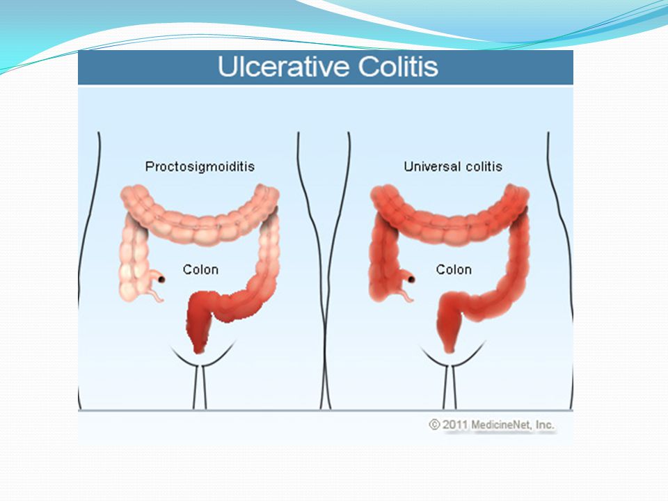
TYPES and SYMPTOMS Ulcerative proctitis - inflammation that is limited to the rectum. Rectal bleeding -rectal pain - rectal inflammation - urgency Proctosigmoiditis - inflammation of the rectum and the sigmoid colon (a short segment of the colon contiguous to the rectum). -rectal bleeding -urgency - diarrhea -cramps
. -rectal bleeding. -urgency. - diarrhea. -cramps.")
99
Left-sided colitis -inflammation that starts at the rectum and extends up the left colon (sigmoid colon and the descending colon). bloody diarrhea abdominal cramps weight loss left-sided abdominal pain Pancolitis or Universal colitis- inflammation affecting the entire colon (right colon, left colon, transverse colon and the rectum). abdominal pain and cramps weight loss, fatigue fever, and night sweats
. abdominal pain and cramps. weight loss, fatigue. fever, and night sweats.")
101
Diagnostic Tests TESTS CROHN’S and ULCERATIVE COLITIS BLOOD TEST
indicate anemia or inflammation gives counts of erythrocytes, protein & c-reactive protein Erythrocyte Sedimentation rate STOOL SAMPLE check for blood and signs of inflammation or infection, including parasites

102
CROHN’S DISEASE and ULCERATIVE COLITIS
TESTS ENDOSCOPY CT Scan (Computed tomography Scan) CROHN’S DISEASE and ULCERATIVE COLITIS a capsule containing miniature video camera is swallowed capsule travels through the small intestine, it sends video images of the lining of the small intestine to a receiver carried on a belt at the waist. images are downloaded and then seen in a computer. Type of X- ray provide cross- sectional view of body Show the body’s internal organs and tissues.
 CROHN’S DISEASE and ULCERATIVE COLITIS. a capsule containing miniature video camera is swallowed. capsule travels through the small intestine, it sends video images of the lining of the small intestine to a receiver carried on a belt at the waist. images are downloaded and then seen in a computer. Type of X- ray provide cross- sectional view of body. Show the body’s internal organs and tissues.")
103
CROHN’S DISEASE and ULCERATIVE COLITIS
TESTS COLONOSCOPY CROHN’S DISEASE and ULCERATIVE COLITIS Detect small ulcers or small areas of inflammation of the colon and terminal ileum Small tissue samples (biopsies) can be obtained Barium X-ray ingested orally, it fills the intestine, and pictures (X-rays) can be taken of the stomach and the small intestines. show ulcerations, narrowing, and, sometimes, fistulae of the bowel when administered through the rectum.
 can be obtained. Barium X-ray. ingested orally, it fills the intestine, and pictures (X-rays) can be taken of the stomach and the small intestines. show ulcerations, narrowing, and, sometimes, fistulae of the bowel when administered through the rectum.")
104
Nutritional care CALORIE- -adequte supply - about kcal PROTEIN -ulcerative colitis patient –losses of 4-8 gm nitrogen per day . -Normal excretion is 2gm daily -serum albumin levels low. CHO -easily absorbable

105
Vitamins -multivitamin preprations should be administered orally or injected Minerals Sodium Potassium Iron- Elemental iron should be given . If anemia is there 100 mg of iron dextrin should be given.

106
COMPARISON ULCERATIVE COLITIS CROHN’S DISEASE Most common sites Rectum
Terminal ileum Distribution Rectum to colon”backwash” ileitis Mouth to anus Spread Continous Discontinuity”skip lesions” Gross features Extensive ulceration Focal appthous ulcers with intervening normal mucosa Pseudo-polyps Linear fissures Cobbelstone apperance Thickened bowel wall

107
ULCERATIVE COLITIS CROHN’S DISEASE Micro No granulomas Granulomas
Inflamation Low More Complication Toxic megacolon Strictures, obstruction Abscess, Fistula Cancer risk 5-25% Slight 1-3% Presentation Bloody diarrhea Variable: Pain ,Diarrhea, Wt. loss Extraintestinal manifestation Common Uncommon

110
DIVERTICULITIS DIVERTICULOSIS - It refers to sac –like herniations of the colonic wall. - Intra colonic pressures and muscular thickening result from complete closure of colonic segments during contractions associated with attempts to small,dry or hard fecal material through the lumen of the bowel. Diverticulitis is a condition in which diverticuli in the colon rupture. The rupture results in infection in the tissues that surround the colon and due to inflamed diverticula.

111
CAUSES Diet- Lack of fiber Lack of exercise Constipation
Inflammation may begin when bacteria or stool are caught in the diverticula

112
SYMPTOMS Abdominal cramping, Constipation Diarrhea
Collection of pus (abscess) in the pelvis, Colon obstruction, Infection of the abdominal cavity Bleeding into the colon
 in the pelvis, Colon obstruction, Infection of the abdominal cavity. Bleeding into the colon.")
113
TREATMENT Clearing up the infection and inflammation with antibiotics.
Resting the colon with a liquid diet plus a pain reliever or a drug such as propantheline (Pro-Banthine) to control muscle spasms. Preventing or minimizing complications with the proper diet.
 to control muscle spasms. Preventing or minimizing complications with the proper diet.")
114
DIAGNOSTIC TESTS: Barium X-rays (barium enemas) can be performed to visualize the colon. Diverticula seen as barium filled pouches protruding from the colon wall. Sigmoidoscopes & colonoscopes Direct visualization of the intestine can be done with flexible tubes inserted through the rectum and advanced into the colon Ultrasoundand CT scan examinations of the abdomen and pelvis can be done to detect collections of pus fluid. In patients having diverticular abscess causing persistent pain and fever.
 can be performed to visualize the colon. Diverticula seen as barium filled pouches protruding from the colon wall. Sigmoidoscopes & colonoscopes. Direct visualization of the intestine can be done with flexible tubes inserted through the rectum and advanced into the colon. Ultrasoundand CT scan examinations of the abdomen and pelvis can be done to detect collections of pus fluid. In patients having diverticular abscess causing persistent pain and fever.")
115
DIET Increase fiber content in food gradually
Avoid refined flour breads and consume whole grain breads Consume fruits such as apples, peaches, bananas, oranges, pears and berries legumes, beans, peas and lentils brown rice, whole-wheat pasta and barley Snack on whole-grain crackers Add wheat bran to salads, casseroles and baked goodies Consume vegetables such as carrots, broccoli, asparagus and salad greens

116
Short bowel syndrome (SBS, also short gut syndrome ) is a malabsorption disorder caused by the surgical removal of the small intestine. Due to the complete dysfunction of a large segment of bowel.

117
CAUSES Crohn's disease Volvulus( a spontaneous twisting of the small intestine that cuts off the blood supply and leads to tissue death) Tumors of the small intestine Injury Necrotizing enterocolitis (premature newborn) Bypass surgery to treat obesity Surgery to remove diseases or damaged portion of the small intestine
 Bypass surgery to treat obesity. Surgery to remove diseases or damaged portion of the small intestine.")
118
SYMPTOMS Abdominal pain Diarrhea and steatorrhea Fluid depletion
Weight loss and malnutrition Fatigue Malabsorption of vitamins and minerals, such as deficiencies in vitamins A, D, E, K, and B12, calcium, magnesium, iron, folic acid, and zinc. Appear as anemia, hyperkeratosis (scaling of the skin), muscle spasms Poor blood clotting Bone pain.
, muscle spasms. Poor blood clotting. Bone pain.")
119
Diagnosis ans TEST Medical history and Physical examination Blood test
-detect anemia Stool Test -absorption of dietary fat CT scan - abnormalities in intestine X-ray - kidney stones - gallstones Endoscopy - inspect esophagus, stomach and duodenum. Colonoscopy - inspect colon and lower small intestine.

120
TREATMENT Anti-diarrheal medicine Vitamin, mineral supplements
H2 blocker and proton pump inhibitors to reduce stomach acid Lactase supplement (to improve the bloating and diarrhoea associated with lactose intolerance) Surgery, including intestinal lengthening, tapering, and small bowel transplant. Parenteral nutrition Nutrition administered via gastrostomy tube.
 Surgery, including intestinal lengthening, tapering, and small bowel transplant. Parenteral nutrition. Nutrition administered via gastrostomy tube.")
121
SYMPTOMS Loss of appetite Nausea Diarrhea Fullness after a meal
Fatty stools Unintentional weight loss weakness

122
FISTULA It can be defined as the connection of two body cavities (such as the rectum and the vagina) or as the connection of a body cavity to the skin (such as the rectum to the skin) which is not a normal occurrence.
 or as the connection of a body cavity to the skin (such as the rectum to the skin) which is not a normal occurrence.")
123
HOW IT DEVELOP? ANAL fistula may form , from an abscess .
The abscess may be constantly filling with body fluids such as stool or urine, which prevents healing. The fistula breaks through to the skin, another body cavity, or an organ. Other causes - IBD - Crohn’s disease - AIDS

124
Rectovaginal fistulas –
Injury to the perineum (the area of tissue between the vagina and the rectum) during child birth IBD Vaginal surgries Rectal surgries
 during child birth. IBD. Vaginal surgries. Rectal surgries.")
125
SYMPTOMS Pain fever tenderness itching Gas through vagina
The fistula may also drain pus or a foul-smelling discharge Painful urination Inflamation of vagina which results Itiching and burning in vagina

126
TYPES Enterocutaneous: This type of fistula is from the intestine to the skin. Enteroenteric or Enterocolic: This is a fistula that involves the large or small intestine. Enterovaginal: This is a fistula that goes to the vagina. Enterovesicular: This type of fistula goes to the bladder. These fistulas may result in frequent urinary tract infections, or the passage of gas from the urethra during urination

127
DIAGNOSIS Physical examination Anoscopy
An anoscop tubular instrument (also called an anal speculum) is inserted a few inches into to the anus in order to evaluate problems of the anal canal. Endoscopy - to examine rectum and the lower part of colon.
 is inserted a few inches into to the anus in order to evaluate problems of the anal canal. Endoscopy. - to examine rectum and the lower part of colon.")
128
ANAL FISSURES These are tears in the skin overlying the anal sphincter, usually due to increased tone of the anal sphincter muscles, and a failure of these muscle to relax. Anal fissures cause pain during defecation and bleeding from the anus.

129
CAUSES Chronic diarrhea Crohn’s disease Immunodeficiency diorders
Constipation Chronic diarrhea Crohn’s disease Immunodeficiency diorders Multiple pregnancies During childbirth Anal cancer Leukemia Chronic use of laxatives Inflammation of the anus and rectum .

130
SYMPTOMS Bright red blood in the toilet bowel
Streak of blood on toilet paper Painful bowel movements Itching DIAGNOSIS Endoscopy Colonscopy Sigmoidoscopy

131
TREATMENT Eating more fiber to create softer stools More fluid
Warm baths of anus In Chronic Anal fissures Anal dilation Lateral internal sphincterotomy (LIS).
.")
132
DIETARY FIBRE(UNAVAILABLE CARBOHYDRATE)
Many Foods Contain Non-digestable Carbohydrates. These Non-digestable Carbohydrate Designated As “Dietary Fibre”or Unavailable Carbohydrates. It used to be believed that fibres could not be used by the human body, as the digestive juices in the body. Our body did not contain enzymes capable of breaking down these compounds. It was overlooked that a part of the fibre is fermented by enzymes found in the micro-organisms of the large intestines.

133
soluble fibres are soluble in water and become sticky in consistency
CLASSIFICATION .. SOLUBLE FIBRES INSOLUBLE FIBRES PECTINS GUMS AND MUCILAGES CELLULOSE HEMICELLULOSE LIGNIN Insoluble fibre doesn’t dissolve or absorb water. it passes through our digestive system in close to its original form soluble fibres are soluble in water and become sticky in consistency

134
IMPORTANCE OF FIBRE Helps maintain bowel integrity and health. A high-fiber diet may lower your risk of developing hemorrhoids, and small pouches in your colon. Lowers blood cholesterol levels. Soluble fiber found in beans, oats, flaxseed and oat bran may help lower total blood cholesterol levels by lowering low-density lipoprotein, or "bad," cholesterol levels. Helps control blood sugar levels. Fiber, particularly soluble fiber, can slow the absorption of sugar, which for people with diabetes can help improve blood sugar levels Aids in weight loss. High-fiber foods generally require more chewing time, which gives your body time to register when you're no longer hungry, so you're less likely to overeat.

Similar presentations


. Absorbing these nutrients into the bloodstream.>")


 Click here to launch video Click here to download print activity.>")


