Download presentation
Presentation is loading. Please wait.

1
Chronic Lymphoproliferative Disorders
Dr.Mitra Heidarpour MD.ACP

2
Definition Chronic lymphoproliferative disorders are a heterogeneous group of malignant clonal proliferations of lymphocytes Classified as sub-types of non-Hodgkin’s lymphoma B-, T- and NK-cell lineages

3
Etiology Requires a number of distinct transforming events to occur within the affected cells Risk factors are Altered immunity Inherited syndromes ataxia telangiectasia Wiskott-Aldrich syndrome common variable immunodeficiency Immunodeficiency due to past medical history long-term immunosuppressive drug therapy, transplant recipients patients with autoimmune diseases Infections (HIV,HTLV-1,HHV8,EBV, H.pylori) Occupational links: herbicides, pesticides, Petrochemical industry, asbestos exposed workers, nickel refinery workers Lifestyles and other exposures: Ionizing radiation,Little conclusive evidence as regards dietary factors and electromagnetic fields
 Occupational links: herbicides, pesticides, Petrochemical industry, asbestos exposed workers, nickel refinery workers. Lifestyles and other exposures: Ionizing radiation,Little conclusive evidence as regards dietary factors and electromagnetic fields.")
4
Clinical Features Annual incidence is approximately 10/100,000
Elderly (median 65 year) M:F ratio 2:1 Chronic B-cell lymphoproliferative disorders account for more than 90% of lymphoid malignancies T-cell and NK-cell neoplasm being relatively uncommon
 M:F ratio 2:1. Chronic B-cell lymphoproliferative disorders account for more than 90% of lymphoid malignancies. T-cell and NK-cell neoplasm being relatively uncommon.")
5
Clinical presentations and natural histories of chronic lymphoproliferative disorders are extremely heterogeneous Many patients are asymptomatic at the time of first presentation, with the diagnosis being made as an incidental finding after a routine medical examination or blood test, for example, CBC

6
Patients may present with lymphadenopathy, systemic symptoms such as weight loss, night sweats and fever or the symptoms of anemia and thrombocytopenia Enlargement of the spleen and, less frequently, the liver Hyper viscosity symptoms Definitive diagnosis is made on the characteristic lymphocyte morphology and immunophenotype usually from samples of peripheral blood or lymph nodes

7
Lymphocytosis Lymphocytosis is defined as an absolute lymphocyte count exceeding 4 x 10⁹/liter (4000/ul) Monoclonal lymphocytosis lymphoproliferative disease (because of an intrinsic defect in the expanded lymphocyte population) Polyclonal lymphocytosis ( secondary to stimulation or reaction to factors extrinsic to lymphocytes, generally infections and/or inflammation)
 Polyclonal lymphocytosis ( secondary to stimulation or reaction to factors extrinsic to lymphocytes, generally infections and/or inflammation)")
8
Characterization of cell surface markers is valuable in distinguishing primary lymphocytosis (leukemic) from secondary lymphocytosis Analysis for immunoglobulin or T cell receptor gene rearrangement also may provide evidence for monoclonal B cell or T cell proliferation, respectively

9
The blood film of patients with lymphocytosis

10
Peripheral smear Small lymphocytes with clump chromatin scant cytoplasm + smudge cells =CLL

11
Atypical CLL-less condensed chromatin and irregular nuclei

12
Medium size cells,round nucleus ,moderately condensed chromatin ,prominent central nucleoli, scant basophilic cytoplasm –prolymphocytic leukemia

13
Small to medium size cell ,oval to indented nuclei ,slightly less clumped chromatin , abundant cytoplasm , circumferential hairy projections Hairy cell leukemia

14
Mature B lymphocytes with pale cytoplasm, irregular cytoplasmic borders, and polar villous projections-SMZL

15
Scant cytoplasm some cell shows clefting –follicular lymphoma

16
Small to medium size cells slightly irregular nuclear contour -like mantle cell lymphoma

17
Large granulocyte - T cell large granular leukemia/lymphoma

18
Large lymphoid cells irregular nuclei ,basophilic cytoplasm (flower cells)-adult T cell leukemia/lymphoma
-adult T cell leukemia/lymphoma")
19
Large lymphocytes with ceribriform nuclei and scant cytoplasm-sezary syndrome

20
Ancillary diagnostic studies
Use of immunologic/molecular techniques Malignant lymphomas reproduce the immunobiology of their benign counterparts This reproduction may be aberrant, and hence distinguishable from normal Expression, normal and aberrant can be used to: Determine lineage, B versus T versus NK Detect clonality Suspect malignancy- loss or aberrant expression of expected antigens Recognize characteristic patterns of antigenic expression associated with certain subtypes of lymphoma

22
Normal lymphoid maturation
Requires two major activities The production of a unique antigenic receptor on it's surface The expression of several surface proteins necessary for antigen recognition, cell activation, cell-cell communication. Antigen receptors are generated through the process of "genetic rearrangement"- the random selection and then juxtaposition of discontinuous genetic segments encoding the antigen receptor genes B cells Immunoglobulin receptor composed of two heavy chains and two light chains Select specific heavy chain gene sequences Select only one of two light chains, kappa or lambda T cells Select one of two heterodimeric receptors Alpha/Beta heterodimer T cell receptor Gamma/Delta heterodimer T cell receptor

23
Surface antigen production
Patholgy of the Lymphoid System Surface antigen production Immune cells require numerous surface molecules for effective immune response, cell-cell communication and regulation Classified into B cell associated, T cell associated, activation associated, cytokine receptors Expression occurs in an orderly sequence in lymphoid maturation Antibodies to these molecules cataloged through the CD - clusters of differentiation - numerical system. Pathology of the Lymhoid System

24
Lymphomas frozen at various stages of antigen dependent B cell maturation and differentiation

25
T cell antigen expression

26
Immunologic Techniques
Flow cytometry Immunohistochemistry Both utilize monoclonal antibodies to detect clonality and unique antigenic patterns

27
B cell lymphoma

28
Definition Mature B cell neoplasms are clonal tumors of mature B cells at various stages of maturation They recapitulate normal stages of maturation.

31
Indolent versus aggressive
Small lymphocytic lymphoma/CLL Follicular lymphoma, Grades 1/2 Extranodal Marginal zone lymphoma of MALT type Nodal marginal zone lymphoma Splenic marginal zone lymphoma Hairy cell leukemia Lymphoplasmacytic lymphoma Plasma cell myeloma Plasmacytoma Cutaneous T cell lymphoma Cutaneous CD30+ anaplastic large cell lymphoma Aggressive Prolymphocytic leukemia Large B cell lymphoma Burkitt lymphoma Mantle cell lymphoma Anaplastic large cell lymphoma All peripheral T cell lymphomas

32
Clinical Characteristics of Patients
Disease Median Age, years Frequency in Children % Male Stage I/II vs III/IV, % B Symptoms, % Bone Marrow Involvement, % % Surviving 5 years CLL/SLL 65 Rare 53 9 vs 91 33 72 51 Mantle cell lymphoma 63 74 20 vs 80 28 64 27 Splenic marginal zone B cell lymphoma 60 48 67 vs 33 19 14 Follicular lymphoma 59 42 33 vs 67 Hairy cell leukemia 50 80 21vs 79 25 100 90

35
SLL/CLL Clinical features
often asymptomatic Non specific : Easy fatigability, Weight loss, anorexia Generalized lymphadenopathy and hepatosplenomegaly in % Hypogammaglobulinomia (>50%) Presented in old ages (>50 years) 35
 Presented in old ages (>50 years) 35.")
36
SLL/CLL Most patients are leukemic at diagnosis 36

37
SLL/CLL Morphology Effacement of normal architecture by Sheets of small round lymphocytes and scattered ill- defined foci of larger actively dividing cells termed prolymphocytes. 37

38
Small Lymphocytic Lymphoma
. 38

39
39

40
SLL/CLL Morphology: The foci of mitotically active cells are called,“ Proliferatin Centers” , their presence are pathognomonic for CLL/SLL Mitosis: rare 40

41
14

42
The larger cells, the prolymphocytes, are the characteristic cells of the proliferation center. In some small lymphocytic lymphomas they are scattered instead of collected into centers.

43
CLL

44
SLL/CLL Transformation of SLL/CLL into “Prolymphocytic Leukemia” or “Diffuse Large B cell Lymphoma” (Richter's syndrome) is rare. The median Survival is less than 1 Year 44

45
SLL/CLL Immunophonotype
Pan B markers CD20, CD19 CD5,CD23 45

47
SLL/CLL Karyotype trisomy 12, del 11q, del 13q Poor prognosis 47

48
Hairy Cell Leukemia Rare chronic lymphoproliferative disorder characterized by circulating B lymphocytes that display prominent cytoplasmic projections Neoplastic B cells infiltrate the marrow(diffusely) and spleen(red pulp) in a characteristic way
 and spleen(red pulp) in a characteristic way.")
49
2 to 3 percent of all adult leukemias
Predominantly a disease of middle-aged males with a median age at presentation of 52 years M:F 4:1

50
Clinical Features Pancytopenia splenomegaly circulating hairy cells
Infection from a wide variety of typical and opportunistic organisms

51
Laboratory Features Anemia, thrombocytopenia, and leukopenia
Absolute neutropenia and monocytopenia(80%) Hairy cells – Mononuclear cells with eccentric or central nuclei Nuclear morphology is variable: round, ovoid, reniform, or convoluted Reticular chromatin pattern Cytoplasm that is blue–gray, exhibiting thin cytoplasmic projections
 Hairy cells – Mononuclear cells with eccentric or central nuclei. Nuclear morphology is variable: round, ovoid, reniform, or convoluted. Reticular chromatin pattern. Cytoplasm that is blue–gray, exhibiting thin cytoplasmic projections.")
52
Marrow Marrow involvement may be diffuse or focal
Hairy cells have monotonous round, oval, or spindle-spaced nuclei that are separated by abundant quantities of pale-staining cytoplasm in a fine fibrillar network The separation of individual hairy cells is characteristic and referred to as the "fried- egg" appearance

53
Marked marrow reticulin fibrosis, the marrow frequently is difficult or impossible to aspirate
Hairy cells synthesize and assemble a fibronectin matrix that likely contributes to the marrow fibrosis characteristic of the disease

54
Spleen, Liver, and Lymph Nodes
The spleen usually is enlarged, with a median weight of 1300 g On section, the spleen has a dark-red, smooth surface On light microscopy, the hairy cells involve the splenic red pulp. Later, the white pulp atrophies and is replaced Red cell lakes, which are blood-filled spaces lined by hairy cells that have disrupted the normal sinus architecture, are characteristic These blood-filled spaces are referred to as pseudosinuses

55
Hepatic infiltration is both sinusoidal and portal
Lymph node involvement is marked by both sinusoidal and interstitial involvement

56
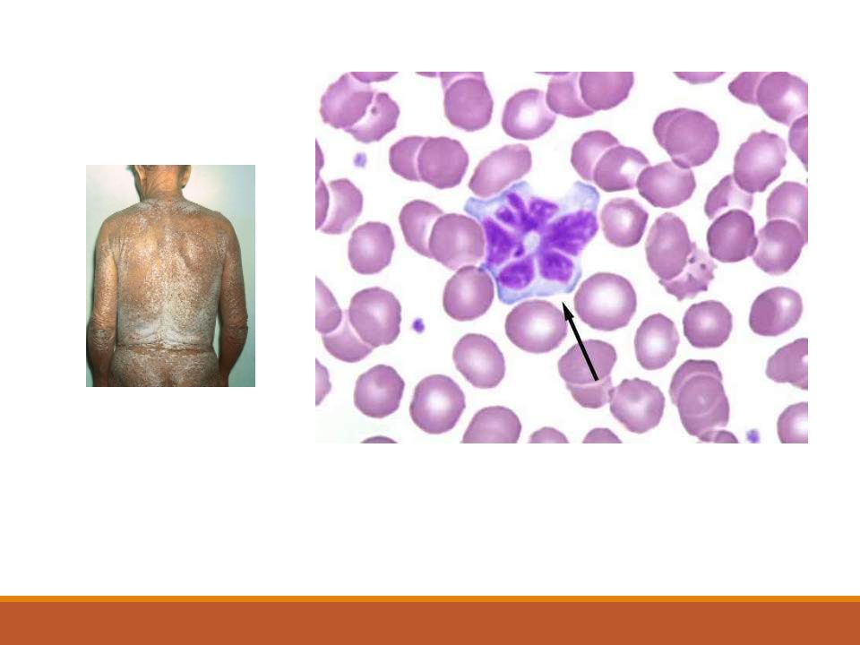
Cytochemistry Strongly positive for TRAP 90 percent of cases
Weak to moderate TRAP staining may occur in other diseases, including prolymphocytic leukemia and lymphoma

57
Electron Microscopy Circumferential cytoplasmic projections
Ribosomal lamella complexes can be in 50 percent of patients

58
Immunophenotypic Profile
Mature B cells, express the pan B cell antigens CD19, CD20, and CD22, but not CD21, an antigen lost in the later stages of B cell development Most distinctively, hairy cells express high levels of CD11c, CD22, CD25, and CD103

60
Annexin A1most specific marker
Always compare with B cell antigen Since it is also expressed in myeloid cells and a proportion of T cells

61
HCL vs HCL-v Compare to HCL HCL-v have Leucocytosis Monocytosis Cell
Prominent nucleoli Blastic or convoluted nuclei Variant immunophenotype (absent CD25,annexin A1 and TRAP)
")
62
Course and Prognosis With treatment survival rate 10 year more than 90% Long term have increased risk of second malignancy

63
Splenic B cell marginal zone lymphoma
B cell neoplasm composed of small lymphocytes which surround and replace the splenic white pulp germinal centers Peripheral blood ,bone marrow and splenic hilar lymph nodes are often involved Lymphoma cell in peripheral blood as villus lymphocytes

64
Clinical features Rare neoplasm ( <2% )
Patient present with splenomegaly ,autoimmune thrombocytopenia or anemia Patient may be positive for hepatitis C

65
Morphology Spleen –central zone of small round lymphocytes replace germinal centre and merge with peripheral zone of marginal zone cell Red pulp –small nodules of larger cells and sheet of small lymphocytes

66
Bone marrow –nodular involvement (compare to diffuse involvement by hairy cell leukemia )
Peripheral blood –cells with short polar villi

67
Immunophenotype Surface Ig M positive and mostly Ig D positive
CD20 and CD 79a positive CD5,CD10,CD23,CD43,annexin A1 negative

68
Prognosis Indolent Response to chemotherapy is poor compare to other lymphoid leukemia

69
Mantle cell lymphoma Rare type of lymphoma(3-10%)
Lymph node involvement is most common Spleen and bone marrow can be involved Peripheral blood 20-60% cases

70
Morphology Monomorphic lymphoid population with nodular,diffuse,mantle,or follicular growth pattern Cells are medium size with irregular nuclear contours ,resembling centrocytes Blastoid,pleomorphic variant can be seen

71
Immnophenotype Intense surface IgM/IgD CD5,FMC-7,CD43 positive
CD10 and BCL 6 negative CD23 negative or weakly positive Cyclin D1 is positive

73
Prognosis Median survival 3-5 years Most of patient can not be cured

74
(CD5 +, CD10-) CLL/SL or MCL? Lack of proliferation centres
Irregular nuclear contours Interspersed histiocytes PAS positive vessels Increased mitoses CD23 and Cyclin D1

75
Follicular lymphoma Most common lymphoma in USA and western Europe
Compose of follicular center cell(centrocytes/centroblasts) with partially follicular pattern
 with partially follicular pattern.")
76
Clinical feature Median age is 6 decade
Mainly involved lymph nodes,spleen,bone marrow, peripheral blood,waldeyer ring Widespread soft tissue involvement can occur

77
Morphology Lymph node –predominately follicular pattern, with closely packed follicles that efface normal architecture Centocytes and blasts are randomly distributed Tingible macrophages are absent

78
Bone marrow- characteristically paratrabecular region may spread to interstial area
Peripheral blood - Scant cytoplasm some cells show clefting

79
Immunophenotype SIg+, B cell marker CD19,CD20,CD22,CD79a positive
Follicular marker BCL6 and CD 10 positive BCL2 positive(variable-higher grade less positive) CD5 and CD 43 negative
 CD5 and CD 43 negative.")
81
CD5-CD10+ Follicular lymphoma?
Morphological features CD10 &Bcl6 expressed in normal and neoplastic follicle centres Presence of large numbers of CD10 &Bcl6 positive cells outside follicles suggests FL Bcl2 distinguishes reactive follicles from FL

83
Prognosis Extent of disease
International prognostic index for FL : Histological grade

84
B cell prolymphocytic leukemia
Affects – peripheral blood, bone marrow and spleen Prolymphocytes must exceeds 55% of lymphoid cells Median age year M:F ratio 1:1

85
Most patients with B symptoms
Massive splenomegaly Absent lymphadenopathy Rapidly raising lymphocyte count( >100x10 9 )
")
86
Morphology Peripheral smear
Majority cells are( usually 90% ) prolymphocytes Medium size Round nucleus Moderately condensed chromatin Prominent central nucleolus Small amount of faintly basophilic cytoplasm
 prolymphocytes. Medium size. Round nucleus. Moderately condensed chromatin. Prominent central nucleolus. Small amount of faintly basophilic cytoplasm.")
87
Bone marrow Interstitial or Nodular involvement

88
D/D Blastic variant of MCL Splenic marginal zone lymphoma
CLL with increase number of prolymphocytes

89
Immunophenotype Surface IgM+/- IgD
B cell antigens –CD 19,CD20,CD22,CD79a CD % CD %

90
Prognosis Respond poorly to therapies for CLL
Median survival months

91
T cell and Nk cell lymphomas

92
Lymphoma Median age Peripheral blood Cytopenia BM spleen Lymph node skin prognosis T cell prolymphocytic leukemia 65 Yes,prolymphocytes Anemia ,thrombocytopenia diffuse Densered pulp Diffuse,paracortical Yes without epidermotrophism Aggressive ,survival < 1 year T cell large granular lymphocytic leukemia Yes,large granular lymphocytes Severe neutropenia+/-anemia Intrasinusoidal,interstial Red pulp invovment no indolant Adult T cell leukemia /lymphoma 50 (20-80) Yes ,flower cell Variable yes Generalized Sezary syndrome 70 Yes,sezary cell No Aggressive CLPD of NK cells 60 Yes rare indolent Agg. NK cell leukemia 42 Yes,variable morphology common uncommon fuminant
 Yes ,flower cell. Variable. yes. Generalized. Sezary syndrome. 70. Yes,sezary cell. No. Aggressive. CLPD of NK cells. 60. Yes. rare. indolent. Agg. NK cell leukemia. 42. Yes,variable morphology. common. uncommon. fuminant.")
93
Mature T cell lymphomas

94
T cell prolymphocytic leukemia
Aggressive lymphoma Proliferation of small to medium sized prolymphocytes in peripheral blood ,bone marrow, lymph node, liver, spleen and skin

95
Clinical features Rare lymphoma (<2%) Median age 55 year
Patient present with hepatosplenomegaly and generalized lymphadenopathy Skin is involved in 20% of cases Lymphocyte count is usually >100x109/l

96
Morphology Peripheral blood Small to medium cell
Non granular basophilic cytoplasm Round to oval or markedly irregular nuclei Visible nucleolus 25% cases no nucleolus (small cell variant) Presence of cytoplasmic blebs or protrusions
 Presence of cytoplasmic blebs or protrusions.")
97
Bone marrow Cutaneous Spleen Lymph node Diffuse involvement
Perivascular or diffuse dermal infiltration without epidermotrophism Spleen Dense red pulp infiltration Lymph node Paracortical infiltration

98
Immunophenotype Positive for CD2,CD3,CD7 Negative for TdT and CD1a

99
Prognosis Aggressive Median survival less than 1 year

100
T cell large granular lymphocytic leukemia
Heterogonous disorder Persistent (more than 6 months) lymphocytosis (2-20x109/l) Large granular lymphocytes
 lymphocytosis (2-20x109/l) Large granular lymphocytes.")
101
Clinical feature 2-3% of lymphoma M:F =1:1 Age range 45-75 year
Involve PB,BM,liver,spleen. Lymphadenopathy is very rare

102
Splenomegaly main physical finding
Varying degree of cytopenia Mainly severe neutropenia Autoimmune diseases common

103
Morphology Peripheral blood
LGL with Moderate to abundant cytoplasm Fine or coarse azurophillic granules Bone marrow –mainly interstitial/sinusoidal involvement Spleen –red pulp cords and sinusoids involvement

104
Immunophenotype Positive for CD3,CD8 and T cell receptor αβ
May be negative for CD5 and CD7

105
Prognosis Indolent course and non progressive
Really neoplasm of uncertain significance or leukemia?

106
Chronic lymphoproliferative disorders of NK cells
Heterogonous disorder Persistent (more than 6 months) lymphocytosis (2-20x109/l) NK cells proliferation Without identifiable cause
 lymphocytosis (2-20x109/l) NK cells proliferation. Without identifiable cause.")
107
Clinical features Median age 60 year Majority are asymptomatic
Some with systemic symptoms and/or cytopenia

108
Morphology NK cells are Bone marrow Intermediate size Round nucleus
Condensed chromatin Moderate amount of slightly basophillic cytoplasm Fine or coarse azurophillic granules Bone marrow Intrasinusoidal and inerstitial infiltration

109
Immunophenotype Surface CD 3 negative CD 16 positive
CD56 weak positive Cytotoxic markers like TIA1,granzyme B and granzyme M positive CD5 CD7 may be lost CD5 and CD8 may be aberrantly expressed

110
Prognosis Indolent clinical course
Patient may die because of cytopenia

111
Aggressive NK-cell leukemia
Systemic neoplastic proliferation of NK cells Almost associated with EB virus

112
Clinical feature Rare form of leukemia Middle age
Peripheral blood, bone marrow liver and spleen involvement Patient present with fever ,constitutional symptoms and leukemic blood picture Varying degree of cytopenia

113
Morphology Range of appearance from cells indistinguishable from large granular lymphocytes to cells with atypical nuclei featuring enlargement , irregular folding, open chromatin or distinct nucleoli Ample amount of basophillic cytoplasm containing fine or azurophillic granules

114
Bone marrow shows massive ,focal or subtle infiltration by the neoplastic cells intermingled with histiocytes and hematophagocytosis

115
Immunophenotype Surface CD3 negative
Cells positive are CD2,CD56 and cytotoxic molecules

116
Prognosis Fulminant course
Complicated by multiorgan failure ,coagulopathy and hemophagocytic syndrome Median survival less than 2 months

117
Adult T cell leukemia/lymphoma
Peripheral T cell neoplasm Highly pleomorphic lymphocytes Caused by human T cell leukemia virus type 1

118
Clinical features Latent course Cumulative risk of ATLL is 2.5%
Involve Lymph nodes Peripheral blood Other Bone marrow Skin (most common extralymphatic site) Spleen,lung ,CNS,GI tract
 Spleen,lung ,CNS,GI tract.")
119
Clinical variants-acute ,lymphomatous ,chronic,smoldering
Acute (most common)-like leukemic phase Increase WBC count,skin rash, generalized lymphadenopathy,hypercalcemia ,lytic bone lesion Lymphomatous variant Prominent lymphadenopathy without PB involvment
-like leukemic phase. Increase WBC count,skin rash, generalized lymphadenopathy,hypercalcemia ,lytic bone lesion. Lymphomatous variant. Prominent lymphadenopathy without PB involvment.")
120
Chronic variant Smoldering variant
Exfoliative skin rash, no numerous atypical lymphocytes Smoldering variant WBC count normal with more than 5% atypical cell

121
Morphology Broad spectrum of cytological features Peripheral blood
Polylobated appearance –flower cells Deeply basophillic cytoplsam Lymph node –Hodgkin lymphoma like picture Dermal –like mycosis fungoides

122
Immunophenotype CD2,CD3,CD5 positive CD7 negative
CD25 positive in all cases

123
Prognosis Clinical subtypes Age Performance status Calcium level
Serum LDH level

124
Sezary syndrome Triad of In addition(one or more criteria)
Erythroderma Generalized lymphadenopathy Clonally related neoplastic T cells with cerebriform nuclei in skin ,lymph nodes and peripheral blood In addition(one or more criteria) Absolte sezary cell count at least 1000 cells per mm3 CD4/CD8 ratio more than 10 Loss of one or more T cell antigen
 Absolte sezary cell count at least 1000 cells per mm3. CD4/CD8 ratio more than 10. Loss of one or more T cell antigen.")
126
Clinical feature Rare .<5%of all cutaneous T cell lymphoma.
Age more than 60 year Generalized disease All visceral organ involved except bone marrow Patient present with erythroderma and generalized lymphadenopathy

127
Morphology Morphological feature similar to mycosis fungoides
Along with infiltration of lymph node and peripheral blood

128
Immunophenotype CD2 CD3 CD5 positive Lack CD7
Skin homing receptor CCR4 positive

129
Prognosis Aggressive disease Overall survival at 5 year is %

130
Thank you.

Similar presentations


>")


>")

i.>")

 DEFINITION CLL is a neoplastic disease characterized by proliferation and accumulation (blood, marrow and lymphoid.>")


:>")
 leukemia Is characterized by an unregulated proliferation of myeloid elements in the bone marrow,>")