Download presentation
Presentation is loading. Please wait.

1
HEART About Disease.co Team

2
Action potential in cardiac muscle fiber is prolonged why ??
Action potential caused by two types of channels: Fast sodium channels Slow calcium channels / calcium- sodium channels --- responsible for plateau portion of action potential –slow to open and remain open for long Immediately after action potential permeability of membrane for potassium decreases thus preventing early return of action potential to resting level

3
SA node action potential
Resting membrane potential millivolts Fast sodium channels are inactivated. Atrial action potential is slower to develop and return to resting level is also slow Sodium ions tend to leak inside and potential rises in positive direction When it reaches to -40mv ,sodium calcium channels become activated Sodium calcium channel inactivate after milliseconds and large number of potassium channels open ---- terminate action potential Potassium channels remain open longer leading to hyperpolarization

4
Draw and label action potential in SA node

5
Draw and label action potential in ventricular muscle ??

6
Action potential in ventricular muscle
Resting membrane potential mv Phase rapid influx of positive sodium ions Phase slight repolarization due to slight efflux of potassium ions Phase opening of slow sodium calcium channels, plateau phase Phase late repolarization phase cessation of influx of sodium and calcium, efflux of potassium Phase cell is at rest and Na K pump will restore resting membrane potential

7
Refractory period of cardiac muscle
Definition The time interval during which a normal cardiac impulse cannot re-excite an already excited area of cardiac muscle fiber. Types Absolute - The time period during which the heart fibers will not contract in response to a stimulus whatever its strength may be. 0.25 to 0.30 sec Relative The myocardial fibers can be stimulated to contract but the stimulus needed will be of much greater intensity. 0.05 sec Refractory period of atria is shorter than for ventricles

8
How impulse spreads in cardiac muscle fibers ??
Self excitation of sinus nodal fibers Transmission of impulse through atria by using anterior, middle and posterior internodal pathways Av nodal delay Rapid transmission through the purkinje system One way conduction through the AV bundle Distribution of purkinje fibers in ventricles – right and the left bundle branch

9
Enlist properties of cardiac muscle.
Functional Syncytium Rhythmicity Conductivity Excitability Or Contractility Contractility All Or None Law Stair Case Phenomena Or Treppe Frank Starling Law Refractory Period Cardiac Muscle Tone

10
Explain Excitation contraction coupling in cardiac muscle fiber

11
Define preload and afterload ??
The degree of tension on the muscle when it begin to contract is called PRELOAD. It is end diastolic pressure. AFTERLOAD The pressure in the aorta leading from the ventricle. This corresponds to systolic pressure.

12
How pumping of heart is regulated ?
Frank Starling Law Within physiological limits the heart pumps all the blood that return to it by veins Nervous regulation Sympathetic Increase in heart rate Strength of contraction Parasympathetic Slows the heart rate Strength of contraction decreases by 20-30%

13
Effect of potassium and calcium ions on heart function
K heart is dilated and flaccid slows the heart rate can block conduction of cardiac impulse Cause high K depolarizes the resting membrane potential ,causing potential to be less negative Membrane potential decreases so intensity of action potential decreases ,making the contraction weaker.

14
CALCIUM calcium spastic contraction calcium cardiac flaccidity

15
Define End Diastolic Volume
During diastole volume of ventricles increases from 70 to 120 ml. This volume is called end diastolic volume. Stroke volume The volume of blood ejected with each beat is called stroke volume . End Systolic Volume The volume of blood remaining in each ventricle at the end of systole is called end systolic volume. It is normally ml. Ejection fraction The fraction of end diastolic volume that is ejected is called ejection fraction. Usually 60%

16
Draw the left ventricular pressure and volume changes ??

17
Mechanical events during cardiac cycle
Isovolumetric contraction Rapid ventricular ejection Slow ventricular ejection Isovolumetric ventricular relaxation Rapid ventricular filling Slow ventricular filling Atrial systole

18
Draw and label right atrial pressure changes ?
Jugular venous pressure (JVP)
")
22
Heart sounds First heart sound
Produced due to closure of AV ( tricuspid and mitral) valves Sound is produced due to vibration of taut valves after closure , along with vibration of walls of heart and major vessels Second heart sound Sudden closure of semilunar valves Third heart sound Due to oscillation of blood back and forth between walls of ventricles Fourth heart sound Contraction of atria and inrush of blood into ventricles
 valves. Sound is produced due to vibration of taut valves after closure , along with vibration of walls of heart and major vessels. Second heart sound. Sudden closure of semilunar valves. Third heart sound. Due to oscillation of blood back and forth between walls of ventricles. Fourth heart sound. Contraction of atria and inrush of blood into ventricles.")
23
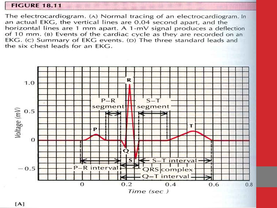
Heart block 1.Prolonged P-R (or P-Q) Interval i.e. First Degree Heart Block PR interval increases to greater than 0.20 sec 2.Second Degree Heart Block PR interval becomes greater than 0.45 sec ,action potential is sometimes strong enough to pass and sometimes not giving dropped beats in ECG. 3.Complete A-V Block (Third degree Block) Complete block of impulse from atria into ventricles . 4.Incomplete Intraventricular block i.e. Electrical Alternans. Impulse conduction blocked in peripheral ventricular purkinje system
 Complete block of impulse from atria into ventricles . 4.Incomplete Intraventricular block i.e. Electrical Alternans. Impulse conduction blocked in peripheral ventricular purkinje system.")
24
Cardiac arrest Definition: Treatment:
Cessation of all rhythmical impulses of the heart. May occur during deep anesthesia or severe myocardial diseases. Treatment: Cardiopulmonary resuscitation is quite successful in re-establishing a normal heart rhythm

25
How blood flow is regulated ?
Acute control Vasodilator theory Oxygen lack /nutrient lack theory Acute metabolic control of blood flow Reactive hyperemia Active hyperemia Autoregulation Metabolic Myogenic theory

26
Write a short note on reactive hyperemia ?
When blood supply to tissue or organ is blocked for few seconds to hours and then unblocked ,blood flow through tissues increases four to seven times normal. This phenomenon is called reactive hyperemia. CAUSE Manifestation of local metabolic blood flow regulation Lack of blood flow causes accumulation of vasodilators Extra blood flow tries to repay the deficit that has occurred

27
Long term regulation Long term regulation Change in tissue vascularity
Role of Oxygen Role of Vascular Endothelial Growth Factor or angiogenic Factors- VEGF Fibroblast growth factor Angiogenin

28
Define edema. Enlist the starling forces
Definition Presence of excess fluid in tissue spaces of body is called edema.

29
Starling forces

30
Short term regulation of blood pressure
Baroreceptor reflex Chemoreceptor reflex Volume reflex Bainbridge reflex CNS ischemic response

31
Long term regulation of blood pressure
Renal blood volume pressure control Renin Angiotensin Aldosterone

32
Renin angiotensin system

33
When person stands from lying position how blood pressure is regulated?
Baroreceptor reflex Receptor Baroreceptors of carotid sinus and aorta Afferents Glossopharyngeal Vagus Centre Tractus solitarius of medulla Efferents Through ANS to the circulation Effect Increase in blood pressure back to normal

34
Define cardiac output. Enlist factors effecting venous return ?
The quantity of blood pumped into the aorta each minute by the heart is called cardiac output. Factors affecting venous return Contraction force of left ventricle Gravity Skeletal muscle pump Venous valves Respiratory pump

35
Vasomotor reflexes Force from front Blood volume

36
Define shock. Give its classification
Generalized inadequate blood flow to through the body, to the extent that tissues are damaged because of lack of oxygen and other nutrients. Classification Circulatory Shock By decreased cardiac output Without decreased cardiac output Hemorrhagic Shock (Hypovolemic Shock) Neurogenic Shock Anaphylactic Shock Septic Shock
 Neurogenic Shock. Anaphylactic Shock. Septic Shock.")
37
What are the compensatory reactions activated by hemorrhage ??

38
Clinical presentation of shock
Fast thready pulse cold sweating on forehead decreased mentation dry tongue and sunken eyes

39
A 45 year old man was brought to accident and emergency department
A 45 year old man was brought to accident and emergency department. He had a road traffic accident. He was bleeding profusely. On examination systolic blood pressure was 60 mmHg, pulse was 108 /min, weak and thready. Skin was pale and extremities were cold. What is the most likely Diagnosis Explain the underlying pathophysiology What is the underlying cause for Tachycardia Cold and pale extremities

40
Hypovolemic shock Diagnosis Hypovolemic shock Pathophysiology
Hemorrhage Hypovolemia Decreased systemic filling pressure Decreased venous return Decreased cardiac output Decreased blood pressure Decreased systemic blood flow

41
Tachycardia Sympathetic stimulation Cold and pale extremities vasoconstriction

42
Write short note on cardiogenic shock ?
Circulatory shock syndrome caused by inadequate cardiac pumping is called cardiogenic shock or cardiac shock. Explanation Damage to heart Inadequate pumping Coronary blood supply also reduced Heart becomes weaker Arterial pressure falls more Shock worsens

43
Thank you For visiting our site
About Disease.co Team

Similar presentations


 –Contracts.>")


