Download presentation
Presentation is loading. Please wait.

1
Physiology Intro: The Integumentary System Unit 8 – Powerpoint #1
Honors Biology Unit 8 – Powerpoint #1 Chapter 35 /36

2
What is Physiology? Physiology is the study of the functions of living organisms and their parts

3
What is Homeostasis The ability to maintain a constant internal environment

4
Eleven Organ Systems For example the Skeletal System which includes your bones, cartilage, ligaments and tendons. Try to think of as many organ systems as you can. Write them on the lines provided.

5
The Eleven Organ Systems
Skeletal Muscular Circulatory Respiratory Digestive Nervous Reproductive Excretory Lymphatic/Immune Endocrine Integumentary

6
Today: Integumentary System
Structures: skin, hair, nails, sweat glands and oil glands. Functions: Barrier against infection and injury (including UV protection) Helps regulate body temperature Prevents H2O loss Waste excretion Vitamin D synthesis
 Helps regulate body temperature. Prevents H2O loss. Waste excretion. Vitamin D synthesis.")
7
Fun Facts Where is your thinnest skin located? Thickest?
Eye lids (thinnest) soles of feet (thickest) How many skin cells do you shed in a minute? About 30,000-50,000 dead cells How many pounds of skin do you loose per year? 8-10 lbs How much of the dust in your home is dead skin cells? 50% (Globally, dead skin accounts for about a billion tons of dust in the atmosphere) Does sweat smell? No, it’s the bacteria How much bacteria is on your body? 1,000 different species AND 1,000,000,000,000 individual bacteria
 soles of feet (thickest) How many skin cells do you shed in a minute About 30,000-50,000 dead cells. How many pounds of skin do you loose per year 8-10 lbs. How much of the dust in your home is dead skin cells 50% (Globally, dead skin accounts for about a billion tons of dust in the atmosphere) Does sweat smell No, it’s the bacteria How much bacteria is on your body 1,000 different species AND 1,000,000,000,000 individual bacteria.")
8
The Largest Organ Skin is 12-15% of body weight
15-20 sq. ft ( sq. meters) Some organisms, such as insects, and some amphibians, use the integumentary system for respiration
 Some organisms, such as insects, and some amphibians, use the integumentary system for respiration.")
9
Epidermis Dermis 2 Layers The skin is made of 2 main layers
Below the dermis is subcutaneous fat (hypodermis) that insulates body
 that insulates body.")
10
Skin: The Layers

11
Epidermis: First layer
Structure Outer layer are flattened, dead cells, inner layer living As lower cells do mitosis they move and push others to surface of skin As move up, become flattened and make keratin (tough, fibrous protein) Takes days for new cells to reach the top Contain nerve endings but no blood vessels
 Takes days for new cells to reach the top. Contain nerve endings but no blood vessels.")
12
Epidermis

14
Epidermis: First layer
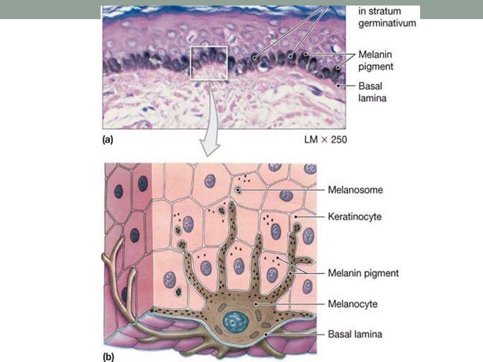
Function 1) Water resistance (keratin filled dead cells) 2) Protective layer against biological/ chemical assault 3) Contains MELANOCYTES (Produce pigment MELANIN which protects from UV) (will talk more about later) 4) Contains Merkel cells – attached to nerves and detect touch 5) Contains Langerhans cells –guard against toxins, microbes and other pathogens that penetrate the skin. If detected, alerts immune system
 Water resistance (keratin filled dead cells) 2) Protective layer against biological/ chemical assault. 3) Contains MELANOCYTES (Produce pigment MELANIN which protects from UV) (will talk more about later) 4) Contains Merkel cells – attached to nerves and detect touch. 5) Contains Langerhans cells –guard against toxins, microbes and other pathogens that penetrate the skin. If detected, alerts immune system.")
15
Epidermis

16
Skin: Dermis Found beneath the Epidermis

17
Dermis: Second layer Structure
Made of strong, flexible connective tissue Contains: Collagen Nerves Blood vessels Lymph vessels Hair follicles Sweat glands Muscles (attached to hair follicles)
")
18
Dermis: Second layer Function
1) Contains Sweat glands (3000/ in2) these: -maintain homeostasis- regulates body temp -Release various wastes from bloodstream -Mammary glands and ear wax glands are modified sweat glands 2) Contains Sebaceous (oil) glands (2 per hair) These glands release SEBUM keeping the epidermis and hair flexible and waterproof 3) Contains many sensory cells 4) Gives skin its elasticity and strength (collagen)
 Contains Sweat glands (3000/ in2) these: -maintain homeostasis- regulates body temp. -Release various wastes from bloodstream. -Mammary glands and ear wax glands are modified sweat glands. 2) Contains Sebaceous (oil) glands (2 per hair) These glands release SEBUM keeping the epidermis and hair flexible and waterproof. 3) Contains many sensory cells. 4) Gives skin its elasticity and strength (collagen)")
19
Skin color 2 types of melanin
Skin color is due mainly to the pigment MELANIN (made by melanocytes in the epidermis) 2 types of melanin Eumelanin (comes in brown and black) Phomelanin (pinkish-red)
 2 types of melanin. Eumelanin (comes in brown and black) Phomelanin (pinkish-red)")
20
Skin Color Darker skin is due to kind and amount of melanin produced (everyone has relatively similar numbers of melanocytes) UV rays stimulate melanocytes to make more melanin (causing tanning) Melanin protects skin cells under it from harmful UV B rays
 Melanin protects skin cells under it from harmful UV B rays.")
21
Melanoctyes

23
Skin Markings Skin Markings: Freckles: Flat, melanized patches Varies with heredity or sun exposure Moles: Elevated patches of melanized skin, with hair

24
This is your hair, and yes that is bacteria

25
Hair Made of Keratin Hair covers almost every exposed surface.
Hair growth is determined by hormones You are born with as many hair follicles as you will ever have Hair is used for protection. From the sun, or dirt Hair color Depends on kind (red, brown, black) and amount of melanin Hair Texture Related to difference in shape of hair Straight = round, Wavy = oval, Tightly curly = flat
 and amount of melanin. Hair Texture. Related to difference in shape of hair. Straight = round, Wavy = oval, Tightly curly = flat.")
26
Hair differences Scanning electron micrograph of a hair fiber from a Caucasian blonde female (above) and an Asian male (below). The overlapping cells of the cuticle are readily apparent on both fibers.
 and an Asian male (below). The overlapping cells of the cuticle are readily apparent on both fibers.")
29
Nails Scale-like modification of the epidermis
Made of thin, dead, scaly cells, packed together Protect the ends of the fingers and toes Produced by the nail root: an area of rapid mitosis Fingernails grow 1mm/week, toenails slower

31
Disorders Basal Cell Carcinoma Squamous Cell Carcinoma
Skin Cancer Basal Cell Carcinoma Most Common – 99% fully cured Squamous Cell Carcinoma Lower layer of epidermis Induced by the sun Malignant Melanoma Cancer of pigment cells = melanocytes Rare – 1% of skin cancers Poor chance of cure Often begins with moles

32
What not to do:

33
Burns: 1st Degree Inflamed, red skin – surface of epidermis is shed

34
2nd Degree Blisters form as fluid builds beneath epidermis
Burns epidermis and part of dermis

35
3rd Degree Epidermis & Dermis is destroyed
Catastrophic loss of fluids (dehydration) Highly susceptible to infection Look up photos online if you would like (they can be pretty graphic)
 Highly susceptible to infection. Look up photos online if you would like (they can be pretty graphic)")
36
Aging Hair Thin and gray as melanocytes die, and mitosis slows
Oil Glands Sebaceous glands atrophy (shrink) = Skin and hair become drier Skin Layers Mitosis slows, Collagen lost from dermis = Skin becomes thin and translucent, Loose and sagging as elastic fibers are lost in dermis Fewer blood vessels = More bruising, slower healing Age Spots – accumulation of pigment cells
 = Skin and hair become drier. Skin Layers. Mitosis slows, Collagen lost from dermis = Skin becomes thin and translucent, Loose and sagging as elastic fibers are lost in dermis. Fewer blood vessels = More bruising, slower healing. Age Spots – accumulation of pigment cells.")
37
Interesting Websites on Integumentary system
Interesting Facts Skin Anatomy and Effects of Aging Skin Cancer Investigation

Similar presentations


 which weighs 7lb (3.2 kg) and has approximately 300 million skin cells. The average.>")


 Barrier to keep water and solutes in Barrier to keep bacterial,>")

 Consists of three major regions.>")


