Download presentation
Presentation is loading. Please wait.

1
GLOMERULONEPHRITIS Division of Nephrology, Internal Medicine

2
Symptoms and Signs Proteinuria Hematuria Edema Hypertension Azotemia
Injury to the glomeruli by various immunologic or non-immunologic mechanism Proteinuria Hematuria Edema Hypertension Azotemia

3
사구체신염의 분류 임상 증상에 따른 분류 ( 5 Syndromes) 1) 무증상성 요이상(AUA) : IgA N
2) 신증후군(NS) : MCD, FSGS, MGN 3) 급성 사구체신염(AGN) : PSGN 4) 만성 사구체신염(CGN) : IgA N, MPGN 5) 급속진행성 사구체신염(RPGN) : crescentic GN
 신증후군(NS) : MCD, FSGS, MGN. 3) 급성 사구체신염(AGN) : PSGN. 4) 만성 사구체신염(CGN) : IgA N, MPGN. 5) 급속진행성 사구체신염(RPGN) : crescentic GN.")
4
원인 질환에 따른 분류 병리학적(형태학적) 분류 1) 속발성 사구체신염 HS nephritis, lupus nephritis,
DM nephropathy Hepatitis B virus associated GN, PSGN 2) 원발성 사구체신염 MCD, FSGS, MGN, MPGN, IgA N, Crescentic GN 병리학적(형태학적) 분류
 원발성 사구체신염. MCD, FSGS, MGN, MPGN, IgA N, Crescentic GN. 병리학적(형태학적) 분류.")
5
NEPHROTIC SYNDROME

6
Definition NS Children : Serum albumin < 2.5g/dl
Proteinuria > 40mg/m2/hr Adult : Massive Proteinuria ( >3g/24h/1.73m2) Hypoalbuminemia Edema Hyperlipidemia
 Hypoalbuminemia. Edema. Hyperlipidemia.")
7
Anti-proteinuric Barrier
NS Anti-proteinuric Barrier Charge selective barrier : negatively charged sialoglycoprotein Size selective barrier : 44A(effective molecular radius)
")
8
Classification Primary NS : MCD FSGS MGN MPGN Secondary NS :
Hepatitis B virus-associated GN Lupus nephritis, Diabetic nephropathy

9
Primary NS according to the age groups
Others FSGS MGN MCD

10
Etiologic Diagnosis of Secondary NS (N=220)
Diabetic nephropathy : m/c Others Renal amyloidosis 5.9% HBGN 46.8% Lupus Nephritis 39.5%

15
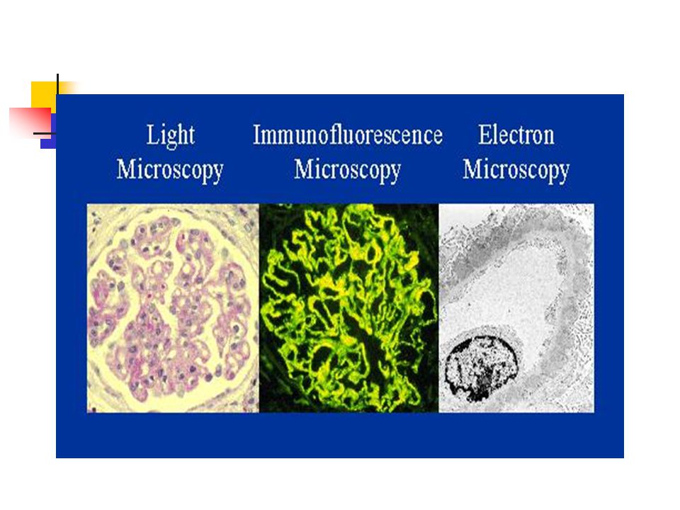
Normal Kidney (1) H&E stained section
 H&E stained section")
16
Normal Kidney (2)
")
18
Minimal Change Disease (MCD)
Synonyms : Nil disease, Lipoid nephrosis Foot process disease Incidence : male>female 80% of NS in children 20-40 % of NS in adults Cause : Drug (NSAIDs, rifampin, interferon a) Hodgkin’s disease, HIV infection Pathology : normal in LM, IF epithelial foot process effacement in EM
 Hodgkin’s disease, HIV infection. Pathology : normal in LM, IF. epithelial foot process effacement in EM.")
19
MCD Treatment Highly steroid responsive, excellent prognosis to glucocorticoid therapy 90% (children) and 50% (adult) : remission after 8weeks of high dose steroid therapy sometimes relapse upon tapering (50-70%) Alkylating agents are reserved for frequent relapsers, cyclosporine for steroid resistant MCNS
 Alkylating agents are reserved for frequent relapsers, cyclosporine for steroid resistant MCNS.")
20
MCD LM EM

22
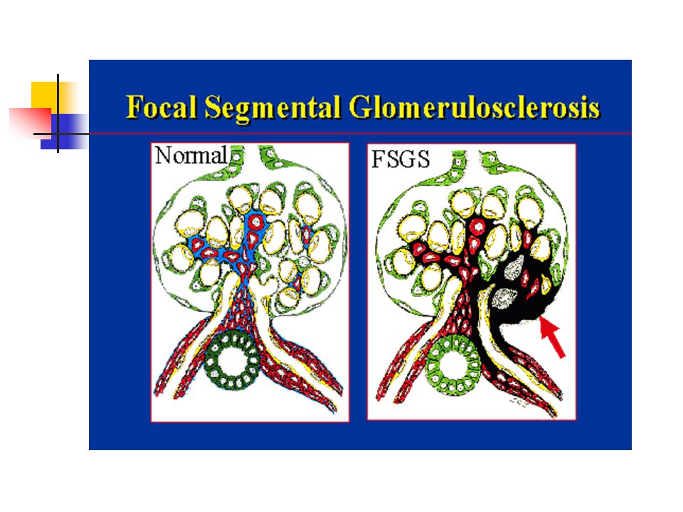
원인 : 특발성, heroin abuse, VUR, AIDS, solitary kidney
FSGS 특발성 신증후군의 10% 정도 차지, 원인 : 특발성, heroin abuse, VUR, AIDS, solitary kidney 임상 양상 : 대부분 전형적인 신증후군(nonselective) 고혈압, 혈뇨, 신기능 저하 병리 : 비증식성, 경화성 변화 (초점성, 분절성) 면역 형광 소견은 비특이적 치료 : 장기간의 steoid 치료, steroid 에 대한 효과가 낮음 예후 : 50% 환자가 7-10년에 ESRD 로 진행 , 신이식 후에도 재발 흔함
 고혈압, 혈뇨, 신기능 저하. 병리 : 비증식성, 경화성 변화. (초점성, 분절성) 면역 형광 소견은 비특이적. 치료 : 장기간의 steoid 치료, steroid 에 대한 효과가 낮음. 예후 : 50% 환자가 7-10년에 ESRD 로 진행 , 신이식 후에도 재발 흔함.")
23
FSGS LM

24
Light micrographs and diagrams depicting patterns of focal segmental glomerulosclerosis. One pattern (A and D) has a predilection for sclerosis in the perihilar regions of the glomeruli. The glomerular tip lesion variant has segmental consolidation confined to the segment adjacent to the origin of the proximal tubule (B and E). The collapsing glomerulopathy variant has segmental collapse of capillaries with hypertrophy and hyperplasia of overlying epithelial cells (C and F) (Jones methenamine silver stain, ×100).
 has a predilection for sclerosis in the perihilar regions of the glomeruli. The glomerular tip lesion variant has segmental consolidation confined to the segment adjacent to the origin of the proximal tubule (B and E). The collapsing glomerulopathy variant has segmental collapse of capillaries with hypertrophy and hyperplasia of overlying epithelial cells (C and F) (Jones methenamine silver stain, ×100). .")
25
Membranous Nephropathy

26
1. Most common idiopathic NS in adults (40%) Rare in children
MGN 1. Most common idiopathic NS in adults (40%) Rare in children Peak incidence between 30 and 50 2. Clinical manifestation Mostly nephrotic (80%), Nonselective proteinuria Microhematuria (50%) HT : less than 30% at initial manifestation but common later with renal progression
 Rare in children. Peak incidence between 30 and Clinical manifestation. Mostly nephrotic (80%), Nonselective proteinuria. Microhematuria (50%) HT : less than 30% at initial manifestation. but common later with renal progression.")
27
Pathology LM : diffuse thickening of the GBM (PAS staining)
MGN Pathology LM : diffuse thickening of the GBM (PAS staining) spike pattern (Silver staining) IF : granular deposit of IgG, IgM, C3 EM : subepithelial electron dense deposit stage I, II, III, IV
 spike pattern (Silver staining) IF : granular deposit of IgG, IgM, C3. EM : subepithelial electron dense deposit. stage I, II, III, IV.")
28
Etiology Idiopathic (majority) Systemic disease or drugs
MGN Etiology Idiopathic (majority) Systemic disease or drugs 1. Infection : hepatitis B, hepatitis C 2. Autoimmune disease : SLE, RA, MCTD 3. Malignancy : carcinoma 4. Drugs : gold, penicillamine, NSAID, captopril 5. Miscellaneous : sarcoidosis, DM,
 Systemic disease or drugs. 1. Infection : hepatitis B, hepatitis C. 2. Autoimmune disease : SLE, RA, MCTD. 3. Malignancy : carcinoma. 4. Drugs : gold, penicillamine, NSAID, captopril. 5. Miscellaneous : sarcoidosis, DM,")
29
Clinical Sx ① Mean onset age: 30 ∼ 50 age (male: female = 2:1)
MGN Clinical Sx ① Mean onset age: 30 ∼ 50 age (male: female = 2:1) ② Older age: correlate with malignancy 20% (>60 age) ③ Massive proteinuria (80%), edema ④ RVT*: 50% - high incidence
 ② Older age: correlate with malignancy 20% (>60 age) ③ Massive proteinuria (80%), edema. ④ RVT*: 50% - high incidence.")
30
Treatment & Prognosis 40% : spontaneous remission
MGN Treatment & Prognosis 40% : spontaneous remission 30-40% : repeated relapse and remission 10-20% : persistent NS and progressive azotemia (ESRD in 20 to 30 years) Risk factors of renal progression : male, older age at onset, heavy proteinuria, hypertension, stage IV lesion, azotemia at initial Bx Tx : no therapeutic effect with steroid alone cyclophosphamide, chlorambucil, cyclosporine
 Risk factors of renal progression : male, older age at onset, heavy proteinuria, hypertension, stage IV lesion, azotemia at initial Bx. Tx : no therapeutic effect with steroid alone. cyclophosphamide, chlorambucil, cyclosporine.")
31
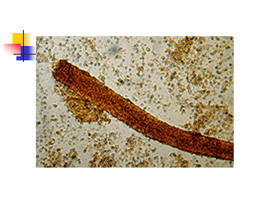
MGN LM

32
IF EM MGN IF with antibody to IgG.
deposition of electron dense material and interposition of lighter GBM material-subepithelial deposition

33
Membranous Proliferative Glomerulonephropathy
Type I: Subendothelial Deposits Type II: Dense Deposit Disease

34
MPGN EM Type I: Subendothelial Deposits Type II: Dense Deposit Disease

35
Clinical Manifestation
MPGN Clinical Manifestation Variable combination of nephritic or nephrotic features Common in ages between 5-30 Decline in GFR, active urine sediment, Proteinuria often in nephrotic range (50%) Type I MPGN : Immune complex disease C3 usually depressed C1q, C4, properdin, factor B : borderline or low Secondary MPGN : associated with infection, SLE, malignancy
 Type I MPGN : Immune complex disease. C3 usually depressed. C1q, C4, properdin, factor B : borderline or low. Secondary MPGN : associated with infection, SLE, malignancy.")
36
Diffuse proliferation of mesangial cells Increased mesangial matrix
MPGN Pathology Diffuse proliferation of mesangial cells Increased mesangial matrix Thickening and reduplication of GBM (Double contour, Tram-tract) Type I and type II MPGN Prognosis Poor, slow progression to ESRD Worse in type II MPGN
 Type I and type II MPGN. Prognosis. Poor, slow progression to ESRD. Worse in type II MPGN.")
37
MPGN LM Normal kidney

38
MPGN EM IF

39
Acute Glomerulonephritis or
Acute Nephritic Syndrome(AGN)
")
40
AGN Acute glomerular inflammation
Sudden onset of acute renal failure and oliguria Obstruction of glomerular capillary lumen GFR falls Na and water retention ECF volume expansion, Edema, Hypertension U/A : RBC cast, dysmorphic RBC, leukocytes, subnephrotic proteinuria Often gross hematuria Azotemia General pathologic feature : proliferative GN (capillary endothelial cell, mesangial cell)
")
42
AGN - PSGN PSGN Acute post-streptococcal GN
Etiology : Pharyngeal or cutaneous infection with group A beta-hemolytic streptococcus, nephritogenic strain Epidemiology : common in children, male > female Latent period : 6-15 days (“post-pharyngitic”) Hematuria (gross or microscopic), Edema, mild HT, Oliguria, Nausea, mild fever, Flank pain ARF of variable degrees
 Hematuria (gross or microscopic), Edema, mild HT, Oliguria, Nausea, mild fever, Flank pain. ARF of variable degrees.")
43
Lab finding & Pathology
PSGN Lab finding & Pathology U/A : hematuria, mild proteinuria GFR reduced Elevated ASO, anti-hyaluronidase, elevated anti-DNase B Culture: Streptococcus in throat or skin Complement level : C3, CH50 markedly reduced, normalized in 8 weeks, C4 mildly reduced LM : Diffuse endocapillary proliferation PMN cell and monocyte infiltration EM : hump (large subepithelial deposit: characteristic) IF : IgG, C3 deposit
 IF : IgG, C3 deposit.")
44
Treatment & prognosis Benign course in children
PSGN Treatment & prognosis Benign course in children Acute symptom : relieved in 1-2 wks U/A abnormality esp. hematuria persists for 2 years Treatment : symptomatic Rest, salt water restriction, diuretics, protein restriction Antibiotics (PCN, EM) needed in limited cases - i.e. incomplete treatment, elevated CRP
 needed in limited cases. - i.e. incomplete treatment, elevated CRP.")
45
PSGN LM IF

46
PSGN EM

47
Asymptomatic Urinary Abnormality (AUA)
Hematuria or subnephrotic proteinuria without HT, renal insufficiency, edema U/A abnormality : persistent or recurrent Hematuria with or without proteinuria Isolated non-nephrotic proteinuria IgA Nephropathy Thin basement membrane disease Alport syndrome Orthostatic proteinuria MN, FSGS, DM, amyloidosis

49
IgA Nephropathy

50
Most common GN worldwide (10-40%)
IgAN Most common GN worldwide (10-40%) Etiology : most cases are idiopathic Clinical spectrum with Henoch-Schonlein Purpura Secondary IgA N due to liver cirrhosis, Crohn’s disease Epidemiology : between 16 and 35 years, male>female Initial manifestation Recurrent gross hematuria, often 24 to 48h after pharyngitis, GI infection (“synpharyngitic”) Or, microscopic hematuria during routine examination HT, nephrotic proteinuria rare at initial presentation
 Etiology : most cases are idiopathic. Clinical spectrum with Henoch-Schonlein Purpura. Secondary IgA N due to liver cirrhosis, Crohn’s disease. Epidemiology : between 16 and 35 years, male>female. Initial manifestation. Recurrent gross hematuria, often 24 to 48h after pharyngitis, GI infection ( synpharyngitic ) Or, microscopic hematuria during routine examination. HT, nephrotic proteinuria rare at initial presentation.")
51
20 to 50% progress to ESRD over 20 years Poor prognosis group :
IgAN Lab : Elevated serum IgA level in 50% cases, circulating IgG or IgA Immune complex Prognosis 20 to 50% progress to ESRD over 20 years Poor prognosis group : - Male sex - Older age at onset - Absence of gross hematuria - heavy proteinuria - Hypertension - azotemia at initial diagnosis

52
IgAN LM mesangial expansion with increased matrix and cellularity

53
Mesangial deposits of IgA
IgAN Mesangial deposits of IgA Subendothelial and subepithelial deposits are rarely seen.

54
AUA : Isolated Non-nephrotic Proteinuria of Glomerular Origin
Orthostatic Proteinuria Persistent Proteinuria Etiology : smoldering GN (mild mes. Prolif. GN, FSGS, focal or diffuse prolif. GN), interstitial nephritis Prognosis : slowly progressive azotemia Dx : separate urine collection(7A-11P, 11P-7A) Prognosis : excellent
, interstitial nephritis. Prognosis : slowly progressive azotemia. Dx : separate urine collection(7A-11P, 11P-7A) Prognosis : excellent.")
55
Glomerulonephritis (RPGN)
Rapidly Progressive Glomerulonephritis (RPGN) Crescent Normal glomerulus
 Crescent. Normal. glomerulus.")
56
Patchy parenchymal consolidations are present, which usually are
bilateral, symmetric perihilar, and bibasilar. The apices and costophrenic angles usually are spared

57
RPGN Subacute glomerular inflammation
Patient develop renal failure over weeks to months Nephritic urinary sediment, subnephrotic proteinuria, oliguria, HT, hypervolemia, edema Renal biopsy almost invariably shows crescents (=Crescentic GN)
")
58
RPGN Crescentic GN Pathology : diffuse crescent formation > 50% glomeruli Clinically : progression to renal failure over weeks to months, clinical features of GN (proteinuria, hematuria, active urinary sediment) If not properly treated, end stage renal disease ensues in 80-90%
 If not properly treated, end stage renal disease ensues in 80-90%")
59
Classification of Crescentic GN
RPGN Classification of Crescentic GN Type I : anti-GBM disease without pulmonary hemorrhage Type II : Immune complex deposition Type III : pauci-immune (no immunoglobulin deposition) ANCA (anti-neutrophil cytoplasm antibody) 양성
 ANCA (anti-neutrophil cytoplasm antibody) 양성.")
60
C-ANCA on ethanol fixed slide
RPGN C-ANCA on ethanol fixed slide C-ANCA is identified as a positive result when there is intense positive granular staining of the cytoplasm that extends to the border of the human granulocyte substrate displaying a 1+ or greater fluorescence and there is absence of nuclear staining P-ANCA on ethanol fixed slide P-ANCA exhibits intense positive perinuclear staining of the multi-lobed nucleus with a poorly defined cell border. A 1+ or greater fluorescence is considered a positive result

61
RPGN LM

62
RPGN IF There is a linear pattern of staining along the glomerular basement membrane with IgG. IF with antibody to fibrinogen.

63
Alport’s Syndrome Genetic defect in type IV collagen Alpha 5 chain
X-linked dominant Ant. lenticonus Sensorineural hearing loss Recurrent hematuria Slowly progress to ESRD EM : thickenend GBM with lamellation, splitting

64
Thin Basement Membrane Disease (= Benign familial hematuria)
")
65
Chronic Glomerulonephritis (CGN)
Persistent proteinuria/hematuria/HT Insiduous onset, slowly progressive renal insufficiency over years Can be a manifestation of virtually all of the major GNs Pathology : renal atrophy, cortical thinning, glomerulosclerosis irrespective of causative GN

66
Secondary GN Lupus Nephritis Systemic Vasculitis Amyloidosis
Cast Nephropathy

67
Lupus Nephritis Lupus Nephritis -- Diffuse Proliferative

68
Amyloidosis Nodular pattern apple green on polarized light

69
Randomly oriented thin fibrils
(EM: x 51,250)
")
70
Cast Nephropathy T-H protein(cast)
")
71
Diagnostic Approach to GN

72
Etiologies of AUA Hematuria with or without proteinuria
Isolated non-nephrotic proteinuria IgA Nephropathy Thin basement membrane disease Alport syndrome Orthostatic proteinuria MN, FSGS, DM, amyloidosis

73
1. Clinical feature가 사구체신염인가?
Proteinuria, Hematuria, Edema, Hypertension, Azotemia(PHEHA) Hematuria of glomerular origin : dark color, no clot formation, RBC cast, dysmorphic RBC, overt albuminuria/proteinuria(>1g) Pathologic proteinuria of glomerular origin : not orthostatic but persistent proteinuria, mainly albuminuria on urine PEP, accompanied by glomerular hematuria
 Hematuria of glomerular origin : dark color, no clot formation, RBC cast, dysmorphic RBC, overt albuminuria/proteinuria(>1g) Pathologic proteinuria of glomerular origin : not orthostatic but persistent proteinuria, mainly albuminuria on urine PEP, accompanied by glomerular hematuria.")
74
2. 사구체신염의 임상증후군 가운데 어디에 해당되는가?
Physical Exam : BP, edema U/A : RBC cast, WBC cast, oval fat body 24H urine collection for protein quantitation Blood Chemistry: protein/albumin cholesterol, BUN/Cr Serial Check of BUN/Cr Kidney sono : renal size, echogenecity NS AGN RPGN CGN AUA

75
3. 사구체신염을 일으키는 원인질환이 있는가? Age/Sex History : URI, family Hx
Physical exam : skin rash, purpura, skin infection, arthritis, oral ulcer X-ray : pulm.hemorrhage, arthritis Hepatitis B, C, HIV, VDRL, FTA-ABS ASO, anti-DNase B, CRP, cryoglobulin, RA factor, FANA, anti-dsDNA, C3/C4 level, ANCA, anti-GBM antibody

76
Serum complement가 감소하는 질환
Idiopathic GN Secondary GN Acute PSGN, MPGN type I, II Lupus nephritis, GN secondary to SBE, Cryoglobulinemia

77
4. 사구체신염 진단을 위해 신생검이 필요한가? GN의 원인적 진단이 이루어진 경우 GN의 원인이 명확하지 않는 경우 진단명
조직형태와 무관하게 원인에 따라 진단명을 붙인다. 예> Lupus nephritis, hepatitis B associated GN 조직 형태에 따라 진단. 예> idiopathic MN, MPGN, IgA N 신생검을 하는 목적 질환의 신침범 정도와 그에 따른 예후 파악 형태학적 진단 및 진단에 따른 예후 파악

78
Renal Biopsy Renal biopsy provides tissues that can be used to determine the diagnosis, indicate the cause, predict the prognosis, direct treatment and collect data for research.

79
Renal Biopsy Indication 1. The cause cannot be determined or predicted by less invasive techniques 2. Signs and symptoms suggest parenchymal disease that requires pathologic evaluation 3. DDx includes diseases that have different treatments or different prognosis

80
Usually Needed in Nephrotic syndrome in adults
Renal Biopsy Usually Needed in Nephrotic syndrome in adults Steroid resistant NS in children GN in adults other than clear-cut PSGN or lupus nephritis ARF of unknown cause RPGN Activity or chronicity determination of lupus nephritis

81
Method Localization of kidney by real-time ultrasound
Renal Biopsy Method Localization of kidney by real-time ultrasound Biopsy needles : Vim-Silverman, Gun biopsy needle (spring loaded disposable gun needle) 1-1.5cm length cortical tissue needed Visualized under LM, FM and EM
 1-1.5cm length cortical tissue needed. Visualized under LM, FM and EM.")
82
Renal Biopsy

83
Renal Biopsy

84
Renal Biopsy

85
Renal Biopsy

86
Complications Small perirenal hematoma : common
Renal Biopsy Complications Small perirenal hematoma : common Gross hematuria (< 10%) AV fistula (< 1%) Massive bleeding that requires surgery (< 1%) Mortality (< 0.1%)
 AV fistula (< 1%) Massive bleeding that requires surgery (< 1%) Mortality (< 0.1%)")
87
Contraindication Uncooperative patient Solitary Kidney
Renal Biopsy Contraindication Uncooperative patient Solitary Kidney Uncontrolled severe hypertension Bleeding tendency Severe anemia or dehydration Cystic kidney Hydronephrosis Multiple renal a. aneurysms APN, perinephric abscess, renal neoplasm Sclerotic kidney, ESRD

Similar presentations


 is the sudden onset of: – Haematuria (macroscopic/microscopic)>")
SC>")
