Download presentation
Presentation is loading. Please wait.

1
Tissue Repair

2
Tissue Repair After the inflammation ?

5
Interphase – 95% of cell cycle
(TG ) Somatic Cell Division - Overview Interphase – 95% of cell cycle Organelle duplication, DNA replication, Growth Somatic Cell Division Interphase – 95% of cell cycle Organelle duplication, DNA replication, Growth
 Somatic Cell Division - Overview. Interphase – 95% of cell cycle. Organelle duplication, DNA replication, Growth. Somatic Cell Division. Interphase – 95% of cell cycle. Organelle duplication, DNA replication, Growth.")
6
Organelle duplication, but no DNA replication
G1 Phase Metabolically active Organelle duplication, but no DNA replication Duration variable – short in embryonic and cancer cells Prepares for S phase Cells that remain in G1 for a long time = G0 (permanent tissues, such as neural tissue) G1 Phase Metabolically active Organelle duplication, but no DNA replication Duration variable – short in embryonic and cancer cells Prepares for S phase Cells that remain in G1 for a long time = G0 (permanent tissues, such as neural tissu
 G1 Phase. Metabolically active. Organelle duplication, but no DNA replication. Duration variable – short in embryonic and. cancer cells. Prepares for S phase. Cells that remain in G1 for a long time = G0. (permanent tissues, such as neural tissu.")
7
Committed to cell division once this starts
S Phase Committed to cell division once this starts DNA and centrosome replication Semi-conservative replication of DNA: two identical daughter genomes S Phase Committed to cell division once this starts DNA and centrosome replication Semi-conservative replication of DNA: two identical daughter genomes

8
Enzymes and proteins synthesized for cell division
G2 Phase Growth continues Enzymes and proteins synthesized for cell division Determining Cell Stage Cells at different stages of the cell cycle can also be distinguished by their DNA content G2 Phase Growth continues Enzymes and proteins synthesized for cell division Determining Cell Stage Cells at different stages of the cell cycle can also be distinguished by their DNA content

9
mitosis plus cytokinesis Mitosis: Prophase Metaphase Anaphase
Mitotic (M) Phase mitosis plus cytokinesis Mitosis: Prophase Metaphase Anaphase Telophase Mitotic (M) Phase mitosis plus cytokinesis Mitosis: Prophase Metaphase Anaphase Telophase
 Phase. mitosis plus cytokinesis. Mitosis: Prophase. Metaphase. Anaphase. Telophase. Mitotic (M) Phase. mitosis plus cytokinesis. Mitosis: Prophase. Metaphase. Anaphase. Telophase.")
10
1-Stem cells (e.g., blood cells and epithelial cells) 2-Sperm cells
Regulation of the Cell Cycle Cell Cycle Lengths Vary by cell type: Embryonic cells 1-Stem cells (e.g., blood cells and epithelial cells) 2-Sperm cells G1 prolonged in stable or permanent cells (called G0) G1 rapid or non-existent in rapidly-dividing cells Regulation of the Cell Cycle Cell Cycle Lengths Vary by cell type: Embryonic cells Stem cells (e.g., blood cells and epithelial cells) Sperm cells G1 prolonged in stable or permanent cells (called G0) G1 rapid or non-existent in rapidly-dividing cells
 2-Sperm cells. G1 prolonged in stable or permanent cells. (called G0) G1 rapid or non-existent in rapidly-dividing cells. Regulation of the Cell. Cycle. Cell Cycle Lengths. Vary by cell type: Embryonic cells. Stem cells (e.g., blood cells and epithelial cells) Sperm cells. G1 prolonged in stable or permanent cells. (called G0) G1 rapid or non-existent in rapidly-dividing cells.")
11
Cell growth not part of cell cycle All energy goes into DNA synthesis
Embryonic cells Cell growth not part of cell cycle All energy goes into DNA synthesis So G1 lacking and G2 quite short Each round of division subdivides original cytoplasm into smaller and smaller cells, Until adult cell size is reached Embryonic cells Cell growth not part of cell cycle All energy goes into DNA synthesis So G1 lacking and G2 quite short Each round of division subdivides original cytoplasm into smaller and smaller cells, Until adult cell size is reached

12
Cell-Cycle Checkpoints G1 checkpoint In yeast, called start
In animal cells, called restriction point G2 checkpoint Located at boundary between G2 and M phase Proper completion of DNA synthesis required before cell can initiate mitosis Spindle Assembly Checkpoint Boundary between metaphase and anaphase All chromosomes must be properly attached to the spindle Cell-Cycle Checkpoints G1 checkpoint In yeast, called start In animal cells, called restriction point G2 checkpoint Located at boundary between G2 and M phase Proper completion of DNA synthesis required before cell can initiate mitosis Spindle Assembly Checkpoint Boundary between metaphase and anaphase All chromosomes must be properly attached to the spindle

13
Mitosis-promoting factor Triggers passage through G2 checkpoint
MPF Cytoplasmic factor Mitosis-promoting factor Triggers passage through G2 checkpoint into M-phase MPF composed of 2 proteins Gene cdc2 codes for a protein kinase Cdk: cyclin-dependent kinase But to be active, must be bound to another group of proteins called cyclins MPF = Cdk-Cyclin complex (cdc2 codes for Cdk1, part of MPF) MPF Cytoplasmic factor(s) first discovered as a promoter of meiosis in frog oocytes Mitosis-promoting factor (M-phase promoting factor, used to be maturationpromoting factor) Triggers passage through G2 checkpoint into M-phase MPF composed of 2 proteins Gene cdc2 codes for a protein kinase Cdk: cyclin-dependent kinase But to be active, must be bound to another group of proteins called cyclins MPF = Cdk-Cyclin complex (cdc2 codes for Cdk1, part of MPF)
 MPF. Cytoplasmic factor(s) first discovered as. a promoter of meiosis in frog oocytes. Mitosis-promoting factor (M-phase. promoting factor, used to be maturationpromoting. factor) Triggers passage through G2 checkpoint. into M-phase. MPF composed of 2. proteins. Gene cdc2 codes for a protein kinase. Cdk: cyclin-dependent kinase. But to be active, must be bound to. another group of proteins called cyclins. MPF = Cdk-Cyclin complex. (cdc2 codes for Cdk1, part of MPF)")
14
Cyclins Mitotic Cyclins (G2 checkpoint) G1 cyclins (with G1 Cdks) Spindle-assembly checkpoint Mitotic Cdk-Cyclin Complex (MPF) and G2 Controls G2 checkpoint by phosphorylating proteins involved in early stages of mitosis Cdk levels constant But mitotic cyclin levels gradually increase – act as cell regulators MPF only active when cyclin levels high enough – triggers passage through G2 Checkpoint Function of G2 checkpoint: Error check: DNA replication must be complete Detects unreplicated DNA, holds cell at G2 Detects damaged DNA, arrests cell in G2 until damage repair
 and G2. Controls G2 checkpoint by phosphorylating. proteins involved in early stages of mitosis. Cdk levels constant. But mitotic cyclin levels gradually increase – act as cell regulators. MPF only active when cyclin levels high. enough – triggers passage through G2. Checkpoint. Function of G2. checkpoint: Error check: DNA replication must be. complete. Detects unreplicated DNA, holds cell at. G2. Detects damaged DNA, arrests cell in G2 until damage repair.")
15
Regulation at Spindle- Assembly Checkpoint
MPF causes activation of anaphase promoting Complex (Complex pathway that promotes anaphase) Regulation at Spindle- Assembly Checkpoint MPF causes activation of anaphasepromoting complex Complex pathway that promotes anaphase
 Regulation at Spindle- Assembly Checkpoint. MPF causes activation of anaphasepromoting. complex. Complex pathway that promotes anaphase.")
16
All wounds result from some kind of trauma to the skin – this may be deliberate
(such as surgery), or accidental (such as the classic garden wound on the anterior lower third of the leg). All wounds must therefore go through the stages of haemostasis and tissue repair. What differentiates wounds is the passage through these events, and those underlying factors which influences this. For example, where nutrition and oxygen supply to tissue is poor, the process of inflammation may be suppressed, and the reconstructive phase significantly delayed. The goal of therapy then becomes focused on addressing the underlying factors affecting or delaying wound healing so the process can be completed. Haemostasis
, or accidental (such as the classic garden wound on the. anterior lower third of the leg). All wounds must therefore go through the. stages of haemostasis and tissue repair. What differentiates wounds is the. passage through these events, and those underlying factors which influences. this. For example, where nutrition and oxygen supply to tissue is poor, the. process of inflammation may be suppressed, and the reconstructive phase. significantly delayed. The goal of therapy then becomes focused on. addressing the underlying factors affecting or delaying wound healing so the. process can be completed. Haemostasis.")
17
The body's ability to replace injured or dead cells and to repair tissues after inflammation is critical to survival When injurious agents damage cells and tissues, the host responds by setting in motion a series of events that serve to eliminate these agents, contain the damage, and prepare the surviving cells for replication. The repair of tissue damage caused by surgical resection, wounds, and diverse types of chronic injury can be broadly separated into two processes, regeneration and healing (Fig. 3-1). Regeneration results in restitution of lost tissues; healing may restore original structures but involves collagen deposition and scar formation.
. Regeneration results in restitution of lost tissues; healing may restore original structures but involves collagen deposition and scar formation.")
19
Regeneration refers to growth of cells and tissues to replace lost structures, such as the growth of an amputated limb in amphibians. In mammals, whole organs and complex tissues rarely regenerate after healing and the term is usually applied to processes such as liver and kidney growth after, respectively, partial hepatectomy and unilateral nephrectomy. These processes consist of compensatory growth rather than true regeneration. Regardless, the term regeneration is well established and is used throughout this book. Tissues with high proliferative capacity, such as the hematopoietic system and the epithelia of the skin and gastrointestinal tract, renew themselves continuously and can regenerate after injury, as long as the stem cells of these tissues are not destroyed.

20
Healing is usually a tissue response to a wound (commonly in the skin), to inflammatory processes in internal organs, or to cell necrosis in organs incapable of regeneration In this broad definition, one should also include conditions such as atherosclerosis, considered to be an attempt to heal injury of the arterial wall. Healing consists of variable proportions of two distinct processes-regeneration, and the laying down of fibrous tissue, or scar formation. Superficial wounds, such as a cutaneous wound that only damages the epithelium, can heal by epithelial regeneration. Incisional and excisional skin wounds that damage the dermis heal through the formation of a collagen scar. Scarring also occurs in the myocardium after infarction, as the original tissue is not reconstituted and is replaced by collagen (see Chapter 12). Inflammatory conditions of the pleura, peritoneum, and pericardium often heal through scar formation, creating adhesions between the visceral and parietal layers of these tissues. The development of a dense, fibrous scar in the pericardium can lead to a serious condition called constrictive pericarditis, discussed in Chapter 12. Persistent, chronic injury and inflammation can also lead to scarring in internal organs, as occurs in stomach ulcers caused by chronic infection with Helicobacter pylori (see Chapter 17). Extensive fibrosis is also present in cirrhosis of the liver and in some forms of coal- and silica-induced lung disease. In parenchymal organs, the replacement of inflammatory infiltrates by granulation tissue (described later) and ultimately fibrosis is called organization.
. Inflammatory conditions of the pleura, peritoneum, and pericardium often heal through scar formation, creating adhesions between the visceral and parietal layers of these tissues. The development of a dense, fibrous scar in the pericardium can lead to a serious condition called constrictive pericarditis, discussed in Chapter 12. Persistent, chronic injury and inflammation can also lead to scarring in internal organs, as occurs in stomach ulcers caused by chronic infection with Helicobacter pylori (see Chapter 17). Extensive fibrosis is also present in cirrhosis of the liver and in some forms of coal- and silica-induced lung disease. In parenchymal organs, the replacement of inflammatory infiltrates by granulation tissue (described later) and ultimately fibrosis is called organization.")
21
Regeneration requires an intact connective tissue scaffold.
By contrast, healing with scar formation occurs if the extracellular matrix (ECM) framework is damaged, causing alterations of the tissue architecture. Regeneration requires an intact connective tissue scaffold. By contrast, healing with scar formation occurs if the extracellular matrix (ECM) framework is damaged, causing alterations of the tissue architecture. An example that clearly illustrates this point is the difference in the outcome of liver injury after a single large dose of a toxic chemical such as carbon tetrachloride (Chapter 1) or the application of multiple, small doses of the compound.3 Acute injury caused by a high dose of the chemical kills more than 50% of hepatocytes. Nevertheless, it is followed by complete regeneration, because the injury does not damage the scaffold of reticular fibers that constitute the framework of the hepatic lobules. In contrast, multiple applications of the chemical in small doses disrupt ECM components, and the repair is by fibrosis. ECM scaffolds are essential for wound healing because they provide the framework for cell migration and maintain the correct cell polarity for the re-assembly of multilayer structures. Furthermore, cells in the ECM (fibroblasts, macrophages, and other cell types) are the source of agents that are critical for tissue repair.
 framework is damaged, causing alterations of the tissue architecture. Regeneration requires an intact connective tissue scaffold. By contrast, healing with scar formation occurs if the extracellular matrix (ECM) framework is damaged, causing alterations of the tissue architecture. An example that clearly illustrates this point is the difference in the outcome of liver injury after a single large dose of a toxic chemical such as carbon tetrachloride (Chapter 1) or the application of multiple, small doses of the compound.3 Acute injury caused by a high dose of the chemical kills more than 50% of hepatocytes. Nevertheless, it is followed by complete regeneration, because the injury does not damage the scaffold of reticular fibers that constitute the framework of the hepatic lobules. In contrast, multiple applications of the chemical in small doses disrupt ECM components, and the repair is by fibrosis. ECM scaffolds are essential for wound healing because they provide the framework for cell migration and maintain the correct cell polarity for the re-assembly of multilayer structures. Furthermore, cells in the ECM (fibroblasts, macrophages, and other cell types) are the source of agents that are critical for tissue repair.")
22
Repair processes are critical for the maintenance of normal structure and function and survival of the organism. The healing of skin wounds is just the most common example of repair processes. However, in healthy tissues, repair, in the form of regeneration or healing, occurs after practically any insult that causes tissue destruction. Repair processes are critical for the maintenance of normal structure and function and survival of the organism. The healing of skin wounds and various other conditions mentioned above are just the most common examples of repair processes. However, in healthy tissues, repair, in the form of regeneration or healing, occurs after practically any insult that causes tissue destruction. In this chapter, we first review the proliferative capacity of tissues and the role of stem cells in maintaining tissue homeostasis. This is followed by an overview of growth factors and cell-signaling mechanisms, a brief introduction to the cell cycle and its main regulatory steps, and a discussion of liver regeneration as a model of organ regeneration. We then consider the important interactions between cells and the ECM, discuss healing and fibrosis, and provide clinical examples, both normal and pathologic, of these conditions. Body_ID: P003005

23
Control of Normal Cell Proliferation and Tissue Growth Body_ID: HC In adult tissues, the size of cell populations is determined by the rates of cell proliferation, differentiation, and death by apoptosis. Figure 3-2 depicts these relationships and shows that increased cell numbers may result from either increased proliferation or decreased cell death. Apoptosis is a physiologic process required for tissue homeostasis, but it can also be induced by a variety of pathologic stimuli4 (see Chapter 1). The impact of differentiation depends on the circumstance under which it occurs. Myocytes and neurons are considered terminally differentiated cells; that is, they are at an end stage of differentiation and are not capable of replicating. In some adult tissues, such as liver and kidney, differentiated cells are normally quiescent but are able to proliferate when needed. In proliferative tissues such as the bone marrow and the multilayered epithelia of the skin and gut, the mature cells are terminally differentiated, short-lived, and incapable of replication, but they may be replaced by new cells arising from stem cells. Thus, in such tissues there is a homeostatic equilibrium between the proliferation of stem cells, their differentiation, and death of mature (differentiated) cells.
. The impact of differentiation depends on the circumstance under which it occurs. Myocytes and neurons are considered terminally differentiated cells; that is, they are at an end stage of differentiation and are not capable of replicating. In some adult tissues, such as liver and kidney, differentiated cells are normally quiescent but are able to proliferate when needed. In proliferative tissues such as the bone marrow and the multilayered epithelia of the skin and gut, the mature cells are terminally differentiated, short-lived, and incapable of replication, but they may be replaced by new cells arising from stem cells. Thus, in such tissues there is a homeostatic equilibrium between the proliferation of stem cells, their differentiation, and death of mature (differentiated) cells..")
24
Figure 3-3 Cell-cycle landmarks
Figure 3-3 Cell-cycle landmarks. The figure shows the cell-cycle phases (G0, G1,G2, S, and M), the location of the G1 restriction point, and the G1/S and G2/M cell-cycle checkpoints. Cells from labile tissues such as the epidermis and the gastrointestinal tract may cycle continuously; stable cells such as hepatocytes are quiescent but can enter the cell cycle; permanent cells such as neurons and cardiac myocytes have lost the capacity to proliferateCell proliferation can be stimulated by physiologic and pathologic conditions. The proliferation of endometrial cells under estrogen stimulation during the menstrual cycle and the thyroid-stimulating hormone-mediated replication of cells of the thyroid that enlarges the gland during pregnancy are examples of physiologic proliferation. Many pathologic conditions such as injury, cell death, and mechanical alterations of tissues also stimulate cell proliferation. Physiologic stimuli may become excessive, creating pathologic conditions such as nodular prostatic hyperplasia resulting from dihydrotestosterone stimulation (Chapter 21) and the development of nodular goiters in the thyroid as a consequence of increased serum levels of thyroid-stimulating hormone (Chapter 24). Cell proliferation is largely controlled by signals (soluble or contact-dependent) from the microenvironment which either stimulate or inhibit cell proliferation. An excess of stimulators or a deficiency of inhibitors leads to net growth and, in the case of cancer, uncontrolled growth.5 Although accelerated growth can be accomplished by shortening the cell cycle, the most important mechanism of growth is the conversion of resting or quiescent cells into proliferating cells by making the cells enter the cell cycle. Both the recruitment of quiescent cells into the cycle and cell-cycle progression require stimulatory signals to overcome the physiologic inhibition of cell proliferation. The cell cycle consists of G1 (presynthetic), S (DNA synthesis), G2 (premitotic), and M (mitotic) phases (Fig. 3-3). Quiescent cells are in a physiologic state called G0. Tissues may be composed primarily of quiescent cells in G0, but most mature tissues contain some combination of continuously dividing cells, terminally differentiated cells, stem cells, and quiescent cells that occasionally enter into the cell cycle. Stem cells have special properties, which are described later. The tissues of the body are divided into three groups on the basis of their proliferative activity.
, the location of the G1 restriction point, and the G1/S and G2/M cell-cycle checkpoints. Cells from labile tissues such as the epidermis and the gastrointestinal tract may cycle continuously; stable cells such as hepatocytes are quiescent but can enter the cell cycle; permanent cells such as neurons and cardiac myocytes have lost the capacity to proliferateCell proliferation can be stimulated by physiologic and pathologic conditions. The proliferation of endometrial cells under estrogen stimulation during the menstrual cycle and the thyroid-stimulating hormone-mediated replication of cells of the thyroid that enlarges the gland during pregnancy are examples of physiologic proliferation. Many pathologic conditions such as injury, cell death, and mechanical alterations of tissues also stimulate cell proliferation. Physiologic stimuli may become excessive, creating pathologic conditions such as nodular prostatic hyperplasia resulting from dihydrotestosterone stimulation (Chapter 21) and the development of nodular goiters in the thyroid as a consequence of increased serum levels of thyroid-stimulating hormone (Chapter 24). Cell proliferation is largely controlled by signals (soluble or contact-dependent) from the microenvironment which either stimulate or inhibit cell proliferation. An excess of stimulators or a deficiency of inhibitors leads to net growth and, in the case of cancer, uncontrolled growth.5 Although accelerated growth can be accomplished by shortening the cell cycle, the most important mechanism of growth is the conversion of resting or quiescent cells into proliferating cells by making the cells enter the cell cycle. Both the recruitment of quiescent cells into the cycle and cell-cycle progression require stimulatory signals to overcome the physiologic inhibition of cell proliferation. The cell cycle consists of G1 (presynthetic), S (DNA synthesis), G2 (premitotic), and M (mitotic) phases (Fig. 3-3). Quiescent cells are in a physiologic state called G0. Tissues may be composed primarily of quiescent cells in G0, but most mature tissues contain some combination of continuously dividing cells, terminally differentiated cells, stem cells, and quiescent cells that occasionally enter into the cell cycle. Stem cells have special properties, which are described later. The tissues of the body are divided into three groups on the basis of their proliferative activity.")
25
Quiescent (or stable) tissues normally have a low level of replication
The tissues of the body are divided into three groups on the basis of their proliferative activity: In continuously dividing tissues (labile tissues) cells proliferate throughout life Quiescent (or stable) tissues normally have a low level of replication Nondividing (permanent) tissues contain cells that have left the cell cycle and cannot undergo mitotic division in postnatal life The tissues of the body are divided into three groups on the basis of their proliferative activity. In continuously dividing tissues (also called labile tissues) cells proliferate throughout life, replacing those that are destroyed. These tissues include surface epithelia, such as stratified squamous surfaces of the skin, oral cavity, vagina, and cervix; the lining mucosa of all the excretory ducts of the glands of the body (e.g., salivary glands, pancreas, biliary tract); the columnar epithelium of the gastrointestinal tract and uterus; the transitional epithelium of the urinary tract, and cells of the bone marrow and hematopoietic tissues. In most of these tissues, mature cells are derived from stem cells, which have an unlimited capacity to proliferate and whose progeny may undergo various streams of differentiation (discussed in more detail below). Quiescent (or stable) tissues normally have a low level of replication; however, cells from these tissues can undergo rapid division in response to stimuli and are thus capable of reconstituting the tissue of origin. They are considered to be in the G0 stage of the cell cycle but can be stimulated to enter G1. In this category are the parenchymal cells of liver, kidneys, and pancreas; mesenchymal cells, such as fibroblasts and smooth muscle; vascular endothelial cells; and resting lymphocytes and other leukocytes. The regenerative capacity of stable cells is best exemplified by the ability of the liver to regenerate after partial hepatectomy and after acute chemical injury. Fibroblasts, endothelial cells, smooth muscle cells, chondrocytes, and osteocytes are quiescent in adult mammals but proliferate in response to injury. Fibroblasts in particular proliferate widely, constituting the connective tissue response to inflammation discussed later in this chapter. Nondividing (permanent) tissues contain cells that have left the cell cycle and cannot undergo mitotic division in postnatal life. To this group belong neurons and skeletal and cardiac muscle cells. If neurons in the central nervous system are destroyed, the tissue is generally replaced by the proliferation of the central nervous system supportive elements, the glial cells. However, recent results demonstrate that neurogenesis from stem cells may occur in adult brains (see below). Although mature skeletal muscle cells do not divide, skeletal muscle does have some regenerative capacity, through the differentiation of the satellite cells that are attached to the endomysial sheaths. If the ends of severed muscle fibers are closely juxtaposed, muscle regeneration in mammals can be excellent, but this is a condition that can rarely be attained under practical conditions. Cardiac muscle has very limited, if any, regenerative capacity, and a large injury to the heart muscle, as may occur in myocardial infarction, is followed by scar formation
 cells proliferate throughout life. Quiescent (or stable) tissues normally have a low level of replication. Nondividing (permanent) tissues contain cells that have left the cell cycle and cannot undergo mitotic division in postnatal life. The tissues of the body are divided into three groups on the basis of their proliferative activity. In continuously dividing tissues (also called labile tissues) cells proliferate throughout life, replacing those that are destroyed. These tissues include surface epithelia, such as stratified squamous surfaces of the skin, oral cavity, vagina, and cervix; the lining mucosa of all the excretory ducts of the glands of the body (e.g., salivary glands, pancreas, biliary tract); the columnar epithelium of the gastrointestinal tract and uterus; the transitional epithelium of the urinary tract, and cells of the bone marrow and hematopoietic tissues. In most of these tissues, mature cells are derived from stem cells, which have an unlimited capacity to proliferate and whose progeny may undergo various streams of differentiation (discussed in more detail below). Quiescent (or stable) tissues normally have a low level of replication; however, cells from these tissues can undergo rapid division in response to stimuli and are thus capable of reconstituting the tissue of origin. They are considered to be in the G0 stage of the cell cycle but can be stimulated to enter G1. In this category are the parenchymal cells of liver, kidneys, and pancreas; mesenchymal cells, such as fibroblasts and smooth muscle; vascular endothelial cells; and resting lymphocytes and other leukocytes. The regenerative capacity of stable cells is best exemplified by the ability of the liver to regenerate after partial hepatectomy and after acute chemical injury. Fibroblasts, endothelial cells, smooth muscle cells, chondrocytes, and osteocytes are quiescent in adult mammals but proliferate in response to injury. Fibroblasts in particular proliferate widely, constituting the connective tissue response to inflammation discussed later in this chapter. Nondividing (permanent) tissues contain cells that have left the cell cycle and cannot undergo mitotic division in postnatal life. To this group belong neurons and skeletal and cardiac muscle cells. If neurons in the central nervous system are destroyed, the tissue is generally replaced by the proliferation of the central nervous system supportive elements, the glial cells. However, recent results demonstrate that neurogenesis from stem cells may occur in adult brains (see below). Although mature skeletal muscle cells do not divide, skeletal muscle does have some regenerative capacity, through the differentiation of the satellite cells that are attached to the endomysial sheaths. If the ends of severed muscle fibers are closely juxtaposed, muscle regeneration in mammals can be excellent, but this is a condition that can rarely be attained under practical conditions. Cardiac muscle has very limited, if any, regenerative capacity, and a large injury to the heart muscle, as may occur in myocardial infarction, is followed by scar formation.")
26
Some self replicate and others differentiate
Stem cell research (regenerative medicine). Challenge to well-established biological concepts and hope that stem cells may one day be used to repair injury in human tissues, including heart, brain, and skeletal muscle. Stem cells are characterized by their prolonged self-renewal capacity and by their asymmetric replication Some self replicate and others differentiate STEM CELLS Body_ID: HC Stem cell research is one of the most exciting topics in modern-day biomedical investigation and stands at the core of a new field called regenerative medicine.6,7 The enthusiasm about stem cell research derives both from data that challenge well-established biological concepts and from the hope that stem cells may one day be used to repair injury in human tissues, including heart, brain, and skeletal muscle.8-10 Body_ID: P Stem cells are characterized by their prolonged self-renewal capacity and by their asymmetric replication. Asymmetric replication describes a special property of stem cells; that is, in every cell division, one of the cells retains its self-renewing capacity while the other enters a differentiation pathway and is converted to a mature, nondividing population.11 This concept has, however, been modified to postulate that asymmetry exists within a whole population of stem cells rather than in every single stem cell division. Thus within a group of stem cells some self replicate and others differentiate. Stem cells were first identified as pluripotent cells in embryos, and these were called embryonic stem cells. It is now clear that stem cells are also present in many tissues in adult animals and contribute to the maintenance of tissue homeostasis.
. Challenge to well-established biological concepts and hope that stem cells may one day be used to repair injury in human tissues, including heart, brain, and skeletal muscle. Stem cells are characterized by their prolonged self-renewal capacity and by their asymmetric replication. Some self replicate and others differentiate. STEM CELLS Body_ID: HC Stem cell research is one of the most exciting topics in modern-day biomedical investigation and stands at the core of a new field called regenerative medicine.6,7 The enthusiasm about stem cell research derives both from data that challenge well-established biological concepts and from the hope that stem cells may one day be used to repair injury in human tissues, including heart, brain, and skeletal muscle.8-10 Body_ID: P Stem cells are characterized by their prolonged self-renewal capacity and by their asymmetric replication. Asymmetric replication describes a special property of stem cells; that is, in every cell division, one of the cells retains its self-renewing capacity while the other enters a differentiation pathway and is converted to a mature, nondividing population.11 This concept has, however, been modified to postulate that asymmetry exists within a whole population of stem cells rather than in every single stem cell division. Thus within a group of stem cells some self replicate and others differentiate. Stem cells were first identified as pluripotent cells in embryos, and these were called embryonic stem cells. It is now clear that stem cells are also present in many tissues in adult animals and contribute to the maintenance of tissue homeostasis.")
27
(1)The identification of stem cells and their niches in various tissues, including the brain, which has been considered a permanent quiescent organ (2) the recognition that stem cells from various tissues and particularly from the bone marrow may have broad developmental plasticity (3) the realization that some stem cells present in tissues of humans and mice may be similar to embryonic stem cells. ES cells have had an enormous impact on biology and medicine: ES cells have been used to study the specific signals and differentiation steps required for the development of many tissues. They have made possible the production of knockout mice. To produce these mice, a specific gene is inactivated or deleted from cultured ES cells. These cells are injected into blastocysts, which are then implanted into the uterus of a surrogate mother. The genetically modified implanted blastocysts develop into full embryos, as long as the gene defect does not cause embryonic lethality. Techniques for the genetic manipulation of ES cells have greatly expanded in scope to produce gene deficiencies that are specific for a single tissue and "conditional gene deficiencies," that is, gene deficiencies that can be turned on and off in adult animals. Knockout mice have become widely used models for the experimental study of human disease and provide essential information about gene function in vivo. ES cells may, in the future, be used to repopulate damaged organs, such as the liver after hepatocyte necrosis and the myocardium after infarction. The generation of some specific cell types from cultured ES cells has already been achieved. Insulin-producing pancreatic cells and nerve cells produced in these cultures have been implanted, respectively, in diabetic animals and in mice with neurologic defects. Although the effectiveness of these procedures for human diseases is still unknown, there is an intense debate about the ethical issues associated with this type of therapy, which is known as therapeutic cloning. The steps involved in therapeutic cloning using ES cells16 are outlined in Figure 3-4.
 the recognition that stem cells from various tissues and particularly from the bone marrow may have broad developmental plasticity. (3) the realization that some stem cells present in tissues of humans and mice may be similar to embryonic stem cells. ES cells have had an enormous impact on biology and medicine: ES cells have been used to study the specific signals and differentiation steps required for the development of many tissues. They have made possible the production of knockout mice. To produce these mice, a specific gene is inactivated or deleted from cultured ES cells. These cells are injected into blastocysts, which are then implanted into the uterus of a surrogate mother. The genetically modified implanted blastocysts develop into full embryos, as long as the gene defect does not cause embryonic lethality. Techniques for the genetic manipulation of ES cells have greatly expanded in scope to produce gene deficiencies that are specific for a single tissue and conditional gene deficiencies, that is, gene deficiencies that can be turned on and off in adult animals. Knockout mice have become widely used models for the experimental study of human disease and provide essential information about gene function in vivo. ES cells may, in the future, be used to repopulate damaged organs, such as the liver after hepatocyte necrosis and the myocardium after infarction. The generation of some specific cell types from cultured ES cells has already been achieved. Insulin-producing pancreatic cells and nerve cells produced in these cultures have been implanted, respectively, in diabetic animals and in mice with neurologic defects. Although the effectiveness of these procedures for human diseases is still unknown, there is an intense debate about the ethical issues associated with this type of therapy, which is known as therapeutic cloning. The steps involved in therapeutic cloning using ES cells16 are outlined in Figure 3-4.")
28
Figure 3-4 Steps involved in therapeutic cloning, using embryonic stem cells (ES cells) for cell therapy. The diploid nucleus of an adult cell from a patient is introduced into an enucleated oocyte. The oocyte is activated, and the zygote divides to become a blastocyst that contains the donor DNA. The blastocyst is dissociated to obtain ES. These cells are capable of differentiating into various tissues, either in culture or after transplantation into the donor. The goal of the procedure is to reconstitute or repopulate damaged organs of a patient, using the cells of the same patient to avoid immunologic rejection. Adult Stem Cells Body_ID: HC Many tissues in adult animals have been shown to contain reservoirs of stem cells, which are called adult stem cells. Compared to ES cells, which are pluripotent, adult stem cells have a more restricted differentiation capacity and are usually lineage-specific. However, stem cell research may have come full circle, as stem cells with broad differentiation potential appear to exist in adult bone marrow and, perhaps, in other tissues as well. Stem cells located outside of the bone marrow as generally referred to as tissue stem cells.

29
Tissue Repair Stem cells are located in sites called niches,17 which differ among various tissues (Fig. 3-5). For instance, in the gastrointestinal tract,18 they are located at the isthmus of stomach glands and at the base of the crypts of the colon (each colonic crypt is the clonal product of a single stem cell). Niches have been identified in other tissues, such as the bulge area of hair follicles and the limbus of the cornea We first consider bone marrow stem cells and then discuss stem cells located in other tissues (tissue stem cells). Body_ID: P Because of the easy accessibility of bone marrow and the need to replace hematopoietic cells in many clinical situations, there has been great interest in studying bone marrow stem cells. It is now recognized that the bone marrow contains hematopoietic stem cells (HSCs) as well as stromal cells capable of differentiation into various lineages. HSCs generate all of the blood cells and can reconstitute the bone marrow after depletion caused by disease or irradiation.22,23 HSCs can be collected directly from the bone marrow, from umbilical cord blood, and from circulating blood of individuals receiving cytokines, such as granulocyte-macrophage colony-stimulating factor, which mobilizes HSCs. Bone marrow stromal cells, depending on the tissue environment, can generate chondrocytes, osteoblasts, adipocytes, myoblasts, and endothelial cell precursors (Fig. 3-6). Body_ID: P A remarkable observation about HSCs is that they may be capable of giving rise to neurons, hepatocytes, and other cell types. Adult bone marrow cells injected into mice can contribute, in variable proportions, to hepatocyte repopulation of injured livers and to muscle cell production in injured muscle.24,25 When injected into the heart, a small proportion of these cells acquire a cardiac myoblast phenotype. In addition, there is some evidence that a small number of hepatocytes in transplanted livers, and cardiac myocytes in transplanted hearts,26 may be derived from cells from the recipient's bone marrow. The vascular bed of these transplants contains a large proportion of endothelial cells generated from bone marrow stromal cells of the recipient. These results challenge the accepted wisdom that cells of adult organisms, including stem cells, are committed to the generation of restricted lineages, and suggest instead that stem cell differentiation programs are not fixed. A change in stem cell differentiation from one cell type to another is called transdifferentiation (Fig. 3-7), and the multiplicity of stem cell differentiation options is known as developmental plasticity.6,7 More recent studies have raised questions about the plasticity of HSCs.27,28 In some situations, transplanted HSCs fuse with host cells and transfer genetic material to them, thus giving the false appearance of having transdifferentiated with generation of new cells in the host.29,30 The relative contribution of true transdifferentiation or cell fusion to the development of various mature cell types from HSC is unclear at present. Also, although HSC may be able to replace cells in damaged tissues, they do not appear to play a role in the maintenance of these tissues under physiologic conditions (steady state).27 Perhaps the generation of tissue cells from HSCs occurs only at sites of injury, where the response to injury recruits stem cells from the bone marrow for local tissue repopulation. It is also possible that the main contribution of bone marrow-derived cells to the repair of non-hematopoietic tissues is not the generation of cells for these tissues. Instead, stem cells may produce growth factors and cytokines that act on the cells of the tissue to which they migrate, promoting injury repair and cell replication. Body_ID: P003021
. For instance, in the gastrointestinal tract,18 they are located at the isthmus of stomach glands and at the base of the crypts of the colon (each colonic crypt is the clonal product of a single stem cell). Niches have been identified in other tissues, such as the bulge area of hair follicles and the limbus of the cornea We first consider bone marrow stem cells and then discuss stem cells located in other tissues (tissue stem cells). Body_ID: P Because of the easy accessibility of bone marrow and the need to replace hematopoietic cells in many clinical situations, there has been great interest in studying bone marrow stem cells. It is now recognized that the bone marrow contains hematopoietic stem cells (HSCs) as well as stromal cells capable of differentiation into various lineages. HSCs generate all of the blood cells and can reconstitute the bone marrow after depletion caused by disease or irradiation.22,23 HSCs can be collected directly from the bone marrow, from umbilical cord blood, and from circulating blood of individuals receiving cytokines, such as granulocyte-macrophage colony-stimulating factor, which mobilizes HSCs. Bone marrow stromal cells, depending on the tissue environment, can generate chondrocytes, osteoblasts, adipocytes, myoblasts, and endothelial cell precursors (Fig. 3-6). Body_ID: P A remarkable observation about HSCs is that they may be capable of giving rise to neurons, hepatocytes, and other cell types. Adult bone marrow cells injected into mice can contribute, in variable proportions, to hepatocyte repopulation of injured livers and to muscle cell production in injured muscle.24,25 When injected into the heart, a small proportion of these cells acquire a cardiac myoblast phenotype. In addition, there is some evidence that a small number of hepatocytes in transplanted livers, and cardiac myocytes in transplanted hearts,26 may be derived from cells from the recipient s bone marrow. The vascular bed of these transplants contains a large proportion of endothelial cells generated from bone marrow stromal cells of the recipient. These results challenge the accepted wisdom that cells of adult organisms, including stem cells, are committed to the generation of restricted lineages, and suggest instead that stem cell differentiation programs are not fixed. A change in stem cell differentiation from one cell type to another is called transdifferentiation (Fig. 3-7), and the multiplicity of stem cell differentiation options is known as developmental plasticity.6,7 More recent studies have raised questions about the plasticity of HSCs.27,28 In some situations, transplanted HSCs fuse with host cells and transfer genetic material to them, thus giving the false appearance of having transdifferentiated with generation of new cells in the host.29,30 The relative contribution of true transdifferentiation or cell fusion to the development of various mature cell types from HSC is unclear at present. Also, although HSC may be able to replace cells in damaged tissues, they do not appear to play a role in the maintenance of these tissues under physiologic conditions (steady state).27 Perhaps the generation of tissue cells from HSCs occurs only at sites of injury, where the response to injury recruits stem cells from the bone marrow for local tissue repopulation. It is also possible that the main contribution of bone marrow-derived cells to the repair of non-hematopoietic tissues is not the generation of cells for these tissues. Instead, stem cells may produce growth factors and cytokines that act on the cells of the tissue to which they migrate, promoting injury repair and cell replication. Body_ID: P")
31
The adult bone marrow also harbors a heterogeneous population of stem cells, which appear to have very broad developmental capabilities.31 These cells, called multipotent adult progenitor cells, or MAPCs, have been isolated from postnatal human and rodent bone marrow. They proliferate in culture without senescence, and can differentiate into mesodermal, endodermal, and neuroectodermal cell types. Interestingly, MAPCs are not confined to the bone marrow. They have been isolated from muscle, brain, and skin, and, similar to bone marrow MAPCs, can be made to differentiate into endothelium, neurons, hepatocytes and other cell types.32,33 MAPCs isolated from bone marrow, muscle, and brain have very similar gene expression profiles, suggesting that they may have a common origin. It has been proposed that MAPCs constitute a population of stem cells derived from, or closely related to, ES cells32 (i.e., they may be the adult counterparts of ES cells). If this view is correct, what has been referred to as "transdifferentiation" and "plasticity" of stem cells in adult tissues may actually represent the process of differentiation of multipotent ES-like cells into specific lineages.34 Indeed, mouse bone marrow MAPCs, injected into blastocysts, contribute to all somatic cell types, a demonstration of their pluripotency.31 It is not known whether a single type of adult bone marrow stem cell is capable of generating all tissue lineages or if, alternatively, there are multiple types of bone marrow stem cells, each committed to differentiate into a specific tissue or a group of related tissues.7
. If this view is correct, what has been referred to as transdifferentiation and plasticity of stem cells in adult tissues may actually represent the process of differentiation of multipotent ES-like cells into specific lineages.34 Indeed, mouse bone marrow MAPCs, injected into blastocysts, contribute to all somatic cell types, a demonstration of their pluripotency.31 It is not known whether a single type of adult bone marrow stem cell is capable of generating all tissue lineages or if, alternatively, there are multiple types of bone marrow stem cells, each committed to differentiate into a specific tissue or a group of related tissues.7.")
32
Liver. After many years of debate, it is now recognized that the liver contains stem cells in the canals of Hering (Fig. 3-5C), the junction between the biliary ductular system and parenchymal hepatocytes (Chapter 18). Cells located in this niche can give rise to a population of precursor cells known as oval cells, which are bipotential progenitors, capable of differentiating into hepatocytes and biliary cells.35 In contrast to stem cells in proliferating tissues, liver stem cells function as a secondary or reserve compartment activated only when hepatocyte proliferation is blocked. In hepatic growth processes such as liver regeneration after partial hepatectomy (discussed later in this chapter), and in liver growth after most types of acute necrotizing injury, hepatocytes themselves readily replicate and the stem cell compartment is not activated. On the other hand, oval cell proliferation and differentiation are prominent in the livers of patients recovering from fulminant hepatic failure, in liver carcinogenesis, and in some cases of chronic hepatitis and advanced liver cirrhosis, situations in which hepatocyte proliferation may be slow or blocked.36 Brain. The brain is the prototype of a nonproliferative tissue in mammals. However, the long established dogma that no new neurons are generated in the brain of normal adult mammals is now known to be incorrect, because neurogenesis does occur in some areas of the adult brain. Neural stem cells (also known as neural precursor cells) have been identified in two areas of adult rodent brains, the olfactory bulb, and the dentate gyrus of the hippocampus The intermediate filament protein nestin can be used as a marker to identify these cells by histochemical methods.40 In some species of birds, particularly in canaries, in which neurogenesis in adult brains was first described, vocal center neurogenesis is required for the bird's ability to sing. The question arises whether, as in birds, newly generated neurons in the adult mammalian brain are functional and, more broadly, what the purpose of adult neurogenesis may be. There is now proof that newly minted neurons in the mammalian hippocampus are functionally integrated into neural circuits.41 However, it remains to be shown that neurogenesis in the adult brain increases "brain power" or improves the ability to sing! Skeletal and cardiac muscle. In contrast to hepatocytes, myocytes of skeletal muscle do not divide, even after injury. Growth and regeneration of injured skeletal muscle occur instead by replication of satellite cells.42 These cells, located beneath the myocyte basal lamina, constitute a reserve pool of stem cells that can generate differentiated myocytes after injury. Placed in different tissue environments, satellite cells can be osteogenic and adipogenic. Stem cells have not been found in cardiac muscle, although it has been proposed that the heart may contain progenitor-like cells.43 Renewal of epithelial tissue. Self-renewing epithelia contain stem cells, highly proliferative intermediate cells that constitute an amplifying compartment, and cells at various stages of differentiation. Terminally differentiated cells do not divide and are continuously lost at the external surface of the epithelium. After injury, self-renewing epithelia reconstitute themselves by following three nonmutually exclusive strategies: (1) increasing the number of actively dividing stem cells, (2) increasing the number of replications of cells in the amplifying compartment, and (3) decreasing the cell-cycle time for cell replication. Figure 3-6 Differentiation pathways for pluripotent bone marrow stromal cells. Activation of key regulatory proteins by growth factors, cytokines, or matrix components leads to commitment of stem cells to differentiate into specific cellular lineages. Differentiation of myotubes requires the combined action of several factors (e.g., myoD, myogenin); fat cells require PPARγ, the osteogenic lineage requires CBFA1 (also known as RUNX2), cartilage formation requires Sox9, and endothelial cells require VEGF and FGF-2.
 have been identified in two areas of adult rodent brains, the olfactory bulb, and the dentate gyrus of the hippocampus The intermediate filament protein nestin can be used as a marker to identify these cells by histochemical methods.40 In some species of birds, particularly in canaries, in which neurogenesis in adult brains was first described, vocal center neurogenesis is required for the bird s ability to sing. The question arises whether, as in birds, newly generated neurons in the adult mammalian brain are functional and, more broadly, what the purpose of adult neurogenesis may be. There is now proof that newly minted neurons in the mammalian hippocampus are functionally integrated into neural circuits.41 However, it remains to be shown that neurogenesis in the adult brain increases brain power or improves the ability to sing! Skeletal and cardiac muscle. In contrast to hepatocytes, myocytes of skeletal muscle do not divide, even after injury. Growth and regeneration of injured skeletal muscle occur instead by replication of satellite cells.42 These cells, located beneath the myocyte basal lamina, constitute a reserve pool of stem cells that can generate differentiated myocytes after injury. Placed in different tissue environments, satellite cells can be osteogenic and adipogenic. Stem cells have not been found in cardiac muscle, although it has been proposed that the heart may contain progenitor-like cells.43. Renewal of epithelial tissue. Self-renewing epithelia contain stem cells, highly proliferative intermediate cells that constitute an amplifying compartment, and cells at various stages of differentiation. Terminally differentiated cells do not divide and are continuously lost at the external surface of the epithelium. After injury, self-renewing epithelia reconstitute themselves by following three nonmutually exclusive strategies: (1) increasing the number of actively dividing stem cells, (2) increasing the number of replications of cells in the amplifying compartment, and (3) decreasing the cell-cycle time for cell replication. Figure 3-6 Differentiation pathways for pluripotent bone marrow stromal cells. Activation of key regulatory proteins by growth factors, cytokines, or matrix components leads to commitment of stem cells to differentiate into specific cellular lineages. Differentiation of myotubes requires the combined action of several factors (e.g., myoD, myogenin); fat cells require PPARγ, the osteogenic lineage requires CBFA1 (also known as RUNX2), cartilage formation requires Sox9, and endothelial cells require VEGF and FGF-2.")
33
Differentiation of embryonic cells and generation of tissue cells by bone marrow precursors. During embryonic development the three germ layers-endoderm, mesoderm, and ectoderm-are formed, generating all tissues of the body. Adult stem cells localized in organs derived from these layers produce cells that are specific for the organs at which they reside. However, some adult bone marrow stem cells, in addition to producing the blood lineages (mesodermal derived), can also generate cells for tissues that originated from the endoderm and ectoderm (indicated by the red lines). (Modified from Korbling M, Estrov Z: Adult stem cells for tissue repair-a new theropeutic concept? N Engl J Med 349: , 2003.)
, can also generate cells for tissues that originated from the endoderm and ectoderm (indicated by the red lines). (Modified from Korbling M, Estrov Z: Adult stem cells for tissue repair-a new theropeutic concept. N Engl J Med 349: , 2003.).")
34
Hepatocyte Growth Factor (HGF)/scatter factor
Epidermal Growth Factor (EGF) and Transforming Growth Factor-α (TGF-α). Hepatocyte Growth Factor (HGF)/scatter factor Vascular Endothelial Growth Factor (VEGF). Platelet-Derived Growth Factor (PDGF). Fibroblast Growth Factor (FGF Wound repair: FGFs participate in macrophage, fibroblast, and endothelial cell migration in damaged tissues and migration of epithelium to form new epidermis TGF-β and Related Growth Hematopoiesis:FGFs have been implicated in the differentiation of specific lineages of blood cells and development of bone marrow stroma. Transforming Growth Factors. Effects of TGF-β on mesenchymal cells it generally stimulates the proliferation of fibroblasts and smooth muscle cells. TGF-β is a potent fibrogenic agent that stimulates fibroblast chemotaxis, enhances the production of collagen, fibronectin, and proteoglycans. It inhibits collagen degradation by decreasing matrix proteases and increasing protease inhibitor activities. TGF-β is involved in the development of fibrosis in a variety of chronic inflammatory conditions particularly in the lungs, kidney, and liver. TGF-β has a strong anti-inflammatory effect. Epidermal Growth Factor (EGF) and Transforming Growth Factor-α (TGF-α). These two factors belong to the EGF family and share a common receptor. EGF was discovered by its ability to cause precocious tooth eruption and eyelid opening in newborn mice. EGF is mitogenic for a variety of epithelial cells, hepatocytes, and fibroblasts. It is widely distributed in tissue secretions and fluids, such as sweat, saliva, urine, and intestinal contents. In healing wounds of the skin, EGF is produced by keratinocytes, macrophages, and other inflammatory cells that migrate into the area. EGF binds to a receptor (EGFR) with intrinsic tyrosine kinase activity, triggering the signal transduction events described later. TGF-α was originally extracted from sarcoma virus-transformed cells and is involved in epithelial cell proliferation in embryos and adults and malignant transformation of normal cells to cancer. TGF-α has homology with EGF, binds to EGFR, and produces most of the biologic activities of EGF. The "EGF receptor" is actually a family of membrane tyrosine kinase receptors that respond to EGF, TGF-α, and other ligands of the EGF family.44 The main EGFR is referred to as EGFR1, or ERB B1. The ERB B2 receptor (also known as HER-2/Neu) has received great attention because it is overexpressed in breast cancers and is a therapeutic target. Body_ID: P Hepatocyte Growth Factor (HGF). HGF was originally isolated from platelets and serum. Subsequent studies demonstrated that it is identical to a previously identified growth factor known as scatter factor (HGF is also referred to as HGF/scatter factor). It has mitogenic effects in most epithelial cells, including hepatocytes and cells of the biliary epithelium in the liver, and epithelial cells of the lungs, mammary gland, skin, and other tissues.45 Besides its mitogenic effects, HGF acts as a morphogen in embryonic development and promotes cell scattering and migration. This factor is produced by fibroblasts, endothelial cells, and liver nonparenchymal cells. The receptor for HGF is the product of the proto-oncogene c-MET, which is frequently overexpressed in human tumors. HGF signaling is required for survival during embryonic development, as demonstrated by the lethality of knockout mice lacking c-MET. Body_ID: P Vascular Endothelial Growth Factor (VEGF). VEGF is a family of peptides that includes VEGF-A (referred throughout as VEGF), VEGF-B, VEGF-C, VEGF-D, and placental growth factor. VEGF is a potent inducer of blood vessel formation in early development (vasculogenesis) and has a central role in the growth of new blood vessels (angiogenesis) in adults (see Table 3-3).46 It promotes angiogenesis in tumors, chronic inflammation, and healing of wounds. Mice that lack a single allele of the gene (heterozygous VEGF knockout mice) die during embryonic development with defective vasculogenesis and hematopoiesis. VEGF family members signal through three tyrosine kinase receptors: VEGFR-1, VEGFR-2, and VEGFR-3. VEGFR-2 is located in endothelial cells and is the main receptor for the vasculogenic and angiogenic effects of VEGF. The role of VEGFR-1 is less well understood, but it may facilitate the mobilization of endothelial stem cells and has a role in inflammation. VEGF-C and VEGF-D bind to VEGFR-3 and act on lymphatic endothelial cells to induce the production of lymphatic vessels (lymphangiogenesis). VEGF-B binds exclusively to VEGFR-1. It is not required for vasculogenesis or angiogenesis, but may play a role in maintenance of myocardial function. Body_ID: P Platelet-Derived Growth Factor (PDGF). PDGF is a family of several closely related proteins, each consisting of two chains designated A and B. All three isoforms of PDGF (AA, AB, and BB) are secreted and are biologically active. Recently, two new isoforms-PDGF-C and PDGF-D-have been identified. PDGF isoforms exert their effects by binding to two cell-surface receptors, designated PDGFR α and β, which have different ligand specificities.47 PDGF is stored in platelet α granules and is released on platelet activation. It can also be produced by a variety of other cells, including activated macrophages, endothelial cells, smooth muscle cells, and many tumor cells. PDGF causes migration and proliferation of fibroblasts, smooth muscle cells, and monocytes, as demonstrated by defects in these functions in mice deficient in either the A or the B chain of PDGF. It also participates in the activation of hepatic stellate cells in the initial steps of liver fibrosis (Chapter 18). Body_ID: P Fibroblast Growth Factor (FGF). This is a family of growth factors containing more than 10 members, of which acidic FGF (aFGF, or FGF-1) and basic FGF (bFGF, or FGF-2) are the best characterized. FGF-1 and FGF-2 are made by a variety of cells. Released FGFs associate with heparan sulfate in the ECM, which can serve as a reservoir for storing inactive factors. FGFs are recognized by a family of cell-surface receptors that have intrinsic tyrosine kinase activity. A large number of functions are attributed to FGFs, including the following: New blood vessel formation (angiogenesis): FGF-2, in particular, has the ability to induce the steps necessary for new blood vessel formation both in vivo and in vitro (see below). Wound repair: FGFs participate in macrophage, fibroblast, and endothelial cell migration in damaged tissues and migration of epithelium to form new epidermis. Development: FGFs play a role in skeletal muscle development and in lung maturation. For example, FGF-6 and its receptor induce myoblast proliferation and suppress myocyte differentiation, providing a supply of proliferating myocytes. FGF-2 is also thought to be involved in the generation of angioblasts during embryogenesis. FGF-1 and FGF-2 are involved in the specification of the liver from endodermal cells.48 Body_ID: P page 96 page 97 Body_ID: P0097 TGF-β and Related Growth Factors. TGF-β belongs to a family of homologous polypeptides that includes three TGF-β isoforms (TGF-β1, TGF-β2, TGF-β3) and factors with wide-ranging functions, such as bone morphogenetic proteins (BMPs), activins, inhibins, and mullerian inhibiting substance.49 TGF-β1 has the most widespread distribution in mammals and will be referred to as TGF-β. It is a homodimeric protein produced by a variety of different cell types, including platelets, endothelial cells, lymphocytes, and macrophages. Native TGF-βs are synthesized as precursor proteins, which are secreted and then proteolytically cleaved to yield the biologically active growth factor and a second latent component. Active TGF-β binds to two cell surface receptors (types I and II) with serine/threonine kinase activity and triggers the phosphorylation of cytoplasmic transcription factors called Smads.50 TGF-β first binds to a type II receptor, which then forms a complex with a type I receptor, leading to the phosphorylation of Smad 2 and 3. Phosphorylated Smad2 and 3 form heterodimers with Smad4, which enter the nucleus and associate with other DNA-binding proteins to activate or inhibit gene transcription. TGF-β has multiple and often opposing effects depending on the tissue and the type of injury. Agents that have multiple effects are called pleiotropic; because of the large diversity of TGF-β effects, it has been said that TGF-β is pleiotropic with a vengeance. TGF-β is a growth inhibitor for most epithelial cell types and for leukocytes.51 It blocks the cell cycle by increasing the expression of cell-cycle inhibitors of the Cip/Kip and INK4/ARF families (see Chapter 7). Loss of TGF-β receptors frequently occurs in human tumors, providing a proliferative advantage to tumor cells. Hematopoiesis:FGFs have been implicated in the differentiation of specific lineages of blood cells and development of bone marrow stroma. TGF-β is a potent fibrogenic agent that stimulates fibroblast chemotaxis, enhances the production of collagen, fibronectin, and proteoglycans. It inhibits collagen degradation by decreasing matrix proteases and increasing protease inhibitor activities. TGF-β is involved in the development of fibrosis in a variety of chronic inflammatory conditions particularly in the lungs, kidney, and liver. The effects of TGF-β on mesenchymal cells depend on concentration and culture conditions, it generally stimulates the proliferation of fibroblasts and smooth muscle cells. TGF-β has a strong anti-inflammatory effect. Knockout mice lacking the TGF-β1 gene have widespread inflammation and abundant lymphocyte proliferation, presumably because of unregulated T-cell proliferation and macrophage activation. Body_ID: P Cytokines. Cytokines have important functions as mediators of inflammation and immune responses (Chapter 6). Some of these proteins can be placed into the larger functional group of polypeptide growth factors because they have growth-promoting activities for a variety of cells. These are discussed in the appropriate chapters.
 and Transforming Growth Factor-α (TGF-α). Hepatocyte Growth Factor (HGF)/scatter factor. Vascular Endothelial Growth Factor (VEGF). Platelet-Derived Growth Factor (PDGF). Fibroblast Growth Factor (FGF. Wound repair: FGFs participate in macrophage, fibroblast, and endothelial cell migration in damaged tissues and migration of epithelium to form new epidermis. TGF-β and Related Growth Hematopoiesis:FGFs have been implicated in the differentiation of specific lineages of blood cells and development of bone marrow stroma. Transforming Growth Factors. Effects of TGF-β on mesenchymal cells it generally stimulates the proliferation of fibroblasts and smooth muscle cells. TGF-β is a potent fibrogenic agent that stimulates fibroblast chemotaxis, enhances the production of collagen, fibronectin, and proteoglycans. It inhibits collagen degradation by decreasing matrix proteases and increasing protease inhibitor activities. TGF-β is involved in the development of fibrosis in a variety of chronic inflammatory conditions particularly in the lungs, kidney, and liver. TGF-β has a strong anti-inflammatory effect. Epidermal Growth Factor (EGF) and Transforming Growth Factor-α (TGF-α). These two factors belong to the EGF family and share a common receptor. EGF was discovered by its ability to cause precocious tooth eruption and eyelid opening in newborn mice. EGF is mitogenic for a variety of epithelial cells, hepatocytes, and fibroblasts. It is widely distributed in tissue secretions and fluids, such as sweat, saliva, urine, and intestinal contents. In healing wounds of the skin, EGF is produced by keratinocytes, macrophages, and other inflammatory cells that migrate into the area. EGF binds to a receptor (EGFR) with intrinsic tyrosine kinase activity, triggering the signal transduction events described later. TGF-α was originally extracted from sarcoma virus-transformed cells and is involved in epithelial cell proliferation in embryos and adults and malignant transformation of normal cells to cancer. TGF-α has homology with EGF, binds to EGFR, and produces most of the biologic activities of EGF. The EGF receptor is actually a family of membrane tyrosine kinase receptors that respond to EGF, TGF-α, and other ligands of the EGF family.44 The main EGFR is referred to as EGFR1, or ERB B1. The ERB B2 receptor (also known as HER-2/Neu) has received great attention because it is overexpressed in breast cancers and is a therapeutic target. Body_ID: P Hepatocyte Growth Factor (HGF). HGF was originally isolated from platelets and serum. Subsequent studies demonstrated that it is identical to a previously identified growth factor known as scatter factor (HGF is also referred to as HGF/scatter factor). It has mitogenic effects in most epithelial cells, including hepatocytes and cells of the biliary epithelium in the liver, and epithelial cells of the lungs, mammary gland, skin, and other tissues.45 Besides its mitogenic effects, HGF acts as a morphogen in embryonic development and promotes cell scattering and migration. This factor is produced by fibroblasts, endothelial cells, and liver nonparenchymal cells. The receptor for HGF is the product of the proto-oncogene c-MET, which is frequently overexpressed in human tumors. HGF signaling is required for survival during embryonic development, as demonstrated by the lethality of knockout mice lacking c-MET. Body_ID: P Vascular Endothelial Growth Factor (VEGF). VEGF is a family of peptides that includes VEGF-A (referred throughout as VEGF), VEGF-B, VEGF-C, VEGF-D, and placental growth factor. VEGF is a potent inducer of blood vessel formation in early development (vasculogenesis) and has a central role in the growth of new blood vessels (angiogenesis) in adults (see Table 3-3).46 It promotes angiogenesis in tumors, chronic inflammation, and healing of wounds. Mice that lack a single allele of the gene (heterozygous VEGF knockout mice) die during embryonic development with defective vasculogenesis and hematopoiesis. VEGF family members signal through three tyrosine kinase receptors: VEGFR-1, VEGFR-2, and VEGFR-3. VEGFR-2 is located in endothelial cells and is the main receptor for the vasculogenic and angiogenic effects of VEGF. The role of VEGFR-1 is less well understood, but it may facilitate the mobilization of endothelial stem cells and has a role in inflammation. VEGF-C and VEGF-D bind to VEGFR-3 and act on lymphatic endothelial cells to induce the production of lymphatic vessels (lymphangiogenesis). VEGF-B binds exclusively to VEGFR-1. It is not required for vasculogenesis or angiogenesis, but may play a role in maintenance of myocardial function. Body_ID: P Platelet-Derived Growth Factor (PDGF). PDGF is a family of several closely related proteins, each consisting of two chains designated A and B. All three isoforms of PDGF (AA, AB, and BB) are secreted and are biologically active. Recently, two new isoforms-PDGF-C and PDGF-D-have been identified. PDGF isoforms exert their effects by binding to two cell-surface receptors, designated PDGFR α and β, which have different ligand specificities.47 PDGF is stored in platelet α granules and is released on platelet activation. It can also be produced by a variety of other cells, including activated macrophages, endothelial cells, smooth muscle cells, and many tumor cells. PDGF causes migration and proliferation of fibroblasts, smooth muscle cells, and monocytes, as demonstrated by defects in these functions in mice deficient in either the A or the B chain of PDGF. It also participates in the activation of hepatic stellate cells in the initial steps of liver fibrosis (Chapter 18). Body_ID: P Fibroblast Growth Factor (FGF). This is a family of growth factors containing more than 10 members, of which acidic FGF (aFGF, or FGF-1) and basic FGF (bFGF, or FGF-2) are the best characterized. FGF-1 and FGF-2 are made by a variety of cells. Released FGFs associate with heparan sulfate in the ECM, which can serve as a reservoir for storing inactive factors. FGFs are recognized by a family of cell-surface receptors that have intrinsic tyrosine kinase activity. A large number of functions are attributed to FGFs, including the following: New blood vessel formation (angiogenesis): FGF-2, in particular, has the ability to induce the steps necessary for new blood vessel formation both in vivo and in vitro (see below). Wound repair: FGFs participate in macrophage, fibroblast, and endothelial cell migration in damaged tissues and migration of epithelium to form new epidermis. Development: FGFs play a role in skeletal muscle development and in lung maturation. For example, FGF-6 and its receptor induce myoblast proliferation and suppress myocyte differentiation, providing a supply of proliferating myocytes. FGF-2 is also thought to be involved in the generation of angioblasts during embryogenesis. FGF-1 and FGF-2 are involved in the specification of the liver from endodermal cells.48. Body_ID: P page 96 page 97 Body_ID: P0097 TGF-β and Related Growth Factors. TGF-β belongs to a family of homologous polypeptides that includes three TGF-β isoforms (TGF-β1, TGF-β2, TGF-β3) and factors with wide-ranging functions, such as bone morphogenetic proteins (BMPs), activins, inhibins, and mullerian inhibiting substance.49 TGF-β1 has the most widespread distribution in mammals and will be referred to as TGF-β. It is a homodimeric protein produced by a variety of different cell types, including platelets, endothelial cells, lymphocytes, and macrophages. Native TGF-βs are synthesized as precursor proteins, which are secreted and then proteolytically cleaved to yield the biologically active growth factor and a second latent component. Active TGF-β binds to two cell surface receptors (types I and II) with serine/threonine kinase activity and triggers the phosphorylation of cytoplasmic transcription factors called Smads.50 TGF-β first binds to a type II receptor, which then forms a complex with a type I receptor, leading to the phosphorylation of Smad 2 and 3. Phosphorylated Smad2 and 3 form heterodimers with Smad4, which enter the nucleus and associate with other DNA-binding proteins to activate or inhibit gene transcription. TGF-β has multiple and often opposing effects depending on the tissue and the type of injury. Agents that have multiple effects are called pleiotropic; because of the large diversity of TGF-β effects, it has been said that TGF-β is pleiotropic with a vengeance. TGF-β is a growth inhibitor for most epithelial cell types and for leukocytes.51 It blocks the cell cycle by increasing the expression of cell-cycle inhibitors of the Cip/Kip and INK4/ARF families (see Chapter 7). Loss of TGF-β receptors frequently occurs in human tumors, providing a proliferative advantage to tumor cells. Hematopoiesis:FGFs have been implicated in the differentiation of specific lineages of blood cells and development of bone marrow stroma. TGF-β is a potent fibrogenic agent that stimulates fibroblast chemotaxis, enhances the production of collagen, fibronectin, and proteoglycans. It inhibits collagen degradation by decreasing matrix proteases and increasing protease inhibitor activities. TGF-β is involved in the development of fibrosis in a variety of chronic inflammatory conditions particularly in the lungs, kidney, and liver. The effects of TGF-β on mesenchymal cells depend on concentration and culture conditions, it generally stimulates the proliferation of fibroblasts and smooth muscle cells. TGF-β has a strong anti-inflammatory effect. Knockout mice lacking the TGF-β1 gene have widespread inflammation and abundant lymphocyte proliferation, presumably because of unregulated T-cell proliferation and macrophage activation. Body_ID: P Cytokines. Cytokines have important functions as mediators of inflammation and immune responses (Chapter 6). Some of these proteins can be placed into the larger functional group of polypeptide growth factors because they have growth-promoting activities for a variety of cells. These are discussed in the appropriate chapters.")
35
Figure 3-8 General patterns of intercellular signaling demonstrating autocrine, paracrine, and endocrine signaling SIGNALING MECHANISMS IN CELL GROWTH Body_ID: HC All growth factors function by binding to specific receptors, which deliver signals to the target cells. These signals have two general effects: (1) they stimulate the transcription of many genes that were silent in the resting cells, and (2) several of these genes regulate the entry of the cells into the cell cycle and their passage through the various stages of the cell cycle. In this section we review the process of receptor-initiated signal transduction as it applies to growth factors and signaling molecules in general, and their role in regulating the cell cycle. Body_ID: P Cell proliferation is a tightly regulated process that involves a large number of molecules and interrelated pathways. The first event that initiates cell proliferation is, usually, the binding of a signaling molecule, the ligand, to a specific cell receptor. As we shall see, typical ligands are growth factors and proteins of the ECM. We describe different classes of receptor molecules and the pathways by which receptor activation initiates a cascade of events leading to expression of specific genes. We end this section with brief comments about transcription factors. Autocrine signaling: Cells respond to the signaling molecules that they themselves secrete, thus establishing an autocrine loop. Several polypeptide growth factors and cytokines act in this manner. Autocrine growth regulation plays a role in liver regeneration, proliferation of antigen-stimulated lymphocytes, and the growth of some tumors. Tumors frequently overproduce growth factors and their receptors, thus stimulating their own proliferation through an autocrine loop. Paracrine signaling: One cell type produces the ligand, which then acts on adjacent target cells that express the appropriate receptors. The responding cells are in close proximity to the ligand-producing cell and are generally of a different type. Paracrine stimulation is common in connective tissue repair of healing wounds, in which a factor produced by one cell type (e.g., a macrophage) has its growth effect on adjacent cells (e.g., a fibroblast). Paracrine signaling is also necessary for hepatocyte replication during liver regeneration (see below). A special type of paracrine signaling, called juxtacrine, occurs when the signaling molecule (e.g., tumor necrosis factor, TGF-α, and heparin-binding epidermal growth factor) is anchored in the cell membrane and binds a receptor in the plasma membrane of another cell. In this type of signaling, receptor-ligand interaction is dependent on and promotes cell-cell adhesion. Endocrine signaling: Hormones are synthesized by cells of endocrine organs and act on target cells distant from their site of synthesis, being usually carried by the blood. Growth factors may also circulate and act at distant sites, as is the case for HGF. Several cytokines, such as those associated with the systemic aspects of inflammation discussed in Chapter 2, also act as endocrine agents.
 has its growth effect on adjacent cells (e.g., a fibroblast). Paracrine signaling is also necessary for hepatocyte replication during liver regeneration (see below). A special type of paracrine signaling, called juxtacrine, occurs when the signaling molecule (e.g., tumor necrosis factor, TGF-α, and heparin-binding epidermal growth factor) is anchored in the cell membrane and binds a receptor in the plasma membrane of another cell. In this type of signaling, receptor-ligand interaction is dependent on and promotes cell-cell adhesion. Endocrine signaling: Hormones are synthesized by cells of endocrine organs and act on target cells distant from their site of synthesis, being usually carried by the blood. Growth factors may also circulate and act at distant sites, as is the case for HGF. Several cytokines, such as those associated with the systemic aspects of inflammation discussed in Chapter 2, also act as endocrine agents.")
36
Examples of signal transduction systems that require cell-surface receptors. Shown are receptors with intrinsic tyrosine kinase activity, seven transmembrane G-protein-coupled receptors, and receptors without intrinsic tyrosine kinase activity. The figure also shows important signaling pathways transduced by the activation of these receptors through ligand

37
The replication of cells is generally stimulated by growth factors or by signaling from ECM components through integrins To enter the cycle, quiescent cells first must go through the transition from G0 to G1, the first decision step, which functions as a gateway to the cell cycle Checkpoint activation delays the cell cycle and triggers DNA repair mechanisms The replication of cells is generally stimulated by growth factors or by signaling from ECM components through integrins

39
Major components of the extracellular matrix (ECM), including collagens, proteoglycans, and adhesive glycoproteins. Both epithelial and mesenchymal cells (e.g., fibroblasts) interact with ECM via integrins. To simplify the diagram, many ECM components (e.g., elastin, fibrillin, hyaluronan, syndecan) are not included.
 interact with ECM via integrins. To simplify the diagram, many ECM components (e.g., elastin, fibrillin, hyaluronan, syndecan) are not included..")
40
Collagen most common protein (framework)
27 different types of collagens encoded by 41 genes dispersed on at least 14 chromosomes Integrins bind both to matrix proteins such as fibronectin and laminin, mediating adhesiveness between cells and ECM, as well as to adhesive proteins in other cells, establishing cell-to-cell contacts (see Box 2-1, Chapter 2). Fibronectin is a large protein that binds to many molecules, such as collagen, fibrin, proteoglycans, and cell-surface receptors. It consists of two glycoprotein chains, held together by disulfide bonds. Fibronectin mRNA has two splice forms, giving rise to tissue fibronectin and plasma fibronectin. The tissue fibronectin forms fibrillar aggregates at wound healing sites. The plasma form binds to fibrin, forming the provisional blood clot that fills the space created by a wound, and serves as a substratum for ECM deposition. Laminin is the most abundant glycoprotein in the basement membrane and has binding domains for both ECM and cell-surface receptors. In the BM, polymers of laminin and collagen type IV form tightly bound networks. Laminin can also mediate the attachment of cells to connective tissue substrates. Body_ID: P Cadherins and integrins link the cell surface with the cytoskeleton through their binding to actin and intermediate filaments. These linkages, particularly for the integrins, provide a mechanism for the transmission of mechanical force and the activation of intracellular signal transduction pathways that respond to these forces. Ligand binding to integrins causes clustering of the receptors in the cell membrane and formation of focal adhesion complexes. The cytoskeletal proteins that colocalize with integrins at the cell focal adhesion complex include talin, vinculin, and paxillin. The integrin-cytoskeleton complexes function as activated receptors and trigger signal transduction pathways, which include the MAP kinase, PKC, and PI-3 kinase pathways. Not only is there functional overlap between integrin and growth factor receptors, but integrins and growth factor receptors interact ("cross-talk") to transmit environmental signals to the cell, which regulate cell proliferation, apoptosis, and differentiation (Fig. 3-16). Integrins and selectins are discussed in more detail in Box 2-1, Chapter 2. Body_ID: P The name cadherin is derived from the term "calcium-dependent adherence protein." This family contains almost 90 members, which, as mentioned, participate in interaction between cells of the same type. These interactions connect the plasma membrane of adjacent cells, forming two types of cell junctions called (1) zonula adherens, small, spotlike junctions located near the apical surface of epithelial cells; and (2) desmosomes, stronger and more extensive junctions, present in epithelial and muscle cells. Linkage of cadherins with the cytoskeleton occurs through two classes of catenins. β-catenin links cadherins with α-catenin, which, in turn, connects to actin, thus completing the connection with the cytoskeleton. Cell-to-cell interactions mediated by cadherins and catenins play a major role in regulating cell motility, proliferation, and differentiation and account for the inhibition of cell proliferation that occurs when cultured normal cells contact each other ("contact inhibition"). Body_ID: P Free β-catenin can act independently of cadherins, functioning as a regulator of nuclear transcription factors in the Wnt signaling pathway described in Chapter 7. Mutation and altered expression of the β-catenin pathway is of major importance for cancer development, particularly in gastrointestinal and liver cancers
. Fibronectin is a large protein that binds to many molecules, such as collagen, fibrin, proteoglycans, and cell-surface receptors. It consists of two glycoprotein chains, held together by disulfide bonds. Fibronectin mRNA has two splice forms, giving rise to tissue fibronectin and plasma fibronectin. The tissue fibronectin forms fibrillar aggregates at wound healing sites. The plasma form binds to fibrin, forming the provisional blood clot that fills the space created by a wound, and serves as a substratum for ECM deposition. Laminin is the most abundant glycoprotein in the basement membrane and has binding domains for both ECM and cell-surface receptors. In the BM, polymers of laminin and collagen type IV form tightly bound networks. Laminin can also mediate the attachment of cells to connective tissue substrates. Body_ID: P Cadherins and integrins link the cell surface with the cytoskeleton through their binding to actin and intermediate filaments. These linkages, particularly for the integrins, provide a mechanism for the transmission of mechanical force and the activation of intracellular signal transduction pathways that respond to these forces. Ligand binding to integrins causes clustering of the receptors in the cell membrane and formation of focal adhesion complexes. The cytoskeletal proteins that colocalize with integrins at the cell focal adhesion complex include talin, vinculin, and paxillin. The integrin-cytoskeleton complexes function as activated receptors and trigger signal transduction pathways, which include the MAP kinase, PKC, and PI-3 kinase pathways. Not only is there functional overlap between integrin and growth factor receptors, but integrins and growth factor receptors interact ( cross-talk ) to transmit environmental signals to the cell, which regulate cell proliferation, apoptosis, and differentiation (Fig. 3-16). Integrins and selectins are discussed in more detail in Box 2-1, Chapter 2. Body_ID: P The name cadherin is derived from the term calcium-dependent adherence protein. This family contains almost 90 members, which, as mentioned, participate in interaction between cells of the same type. These interactions connect the plasma membrane of adjacent cells, forming two types of cell junctions called (1) zonula adherens, small, spotlike junctions located near the apical surface of epithelial cells; and (2) desmosomes, stronger and more extensive junctions, present in epithelial and muscle cells. Linkage of cadherins with the cytoskeleton occurs through two classes of catenins. β-catenin links cadherins with α-catenin, which, in turn, connects to actin, thus completing the connection with the cytoskeleton. Cell-to-cell interactions mediated by cadherins and catenins play a major role in regulating cell motility, proliferation, and differentiation and account for the inhibition of cell proliferation that occurs when cultured normal cells contact each other ( contact inhibition ). Body_ID: P Free β-catenin can act independently of cadherins, functioning as a regulator of nuclear transcription factors in the Wnt signaling pathway described in Chapter 7. Mutation and altered expression of the β-catenin pathway is of major importance for cancer development, particularly in gastrointestinal and liver cancers.")
41
Mechanisms by which ECM (e. g
Mechanisms by which ECM (e.g., fibronectin and laminin) and growth factors can influence cell growth, motility, differentiation, and protein synthesis. Integrins bind ECM components and interact with the cytoskeleton at focal adhesion complexes (protein aggregates that include vinculin, α-actin, and talin). This can initiate the production of intracellular messengers or can directly mediate nuclear signals. Cell-surface receptors for growth factors may activate signal transduction pathways that overlap with those activated by integrins. Collectively, these are integrated by the cell to yield various responses, including changes in cell growth, locomotion, and differentiation.
 and growth factors can influence cell growth, motility, differentiation, and protein synthesis. Integrins bind ECM components and interact with the cytoskeleton at focal adhesion complexes (protein aggregates that include vinculin, α-actin, and talin). This can initiate the production of intracellular messengers or can directly mediate nuclear signals. Cell-surface receptors for growth factors may activate signal transduction pathways that overlap with those activated by integrins. Collectively, these are integrated by the cell to yield various responses, including changes in cell growth, locomotion, and differentiation.")
42
Repair by Healing, Scar Formation, and Fibrosis :
Regeneration involves the restitution of tissue components identical to those removed or killed. Healing is a fibroproliferative response that "patches" rather than restores a tissue. 1-Inflammatory process in response to the initial injury, with removal of damaged and dead tissue 2-Proliferation and migration of parenchymal and connective tissue cells 3-Formation of new blood vessels (angiogenesis) and granulation tissue 4-Synthesis of ECM proteins and collagen deposition 5-Tissue remodeling 6-Wound contraction 7-Acquisition of wound strength
 and granulation tissue. 4-Synthesis of ECM proteins and collagen deposition. 5-Tissue remodeling. 6-Wound contraction. 7-Acquisition of wound strength.")
43
Repair process is influenced by many factors
The tissue environment and the extent of tissue damage The intensity and duration of the stimulus Conditions that inhibit repair, such as the presence of foreign bodies or inadequate blood supply Various diseases that inhibit repair (diabetes in particular), and treatment with steroids.
, and treatment with steroids.")
44
The goal of the repair process is to restore the tissue to its original state. The inflammatory reaction set in motion by the injury contains the damage, eliminates the damaging stimulus, removes injured tissue Some tissues can be completely reconstituted after injury, such as the repair of bone after a fracture or the regeneration of the surface epithelium in a cutaneous wound. For tissues that are incapable of regeneration, repair is accomplished by connective tissue deposition, producing a scar. This term is most often used in connection to wound healing in the skin, but it is also used to describe the replacement of parenchymal cells by connective tissue, as in the heart after myocardial infarction. If damage persists, inflammation becomes chronic, and tissue damage and repair may occur concurrently. Connective tissue deposition in these conditions is usually referred to as fibrosis. The goal of the repair process is to restore the tissue to its original state. The inflammatory reaction set in motion by the injury contains the damage, eliminates the damaging stimulus, removes injured tissue, and initiates the deposition of ECM components in the area of injury. Some tissues can be completely reconstituted after injury, such as the repair of bone after a fracture or the regeneration of the surface epithelium in a cutaneous wound. For tissues that are incapable of regeneration, repair is accomplished by connective tissue deposition, producing a scar. This term is most often used in connection to wound healing in the skin, but it is also used to describe the replacement of parenchymal cells by connective tissue, as in the heart after myocardial infarction. If damage persists, inflammation becomes chronic, and tissue damage and repair may occur concurrently. Connective tissue deposition in these conditions is usually referred to as fibrosis. In the broader sense, the term fibrosis applies to any abnormal deposition of connective tissue, regardless of cause.

45
CELL ADHESION PROTEINS : CAMs
(cell adhesion molecules) Four main families: Immunoglobulin family CAMs, cadherins, integrins, and selectins.
 Four main families: Immunoglobulin family CAMs, cadherins, integrins, and selectins.")
46
Repair begins early in inflammation.
Fibroblasts and vascular endothelial cells begin proliferating to form a specialized type of tissue that is the hallmark of healing, granulation tissue. Pink, soft, granular appearance on the surface of wounds, but it is the histologic features that are characteristic: The formation of new small blood vessels (angiogenesis) and the proliferation of fibroblasts New granulation tissue is often edematous. Repair begins early in inflammation. Sometimes as early as 24 hours after injury, if resolution has not occurred, fibroblasts and vascular endothelial cells begin proliferating to form a specialized type of tissue that is the hallmark of healing, called granulation tissue. The term derives from its pink, soft, granular appearance on the surface of wounds, but it is the histologic features that are characteristic: the formation of new small blood vessels (angiogenesis) and the proliferation of fibroblasts (Fig. 3-17). These new vessels are leaky, allowing the passage of proteins and red cells into the extravascular space. Thus, new granulation tissue is often edematous. We next discuss mechanisms of angiogenesis and scar formation, the main components of the healing process.
 and the proliferation of fibroblasts. New granulation tissue is often edematous. Repair begins early in inflammation. Sometimes as early as 24 hours after injury, if resolution has not occurred, fibroblasts and vascular endothelial cells begin proliferating to form a specialized type of tissue that is the hallmark of healing, called granulation tissue. The term derives from its pink, soft, granular appearance on the surface of wounds, but it is the histologic features that are characteristic: the formation of new small blood vessels (angiogenesis) and the proliferation of fibroblasts (Fig. 3-17). These new vessels are leaky, allowing the passage of proteins and red cells into the extravascular space. Thus, new granulation tissue is often edematous. We next discuss mechanisms of angiogenesis and scar formation, the main components of the healing process.")
47
Granulation tissue showing numerous blood vessels, edema, and a loose ECM containing occasional inflammatory cells. This is a trichrome stain that stains collagen blue; minimal mature collagen can be seen at this point. B, Trichrome stain of mature scar, showing dense collagen, with only scattered vascular channels.

48
Angiogenesis by mobilization of endothelial precursor cells (EPCs) from the bone marrow and from pre-existing vessels (capillary growth). EPCs are mobilized from the bone marrow and may migrate to a site of injury or tumor growth (upper panel). The homing mechanisms have not yet been defined. At these sites, EPCs differentiate and form a mature network by linking with existing vessels. In angiogenesis from pre-existing vessels, endothelial cells from these vessels become motile and proliferate to form capillary sprouts (lower panel). Regardless of the initiating mechanism, vessel maturation (stabilization) involves the recruitment of pericytes and smooth muscle cells to form the periendothelial layer. (Modified from Conway EM, Collen D, Carmeliet P: Molecular mechanisms of blood vessel growth.
. The homing mechanisms have not yet been defined. At these sites, EPCs differentiate and form a mature network by linking with existing vessels. In angiogenesis from pre-existing vessels, endothelial cells from these vessels become motile and proliferate to form capillary sprouts (lower panel). Regardless of the initiating mechanism, vessel maturation (stabilization) involves the recruitment of pericytes and smooth muscle cells to form the periendothelial layer. (Modified from Conway EM, Collen D, Carmeliet P: Molecular mechanisms of blood vessel growth..")
49
Angiogenesis by mobilization of endothelial precursor cells (EPCs) from the bone marrow and from pre-existing vessels (capillary growth). EPCs are mobilized from the bone marrow and may migrate to a site of injury or tumor growth (upper panel). The homing mechanisms have not yet been defined. At these sites, EPCs differentiate and form a mature network by linking with existing vessels. In angiogenesis from pre-existing vessels, endothelial cells from these vessels become motile and proliferate to form capillary sprouts (lower panel). Regardless of the initiating mechanism, vessel maturation (stabilization) involves the recruitment of pericytes and smooth muscle cells to form the periendothelial layer.
. The homing mechanisms have not yet been defined. At these sites, EPCs differentiate and form a mature network by linking with existing vessels. In angiogenesis from pre-existing vessels, endothelial cells from these vessels become motile and proliferate to form capillary sprouts (lower panel). Regardless of the initiating mechanism, vessel maturation (stabilization) involves the recruitment of pericytes and smooth muscle cells to form the periendothelial layer..")
50
Angiogenesis from Pre-Existing Vessels
Angiogenesis from Endothelial Precursor Cells Growth Factors and Receptors Involved in Angiogenesis

51
Vasodilation in response to nitric oxide and VEGF-induced increased permeability of the pre-existing vessel Proteolytic degradation of the BM of the parent vessel by metalloproteinases and disruption of cell-to-cell contact between endothelial cells of the vessel by plasminogen activator Migration of endothelial cells toward the angiogenic stimulus Proliferation of endothelial cells, just behind the leading front of migrating cells Maturation of endothelial cells, which includes inhibition of growth and remodeling into capillary tubes Recruitment of periendothelial cells (including pericytes for small capillaries and vascular smooth muscle cells for larger vessels) to support the endothelial tubes and form the mature vessel.
 to support the endothelial tubes and form the mature vessel.")
52
(1) emigration and proliferation of fibroblasts in the site of injury
SCAR FORMATION Growth factors and cytokines released at the site of injury induce fibroblast proliferation and migration into the granulation tissue framework of new blood vessels and loose ECM that initially forms at the repair site. (1) emigration and proliferation of fibroblasts in the site of injury (2) deposition of ECM, and (3) tissue remodeling. Fibroblast Migration and Proliferation Migration of fibroblasts to the site of injury and their subsequent proliferation are triggered by multiple growth factors, including TGF-β, PDGF, EGF, FGF, and the cytokines IL-1 and TNF Macrophages are important cellular constituents of granulation tissue, clearing extracellular debris, fibrin, and other foreign material at the site of repair. Fibroblast migration and proliferation, increased synthesis of collagen and fibronectin, and decreased degradation of ECM by metalloproteinases TGF-β is also chemotactic for monocytes and causes angiogenesis in vivo, possibly by inducing macrophage influx. ECM Deposition and Scar Formation : As repair continues, the number of proliferating endothelial cells and fibroblasts decreases. (PDGF, FGF, TGF-β) and cytokines (IL-1, IL-13) Net collagen accumulation, however, depends not only on increased collagen synthesis but also on decreased degradation. The replacement of granulation tissue with a scar involves transitions in the composition of the ECM. The balance between ECM synthesis and degradation results in remodeling of the connective tissue framework-an important feature of both chronic inflammation and wound repair. SCAR FORMATION Body_ID: HC Growth factors and cytokines released at the site of injury induce fibroblast proliferation and migration into the granulation tissue framework of new blood vessels and loose ECM that initially forms at the repair site. We discuss three processes that participate in the formation of a scar: (1) emigration and proliferation of fibroblasts in the site of injury, (2) deposition of ECM, and (3) tissue remodeling. Body_ID: P Fibroblast Migration and Proliferation Body_ID: HC Granulation tissue contains numerous newly formed blood vessels. As discussed previously, VEGF promotes angiogenesis but is also responsible for a marked increase in vascular permeability (VEGF was first named vascular permeability factor).107 The latter activity leads to exudation and deposition of plasma proteins, such as fibrinogen and plasma fibronectin, in the ECM and provides a provisional stroma for fibroblast and endothelial cell ingrowth. Migration of fibroblasts to the site of injury and their subsequent proliferation are triggered by multiple growth factors, including TGF-β, PDGF, EGF, FGF, and the cytokines IL-1 and TNF (see Table 3-5). The sources of these growth factors and cytokines include platelets, a variety of inflammatory cells (notably macrophages), and activated endothelium. Macrophages are important cellular constituents of granulation tissue, clearing extracellular debris, fibrin, and other foreign material at the site of repair. These cells also elaborate TGF-β, PDGF, and FGF and therefore promote fibroblast migration and proliferation.108 If the appropriate chemotactic stimuli are present, mast cells, eosinophils, and lymphocytes may also accumulate. Each of these cells can contribute directly or indirectly to fibroblast migration and proliferation. Of the growth factors involved in inflammatory fibrosis, TGF-β appears to be the most important because of the multitude of effects that favor fibrous tissue deposition. TGF-β is produced by most of the cells in granulation tissue and causes fibroblast migration and proliferation, increased synthesis of collagen and fibronectin, and decreased degradation of ECM by metalloproteinases (discussed later). TGF-β is also chemotactic for monocytes and causes angiogenesis in vivo, possibly by inducing macrophage influx. TGF-β expression is increased in tissues in a number of chronic fibrotic diseases in humans and experimental animals. Body_ID: P ECM Deposition and Scar Formation Body_ID: HC As repair continues, the number of proliferating endothelial cells and fibroblasts decreases. Fibroblasts progressively deposit increased amounts of ECM. Fibrillar collagens form a major portion of the connective tissue in repair sites and are important for the development of strength in healing wounds. As described later in the discussion of cutaneous wound healing, collagen synthesis by fibroblasts begins within 3 to 5 days after injury and continues for several weeks, depending on the size of wound. Many of the same growth factors that regulate fibroblast proliferation also stimulate ECM synthesis (see Table 3-4). For example, collagen synthesis is enhanced by several factors, including growth factors (PDGF, FGF, TGF-β) and cytokines (IL-1, IL-13), which are secreted by leukocytes and fibroblasts in healing wounds. Net collagen accumulation, however, depends not only on increased collagen synthesis but also on decreased degradation. Ultimately, the granulation tissue scaffolding is converted into a scar composed of spindle-shaped fibroblasts, dense collagen, fragments of elastic tissue, and other ECM components. As the scar matures, vascular regression continues, eventually transforming the richly vascularized granulation tissue into a pale, avascular scar. Body_ID: P Tissue Remodeling Body_ID: HC The replacement of granulation tissue with a scar involves transitions in the composition of the ECM. Some of the growth factors that stimulate synthesis of collagen and other connective tissue molecules also modulate the synthesis and activation of metalloproteinases, enzymes that degrade these ECM components. The balance between ECM synthesis and degradation results in remodeling of the connective tissue framework-an important feature of both chronic inflammation and wound repair.
 emigration and proliferation of fibroblasts in the site of injury. (2) deposition of ECM, and (3) tissue remodeling. Fibroblast Migration and Proliferation. Migration of fibroblasts to the site of injury and their subsequent proliferation are triggered by multiple growth factors, including TGF-β, PDGF, EGF, FGF, and the cytokines IL-1 and TNF. Macrophages are important cellular constituents of granulation tissue, clearing extracellular debris, fibrin, and other foreign material at the site of repair. Fibroblast migration and proliferation, increased synthesis of collagen and fibronectin, and decreased degradation of ECM by metalloproteinases TGF-β is also chemotactic for monocytes and causes angiogenesis in vivo, possibly by inducing macrophage influx. ECM Deposition and Scar Formation : As repair continues, the number of proliferating endothelial cells and fibroblasts decreases. (PDGF, FGF, TGF-β) and cytokines (IL-1, IL-13) Net collagen accumulation, however, depends not only on increased collagen synthesis but also on decreased degradation. The replacement of granulation tissue with a scar involves transitions in the composition of the ECM. The balance between ECM synthesis and degradation results in remodeling of the connective tissue framework-an important feature of both chronic inflammation and wound repair. SCAR FORMATION Body_ID: HC Growth factors and cytokines released at the site of injury induce fibroblast proliferation and migration into the granulation tissue framework of new blood vessels and loose ECM that initially forms at the repair site. We discuss three processes that participate in the formation of a scar: (1) emigration and proliferation of fibroblasts in the site of injury, (2) deposition of ECM, and (3) tissue remodeling. Body_ID: P Fibroblast Migration and Proliferation Body_ID: HC Granulation tissue contains numerous newly formed blood vessels. As discussed previously, VEGF promotes angiogenesis but is also responsible for a marked increase in vascular permeability (VEGF was first named vascular permeability factor).107 The latter activity leads to exudation and deposition of plasma proteins, such as fibrinogen and plasma fibronectin, in the ECM and provides a provisional stroma for fibroblast and endothelial cell ingrowth. Migration of fibroblasts to the site of injury and their subsequent proliferation are triggered by multiple growth factors, including TGF-β, PDGF, EGF, FGF, and the cytokines IL-1 and TNF (see Table 3-5). The sources of these growth factors and cytokines include platelets, a variety of inflammatory cells (notably macrophages), and activated endothelium. Macrophages are important cellular constituents of granulation tissue, clearing extracellular debris, fibrin, and other foreign material at the site of repair. These cells also elaborate TGF-β, PDGF, and FGF and therefore promote fibroblast migration and proliferation.108 If the appropriate chemotactic stimuli are present, mast cells, eosinophils, and lymphocytes may also accumulate. Each of these cells can contribute directly or indirectly to fibroblast migration and proliferation. Of the growth factors involved in inflammatory fibrosis, TGF-β appears to be the most important because of the multitude of effects that favor fibrous tissue deposition. TGF-β is produced by most of the cells in granulation tissue and causes fibroblast migration and proliferation, increased synthesis of collagen and fibronectin, and decreased degradation of ECM by metalloproteinases (discussed later). TGF-β is also chemotactic for monocytes and causes angiogenesis in vivo, possibly by inducing macrophage influx. TGF-β expression is increased in tissues in a number of chronic fibrotic diseases in humans and experimental animals. Body_ID: P ECM Deposition and Scar Formation Body_ID: HC As repair continues, the number of proliferating endothelial cells and fibroblasts decreases. Fibroblasts progressively deposit increased amounts of ECM. Fibrillar collagens form a major portion of the connective tissue in repair sites and are important for the development of strength in healing wounds. As described later in the discussion of cutaneous wound healing, collagen synthesis by fibroblasts begins within 3 to 5 days after injury and continues for several weeks, depending on the size of wound. Many of the same growth factors that regulate fibroblast proliferation also stimulate ECM synthesis (see Table 3-4). For example, collagen synthesis is enhanced by several factors, including growth factors (PDGF, FGF, TGF-β) and cytokines (IL-1, IL-13), which are secreted by leukocytes and fibroblasts in healing wounds. Net collagen accumulation, however, depends not only on increased collagen synthesis but also on decreased degradation. Ultimately, the granulation tissue scaffolding is converted into a scar composed of spindle-shaped fibroblasts, dense collagen, fragments of elastic tissue, and other ECM components. As the scar matures, vascular regression continues, eventually transforming the richly vascularized granulation tissue into a pale, avascular scar. Body_ID: P Tissue Remodeling Body_ID: HC The replacement of granulation tissue with a scar involves transitions in the composition of the ECM. Some of the growth factors that stimulate synthesis of collagen and other connective tissue molecules also modulate the synthesis and activation of metalloproteinases, enzymes that degrade these ECM components. The balance between ECM synthesis and degradation results in remodeling of the connective tissue framework-an important feature of both chronic inflammation and wound repair.")
53
Zn dependent Matrix metalloproteinase regulation
Zn dependent Matrix metalloproteinase regulation. Four mechanisms are shown: (1) regulation of synthesis by growth factors or cytokines, (2) inhibition of synthesis by corticosteroids or TGF-β, (3) regulation of the activation of the secreted but inactive precursors, and (4) blockage of the enzymes by specific tissue inhibitors of metalloproteinase (TIMPs).
 regulation of synthesis by growth factors or cytokines, (2) inhibition of synthesis by corticosteroids or TGF-β, (3) regulation of the activation of the secreted but inactive precursors, and (4) blockage of the enzymes by specific tissue inhibitors of metalloproteinase (TIMPs).")
54
We discuss wound healing in the skin to illustrate general principles of repair that apply to most tissues. Although most skin lesions heal efficiently, the end product may not be functionally perfect. Epidermal appendages do not regenerate, and there remains a connective tissue scar in place of the mechanically efficient meshwork of collagen in the unwounded dermis. In very superficial wounds, the epithelium is reconstituted and there may be little scar formation.112 In marked contrast with wound healing in adults, fetal cutaneous wounds heal without scar formation, up to mid-gestation age in some animals.113 These wounds show little inflammation and practically no fibrosis, probably because of the differential expression of TGF-β isoforms that are less fibrogenic than TGF-β1. Body_ID: P Cutaneous wound healing is generally divided into three phases: (1) inflammation (early and late); (2) granulation tissue formation and reepithelialization; and (3) wound contraction, ECM deposition, and remodeling (Fig. 3-20). These phases overlap, and their separation is somewhat arbitrary. Nevertheless, they help understand the sequence of events in wound healing. As already indicated, wound healing is a fibroproliferative response that is mediated through growth factors and cytokines. Growth factors and cytokines involved in cutaneous wound healing and their effects are listed in Table 3-1. Table 3-4 lists the main growth factors and cytokines affecting various steps in wound healing Body_ID: P Skin wounds are classically described to heal by primary or secondary intention. As will be discussed below, this distinction is based on the nature of the wound rather than the healing process itself.
 inflammation (early and late); (2) granulation tissue formation and reepithelialization; and (3) wound contraction, ECM deposition, and remodeling (Fig. 3-20). These phases overlap, and their separation is somewhat arbitrary. Nevertheless, they help understand the sequence of events in wound healing. As already indicated, wound healing is a fibroproliferative response that is mediated through growth factors and cytokines. Growth factors and cytokines involved in cutaneous wound healing and their effects are listed in Table 3-1. Table 3-4 lists the main growth factors and cytokines affecting various steps in wound healing Body_ID: P Skin wounds are classically described to heal by primary or secondary intention. As will be discussed below, this distinction is based on the nature of the wound rather than the healing process itself..")
55
Growth Factors and Cytokines
Monocyte chemotaxis PDGF, FGF, TGF-β Fibroblast migration PDGF, EGF, FGF, TGF-β, TNF, IL-1 Fibroblast proliferation PDGF, EGF, FGF, TNF Angiogenesis VEGF, Ang, FGF Collagen synthesis TGF-β, PDGF Collagenase secretion PDGF, FGF, EGF, TNF, TGF-β inhibits

57
Within 24 hours, neutrophils appear at the margins of the incision, moving toward the fibrin clot. In 24 to 48 hours, spurs of epithelial cells move from the wound edges along the cut margins of the dermis, depositing basement membrane components as they move. They fuse in the midline beneath the surface scab, producing a continuous but thin epithelial layer that closes the wound. By day 3, the neutrophils have been largely replaced by macrophages. Granulation tissue progressively invades the incision space. Collagen fibers are now present in the margins of the incision, but at first these are vertically oriented and do not bridge the incision. Epithelial cell proliferation thickens the epidermal layer. By day 5, the incisional space is filled with granulation tissue. Collagen fibrils become more abundant and begin to bridge the incision. The epidermis recovers its normal thickness, and differentiation of surface cells yields a mature epidermal architecture with surface keratinization. During the second week, there is continued accumulation of collagen and proliferation of fibroblasts. The leukocytic infiltrate, edema, and increased vascularity have largely disappeared. At this time, the long process of blanching begins, accomplished by the increased accumulation of collagen within the incisional scar, accompanied by regression of vascular channels. By the end of the first month, the scar is made up of a cellular connective tissue, covered now by intact epidermis. The dermal appendages that have been destroyed in the line of the incision are permanently lost. Tensile strength of the wound increases thereafter, but it may take months for the wounded area to obtain its maximal strength. Within 24 hours, neutrophils appear at the margins of the incision, moving toward the fibrin clot. In 24 to 48 hours, spurs of epithelial cells move from the wound edges (with little cell proliferation) along the cut margins of the dermis, depositing basement membrane components as they move.114 They fuse in the midline beneath the surface scab, producing a continuous but thin epithelial layer that closes the wound. By day 3, the neutrophils have been largely replaced by macrophages. Granulation tissue progressively invades the incision space. Collagen fibers are now present in the margins of the incision, but at first these are vertically oriented and do not bridge the incision. Epithelial cell proliferation thickens the epidermal layer. By day 5, the incisional space is filled with granulation tissue. Neovascularization is maximal. Collagen fibrils become more abundant and begin to bridge the incision. The epidermis recovers its normal thickness, and differentiation of surface cells yields a mature epidermal architecture with surface keratinization. During the second week, there is continued accumulation of collagen and proliferation of fibroblasts. The leukocytic infiltrate, edema, and increased vascularity have largely disappeared. At this time, the long process of blanching begins, accomplished by the increased accumulation of collagen within the incisional scar, accompanied by regression of vascular channels. By the end of the first month, the scar is made up of a cellular connective tissue devoid of inflammatory infiltrate, covered now by intact epidermis. The dermal appendages that have been destroyed in the line of the incision are permanently lost. Tensile strength of the wound increases thereafter, but it may take months for the wounded area to obtain its maximal strength.
 along the cut margins of the dermis, depositing basement membrane components as they move.114 They fuse in the midline beneath the surface scab, producing a continuous but thin epithelial layer that closes the wound. By day 3, the neutrophils have been largely replaced by macrophages. Granulation tissue progressively invades the incision space. Collagen fibers are now present in the margins of the incision, but at first these are vertically oriented and do not bridge the incision. Epithelial cell proliferation thickens the epidermal layer. By day 5, the incisional space is filled with granulation tissue. Neovascularization is maximal. Collagen fibrils become more abundant and begin to bridge the incision. The epidermis recovers its normal thickness, and differentiation of surface cells yields a mature epidermal architecture with surface keratinization. During the second week, there is continued accumulation of collagen and proliferation of fibroblasts. The leukocytic infiltrate, edema, and increased vascularity have largely disappeared. At this time, the long process of blanching begins, accomplished by the increased accumulation of collagen within the incisional scar, accompanied by regression of vascular channels. By the end of the first month, the scar is made up of a cellular connective tissue devoid of inflammatory infiltrate, covered now by intact epidermis. The dermal appendages that have been destroyed in the line of the incision are permanently lost. Tensile strength of the wound increases thereafter, but it may take months for the wounded area to obtain its maximal strength.")
60
The inflammatory reaction is more intense.
Large tissue defects generate a larger fibrin clot that fills the defect and more necrotic debris and exudate. The inflammatory reaction is more intense. Much larger amounts of granulation tissue are formed. Perhaps the feature that most clearly differentiates primary from secondary healing is the phenomenon of wound contraction, which occurs in large surface wounds. Large defects in the skin of a rabbit are reduced in approximately 6 weeks, to 5% to 10% of their original size, largely by contraction. The formation of a network of actin-containing fibroblasts at the edge of the wound. Permanent wound contraction requires the action of myofibroblasts-altered fibroblasts that have the ultrastructural characteristics of smooth muscle cells. Contraction of these cells at the wound site decreases the gap between the dermal edges of the wound. Substantial scar formation and thinning of the epidermis. Inevitably, large tissue defects generate a larger fibrin clot that fills the defect and more necrotic debris and exudate that must be removed. Consequently the inflammatory reaction is more intense. Much larger amounts of granulation tissue are formed. Perhaps the feature that most clearly differentiates primary from secondary healing is the phenomenon of wound contraction, which occurs in large surface wounds. Large defects in the skin of a rabbit are reduced in approximately 6 weeks, to 5% to 10% of their original size, largely by contraction. The initial steps of wound contraction involve the formation of a network of actin-containing fibroblasts at the edge of the wound. Permanent wound contraction requires the action of myofibroblasts-altered fibroblasts that have the ultrastructural characteristics of smooth muscle cells.118 Contraction of these cells at the wound site decreases the gap between the dermal edges of the wound. Substantial scar formation and thinning of the epidermis.

61
Protection (dressings)
Local Factors Blood supply Mechanical stress Denervation Necrotic tissue Local infection Protection (dressings) Foreign body Surgical techniques Hematoma Type of tissue WOUND STRENGTH Body_ID: HC page 113 page 114 Body_ID: P0114 How long does it take for a skin wound to achieve its maximal strength, and what substances contribute to this strength? When sutures are removed, usually at the end of the first week, wound strength is approximately 10% that of unwounded skin, but strength increases rapidly over the next 4 weeks. This rate of increase then slows at approximately the third month after the original incision, and reaches a plateau at about 70% to 80% of the tensile strength of unwounded skin, a condition that may persist for life. The recovery of tensile strength results from the excess of collagen synthesis over collagen degradation during the first 2 months of healing, and, at later times, from structural modifications of collagen fibers (cross-linking, increased fiber size) after collagen synthesis ceases.119
 Foreign body. Surgical techniques. Hematoma. Type of tissue. WOUND STRENGTH Body_ID: HC page 113 page 114 Body_ID: P0114 How long does it take for a skin wound to achieve its maximal strength, and what substances contribute to this strength When sutures are removed, usually at the end of the first week, wound strength is approximately 10% that of unwounded skin, but strength increases rapidly over the next 4 weeks. This rate of increase then slows at approximately the third month after the original incision, and reaches a plateau at about 70% to 80% of the tensile strength of unwounded skin, a condition that may persist for life. The recovery of tensile strength results from the excess of collagen synthesis over collagen degradation during the first 2 months of healing, and, at later times, from structural modifications of collagen fibers (cross-linking, increased fiber size) after collagen synthesis ceases.119.")
62
Malnutrition ,Anemia, Obesity
Systemic Factors Age Malnutrition ,Anemia, Obesity Drugs (steroids, cytotoxic medications, intensive antibiotic therapy) Systemic infection Temperature Trauma, hypovolemia, and hypoxia Genetic disorders (osteogenesis imperfecta, Ehlers-Danlos syndromes, Marfan syndrome) Uremia Vitamin deficiency (vitamin C) Hormones Trace metal deficiency (zinc, copper) Diabetes Malignant disease We have discussed the usual manifestations of repair and reviewed the orderly healing of wounds in healthy persons. Healing is modified by a number of known influences and some unknown ones, frequently impairing the quality and adequacy of both inflammation and repair. These influences include both systemic and local host factors (Table 3-5). Systemic factors include the following: Nutrition has profound effects on wound healing. Protein deficiency, for example, and particularly vitamin C deficiency, inhibit collagen synthesis and retard healing. Metabolic status can change wound healing. Diabetes mellitus, for example, is associated with delayed healing, as a consequence of the microangiopathy that is a frequent feature of this disease (Chapter 24). Circulatory status can modulate wound healing. Inadequate blood supply, usually caused by arteriosclerosis or venous abnormalities (e.g. varicose veins) that retard venous drainage, also impair healing. Hormones,such as glucocorticoids, have well-documented anti-inflammatory effects that influence various components of inflammation. These agents also inhibit collagen synthesis. Local factors that influence healing include the following: Infection is the single most important cause of delay in healing because it results in persistent tissue injury and inflammation. Mechanical factors, such as early motion of wounds, can delay healing, by compressing blood vessels and separating the edges of the wound. Foreign bodies, such as unnecessary sutures or fragments of steel, glass, or even bone, constitute impediments to healing. Size, location, and type of wound influence healing. Wounds in richly vascularized areas, such as the face, heal faster than those in poorly vascularized ones, such as the foot. As we have discussed, small incisional injuries heal faster and with less scar formation than large excisional wounds or wounds caused by blunt trauma.
 Systemic infection. Temperature. Trauma, hypovolemia, and hypoxia. Genetic disorders (osteogenesis imperfecta, Ehlers-Danlos syndromes, Marfan syndrome) Uremia. Vitamin deficiency (vitamin C) Hormones. Trace metal deficiency (zinc, copper) Diabetes. Malignant disease We have discussed the usual manifestations of repair and reviewed the orderly healing of wounds in healthy persons. Healing is modified by a number of known influences and some unknown ones, frequently impairing the quality and adequacy of both inflammation and repair. These influences include both systemic and local host factors (Table 3-5). Systemic factors include the following: Nutrition has profound effects on wound healing. Protein deficiency, for example, and particularly vitamin C deficiency, inhibit collagen synthesis and retard healing. Metabolic status can change wound healing. Diabetes mellitus, for example, is associated with delayed healing, as a consequence of the microangiopathy that is a frequent feature of this disease (Chapter 24). Circulatory status can modulate wound healing. Inadequate blood supply, usually caused by arteriosclerosis or venous abnormalities (e.g. varicose veins) that retard venous drainage, also impair healing. Hormones,such as glucocorticoids, have well-documented anti-inflammatory effects that influence various components of inflammation. These agents also inhibit collagen synthesis. Local factors that influence healing include the following: Infection is the single most important cause of delay in healing because it results in persistent tissue injury and inflammation. Mechanical factors, such as early motion of wounds, can delay healing, by compressing blood vessels and separating the edges of the wound. Foreign bodies, such as unnecessary sutures or fragments of steel, glass, or even bone, constitute impediments to healing. Size, location, and type of wound influence healing. Wounds in richly vascularized areas, such as the face, heal faster than those in poorly vascularized ones, such as the foot. As we have discussed, small incisional injuries heal faster and with less scar formation than large excisional wounds or wounds caused by blunt trauma.")
63
(1) deficient scar formation
(2) excessive formation of the repair components (3) formation of contractures Inadequate formation of granulation tissue or assembly of a scar can lead to two types of complications: wound dehiscence and ulceration. Dehiscence or rupture of a wound is most common after abdominal surgery and is due to increased abdominal pressure. This mechanical stress on the abdominal wound can be generated by vomiting, coughing, or ileus. Wounds can ulcerate because of inadequate vascularization during healing. For example, lower extremity wounds in individuals with atherosclerotic peripheral vascular disease typically ulcerate (Chapter 11). Nonhealing wounds also form in areas devoid of sensation. These neuropathic ulcers are occasionally seen in patients with diabetic peripheral neuropathy (Chapters 24 and 27). Body_ID: P page 114 page 115 Body_ID: P0115 Excessive formation of the components of the repair process can also complicate wound healing. Aberrations of growth may occur even in what may begin initially as normal wound healing. The accumulation of excessive amounts of collagen may give rise to a raised scar known as a hypertrophic scar; if the scar tissue grows beyond the boundaries of the original wound and does not regress, it is called a keloid (Fig. 3-23). Keloid formation appears to be an individual predisposition, and for unknown reasons this aberration is somewhat more common in African-Americans. The mechanisms of keloid formation are still unknown. Another deviation in wound healing is the formation of excessive amounts of granulation tissue, which protrudes above the level of the surrounding skin and blocks re-epithelialization. This has been called exuberant granulation (or, with more literary fervor, proud flesh). Excessive granulation must be removed by cautery or surgical excision to permit restoration of the continuity of the epithelium. Finally (fortunately rarely), incisional scars or traumatic injuries may be followed by exuberant proliferation of fibroblasts and other connective tissue elements that may, in fact, recur after excision. Called desmoids, or aggressive fibromatoses, these lie in the interface between benign proliferations and malignant (though low-grade) tumors. The line between the benign hyperplasias characteristic of repair and neoplasia is frequently finely drawn (Chapter 7). Body_ID: P Contraction in the size of a wound is an important part of the normal healing process. An exaggeration of this process is called a contracture and results in deformities of the wound and the surrounding tissues. Contractures are particularly prone to develop on the palms, the soles, and the anterior aspect of the thorax. Contractures are commonly seen after serious burns and can compromise the movement of joints. Impaired wound contraction occurs in stromelysin-1 (MMP3)-deficient mice, suggesting that proteolysis by this metalloproteinase is required for the assembly
 excessive formation of the repair components. (3) formation of contractures. Inadequate formation of granulation tissue or assembly of a scar can lead to two types of complications: wound dehiscence and ulceration. Dehiscence or rupture of a wound is most common after abdominal surgery and is due to increased abdominal pressure. This mechanical stress on the abdominal wound can be generated by vomiting, coughing, or ileus. Wounds can ulcerate because of inadequate vascularization during healing. For example, lower extremity wounds in individuals with atherosclerotic peripheral vascular disease typically ulcerate (Chapter 11). Nonhealing wounds also form in areas devoid of sensation. These neuropathic ulcers are occasionally seen in patients with diabetic peripheral neuropathy (Chapters 24 and 27). Body_ID: P page 114 page 115 Body_ID: P0115 Excessive formation of the components of the repair process can also complicate wound healing. Aberrations of growth may occur even in what may begin initially as normal wound healing. The accumulation of excessive amounts of collagen may give rise to a raised scar known as a hypertrophic scar; if the scar tissue grows beyond the boundaries of the original wound and does not regress, it is called a keloid (Fig. 3-23). Keloid formation appears to be an individual predisposition, and for unknown reasons this aberration is somewhat more common in African-Americans. The mechanisms of keloid formation are still unknown. Another deviation in wound healing is the formation of excessive amounts of granulation tissue, which protrudes above the level of the surrounding skin and blocks re-epithelialization. This has been called exuberant granulation (or, with more literary fervor, proud flesh). Excessive granulation must be removed by cautery or surgical excision to permit restoration of the continuity of the epithelium. Finally (fortunately rarely), incisional scars or traumatic injuries may be followed by exuberant proliferation of fibroblasts and other connective tissue elements that may, in fact, recur after excision. Called desmoids, or aggressive fibromatoses, these lie in the interface between benign proliferations and malignant (though low-grade) tumors. The line between the benign hyperplasias characteristic of repair and neoplasia is frequently finely drawn (Chapter 7). Body_ID: P Contraction in the size of a wound is an important part of the normal healing process. An exaggeration of this process is called a contracture and results in deformities of the wound and the surrounding tissues. Contractures are particularly prone to develop on the palms, the soles, and the anterior aspect of the thorax. Contractures are commonly seen after serious burns and can compromise the movement of joints. Impaired wound contraction occurs in stromelysin-1 (MMP3)-deficient mice, suggesting that proteolysis by this metalloproteinase is required for the assembly.")
65
The mechanisms underlying the formation of a cutaneous scar-cell proliferation, cell-cell interactions, cell-matrix interactions, and ECM deposition-are similar to those that occur in the fibrosis associated with chronic inflammatory diseases such as rheumatoid arthritis, lung fibrosis, and hepatic cirrhosis. In contrast to orderly wound healing, however, these diseases are associated with persistence of the initial stimuli for fibrosis or the development of immune and autoimmune reactions (Fig. 3-24). In such reactions, lymphocyte-monocyte interactions sustain the synthesis and secretion of growth factors and fibrogenic cytokines, proteolytic enzymes, and other biologically active molecules. Collagen degradation by collagenases, for example, which is important in the normal remodeling of healing wounds, causes much of the joint destruction in rheumatoid arthritis (Chapter 26). In liver cirrhosis caused by chronic alcoholism or infection with hepatitis B or C viruses, hepatic stellate cells are activated and produce collagen. The deposition of collagen in the liver parenchyma and the changes in the structure of sinusoids caused by deposition of ECM components greatly alter liver structure and function, leading to cirrhosis (Chapter 18). Fibrosis is a hallmark of the chronic interstitial lung diseases known as pneumoconiosis, caused by the inhalation of mineral dusts (discussed in Chapter 15). In such conditions, which are usually the consequence of inhalation of coal, asbestos or silica, macrophages play an important role in fibrogenesis. These cells became activated by engulfing mineral particles and secrete cytokines and growth factors that promote fibrosis. A common cause of organ fibrosis is ionizing radiation, used for cancer treatment. Delayed fibrosis may occur in the lungs, gastrointestinal tract, kidney, and other organs after radiation therapy (see Chapter 9). In chronic pancreatitis, repeated bouts of acute pancreatic inflammation lead to loss of pancreatic acinar cells and replacement by fibrous tissue (Chapter 19).
. In such reactions, lymphocyte-monocyte interactions sustain the synthesis and secretion of growth factors and fibrogenic cytokines, proteolytic enzymes, and other biologically active molecules. Collagen degradation by collagenases, for example, which is important in the normal remodeling of healing wounds, causes much of the joint destruction in rheumatoid arthritis (Chapter 26). In liver cirrhosis caused by chronic alcoholism or infection with hepatitis B or C viruses, hepatic stellate cells are activated and produce collagen. The deposition of collagen in the liver parenchyma and the changes in the structure of sinusoids caused by deposition of ECM components greatly alter liver structure and function, leading to cirrhosis (Chapter 18). Fibrosis is a hallmark of the chronic interstitial lung diseases known as pneumoconiosis, caused by the inhalation of mineral dusts (discussed in Chapter 15). In such conditions, which are usually the consequence of inhalation of coal, asbestos or silica, macrophages play an important role in fibrogenesis. These cells became activated by engulfing mineral particles and secrete cytokines and growth factors that promote fibrosis. A common cause of organ fibrosis is ionizing radiation, used for cancer treatment. Delayed fibrosis may occur in the lungs, gastrointestinal tract, kidney, and other organs after radiation therapy (see Chapter 9). In chronic pancreatitis, repeated bouts of acute pancreatic inflammation lead to loss of pancreatic acinar cells and replacement by fibrous tissue (Chapter 19)..")
67
Components of the ECM • Collagens for tensile strength. • Elastin important in large vessels, uterus, skin and ligament • Proteoglycans and hyaluronan forms hydrated gels. Proteoglycans also store bFGF • Adhesive Proteoglycans connects ECM-ECM and ECM-cell integrins . The ECM -cell interaction can activate the same pathways as growth factors

68
The surgeon told his patient that woke up after having been operated: "I'm afraid we're going to have to operate you again. Because, you see, I forgot my rubber gloves inside you." "Well, if it's just because of them, I'd rather pay for them if you just leave me alone."

69
Questions

75
Examples of signal transduction systems that require cell-surface receptors. Shown are receptors with intrinsic tyrosine kinase activity, seven transmembrane G-protein-coupled receptors, and receptors without intrinsic tyrosine kinase activity. The figure also shows important signaling pathways transduced by the activation of these receptors through ligand

80
Collection of fluid in a space is a transudate
Collection of fluid in a space is a transudate. If this fluid is protein-rich or has many cells then it becomes an exudate. The large amount of fibrin in such fluid can form a fibrinous exudate on body cavity surfaces. Here, the pericardial cavity has been opened to reveal a fibrinous pericarditis with strands of stringy pale fibrin between visceral and parietal pericardium. Microscopically, the fibrinous exudate is seen to consist of pink strands of fibrin jutting from the pericardial surface at the upper left. Below this, there are a few scattered inflammatory cells. Here is a purulent exudate in which the exuded fluid also contains a large number of acute inflammatory cells. Thus, the yellowish fluid in this opened pericardial cavity is a purulent exudate.

81
Acute bronchopneumonia

82
1) Which of the following is associated with acute inflammation?
a) Neutophils b) Macrophages c) Lymphocytes d) Tissue fibrosis e) Tissue necrosis 1) A
 Neutophils. b) Macrophages. c) Lymphocytes. d) Tissue fibrosis. e) Tissue necrosis. 1) A.")
83
4-7Mediators such as histamine, thrombin, and platelet activating factor (PAF) stimulate the redistribution of which of the following from its normal intracellular stores in granules (Weibel-Palade bodies) to the cell surface? a) P-selectin b) E-selectin c) ICAM-1 d) VCAM-1 e) CD31 4-7A 4.6) Lymphocyte homing to high endothelial venules
 P-selectin. b) E-selectin. c) ICAM-1. d) VCAM-1. e) CD A. 4.6) Lymphocyte homing to high endothelial venules.")
84
a) Alterations in vascular caliber b) Decrease in blood flow
2) Acute inflammation may be triggered by infections, trauma, physical or chemical agents, tissue necrosis, foreign bodies, and immune reactions. Which of the following is NOT seen in acute inflammation? a) Alterations in vascular caliber b) Decrease in blood flow c) Structural changes in the microvasculature (edema) d) Plasma proteins and leukocytes leaving the circulation e) Leukocyte focusing to eliminate the offending agent 2)B
 Acute inflammation may be triggered by infections, trauma, physical or chemical agents, tissue necrosis, foreign bodies, and immune reactions. Which of the following is NOT seen in acute inflammation a) Alterations in vascular caliber. b) Decrease in blood flow. c) Structural changes in the microvasculature (edema) d) Plasma proteins and leukocytes leaving the circulation. e) Leukocyte focusing to eliminate the offending agent. 2)B.")
85
4.1) Rolling, adhesion to activated endothelium (neutrophils, T cells)
Match the endothelial molecule (leukocyte adhesion molecule) with its major role: 4.1) Rolling, adhesion to activated endothelium (neutrophils, T cells) 4.2) Adhesion (eosinophils, monocytes, lymphocytes) 4.3) Leukocyte migration through endothelium 4.4) Rolling (neutrophils, monocytes, lymphocytes) 4.5) Adhesion, arrest, transmigration (all leukocytes) 4.6) Lymphocyte homing to high endothelial venules a) ICAM-1 b) P-selectin c) E-selectin d) VCAM-1 e) GlyCam-1 f) CD31 (PECAM) 4-1C/ 4-2D/ 4-3F/ 4-4B/4-5A/4-6E.
 with its major role: 4.1) Rolling, adhesion to activated endothelium (neutrophils, T cells) 4.2) Adhesion (eosinophils, monocytes, lymphocytes) 4.3) Leukocyte migration through endothelium. 4.4) Rolling (neutrophils, monocytes, lymphocytes) 4.5) Adhesion, arrest, transmigration (all leukocytes) 4.6) Lymphocyte homing to high endothelial venules. a) ICAM-1. b) P-selectin. c) E-selectin. d) VCAM-1. e) GlyCam-1. f) CD31 (PECAM) 4-1C/ 4-2D/ 4-3F/ 4-4B/4-5A/4-6E.")
86
d) TH-1 cells and TH-2 cells e) IL-1 and TNF
4-8 Cytokine activation of endothelium (with neutrophils) involves which of the following? a) IL-1 and IL-2 b) IL-3 c) IL-4 and IL-5 d) TH-1 cells and TH-2 cells e) IL-1 and TNF 4-8 E
 involves which of the following a) IL-1 and IL-2. b) IL-3. c) IL-4 and IL-5. d) TH-1 cells and TH-2 cells. e) IL-1 and TNF. 4-8 E.")
87
a) Mediators originate either from plasma or from cells
5-Which of the following is NOT a general principle of the chemical mediators of inflammation? a) Mediators originate either from plasma or from cells b) The production of active mediators is triggered by microbial products or by host proteins c) Most mediators perform their biologic activity by initially binding to specific receptors on target cells d) One mediator can stimulate the release of other mediators by target cells themselves e) Mediators can act on one or few target cell types f) Once activated and released from the cell, most of these mediators last a long time (long-lived) g) Most mediators have the potential to cause harmful effects 5-F
 Mediators originate either from plasma or from cells. b) The production of active mediators is triggered by microbial products or by. host proteins. c) Most mediators perform their biologic activity by initially binding to specific. receptors on target cells. d) One mediator can stimulate the release of other mediators by target cells. themselves. e) Mediators can act on one or few target cell types. f) Once activated and released from the cell, most of these mediators last a long. time (long-lived) g) Most mediators have the potential to cause harmful effects. 5-F.")
88
7-2-NO causes vasodilation by what mechanism?
a) Constriction of smooth muscle b) Relaxation of smooth muscle c) Constriction of striated muscle d) Relaxation of striated muscle 7-2 B
 Constriction of smooth muscle. b) Relaxation of smooth muscle. c) Constriction of striated muscle. d) Relaxation of striated muscle. 7-2 B.")
89
c) Neuropeptides help initiate and propagate the inflammatory response
8- Which of the following is NOT true regarding contribution to inflammation? a) Lysosomal constituents increase vascular permeability and tissue damage b) Oxygen free radicals amplify the cascade that elicits the inflammatory response c) Neuropeptides help initiate and propagate the inflammatory response d) The response to hypoxia decreases vascular permeability e) The response to necrotic cells is pro-inflammatory 8-D
 Lysosomal constituents increase vascular permeability and tissue damage. b) Oxygen free radicals amplify the cascade that elicits the inflammatory response. c) Neuropeptides help initiate and propagate the inflammatory response. d) The response to hypoxia decreases vascular permeability. e) The response to necrotic cells is pro-inflammatory. 8-D.")
90
a) Serous inflammation b) Fibrinous inflammation
10-1Which of the following appears, histologically, as an eosinophilic meshwork of threads or sometimes as an amorphous coagulum? a) Serous inflammation b) Fibrinous inflammation c) Suppurative or purulent inflammation d) Ulcers 10-1 B
 Serous inflammation. b) Fibrinous inflammation. c) Suppurative or purulent inflammation. d) Ulcers B.")
91
a) Serous inflammation b) Fibrinous inflammation
10-2Which of the following is characterized by the production of large amounts of pus consisting of neutrophils, necrotic cells, and edema fluid? a) Serous inflammation b) Fibrinous inflammation c) Suppurative or purulent inflammation d) Ulcers 10-2 C
 Serous inflammation. b) Fibrinous inflammation. c) Suppurative or purulent inflammation. d) Ulcers C.")
92
a) Serous inflammation b) Fibrinous inflammation
10-3Which of the following is marked by the outpouring of a thin fluid that, depending on the size of injury, is derived from either the plasma or the secretions of mesothelial cells lining the peritoneal, pleural, and pericardial cavities? a) Serous inflammation b) Fibrinous inflammation c) Suppurative or purulent inflammation d) Ulcers 10-3 A
 Serous inflammation. b) Fibrinous inflammation. c) Suppurative or purulent inflammation. d) Ulcers A.")
93
1-Which of the following is NOT capable of regeneration?
a) Epithelial tissue b) Cardiac tissue c) Skin d) Liver e) Kidney 1-B
 Epithelial tissue. b) Cardiac tissue. c) Skin. d) Liver. e) Kidney. 1-B.")
94
2-1 Which of the following is the correct order of the cell cycle?
a) G1 => S => G2 => M b) G2 => S => G1 => M c) G1 => M => G2 => S d) G2 => M => G1 => S e) G1 => G2 => S => M f) G1 => G2 => M => S 2-1 A
 G1 => S => G2 => M. b) G2 => S => G1 => M. c) G1 => M => G2 => S. d) G2 => M => G1 => S. e) G1 => G2 => S => M. f) G1 => G2 => M => S. 2-1 A.")
95
2-2 Which part of the cell cycle has the most redundancies, is tightly regulated by proteins called cyclins, and associated enzymes called cyclin-dependent kinases (CDKs)? a) Between G0 and G1 b) Between G1 and S c) Between S and G2 d) Between G2 and M e) Between M and G1 2-2 B
 Between G0 and G1. b) Between G1 and S. c) Between S and G2. d) Between G2 and M. e) Between M and G B.")
96
Match the following cell types with their proliferative activity:
3.1) Epidermis & GI tract epithelium 3.2) Neurons & cardiac myocytes 3.3) Hepatocytes 3-1 B 3-2 C 3-3 A a) Stable cells (quiescent) b) Labile cells (cycling) c) Permanent cells (nondividing)
 Epidermis & GI tract epithelium. 3.2) Neurons & cardiac myocytes. 3.3) Hepatocytes. 3-1 B. 3-2 C. 3-3 A. a) Stable cells (quiescent) b) Labile cells (cycling) c) Permanent cells (nondividing)")
97
4-Which of the following cell types is pluripotent? a) Quiescent
b) Labile c) Permanent d) Adult stem cell e) Embryonic stem cell 4-E
 Labile. c) Permanent. d) Adult stem cell. e) Embryonic stem cell. 4-E.")
98
5-Pluripotent cells can give rise to all types of tissues in the body.
a) True b) False 6-1 Which of the following growth factors comes from mesenchymal cells and functions to replicate hepatocytes? a) Epidermal Growth Factor (EGF) b) Transforming Growth Factor- (TGF-) c) Hepatocyte Growth Factor (HGF) d) Vascular Endothelial Growth Factor (VEGF) e) Platelet-Derived Growth Factor (PDGF) f) Fibroblast Growth Factor (FGF) 5-A 6-1 B
 True. b) False. 6-1 Which of the following growth factors comes from mesenchymal cells and functions to replicate hepatocytes a) Epidermal Growth Factor (EGF) b) Transforming Growth Factor- (TGF-) c) Hepatocyte Growth Factor (HGF) d) Vascular Endothelial Growth Factor (VEGF) e) Platelet-Derived Growth Factor (PDGF) f) Fibroblast Growth Factor (FGF) 5-A. 6-1 B.")
99
a) Epidermal Growth Factor (EGF) b) Transforming Growth Factor- (TGF-)
6-3 Which of the following growth factors comes from macrophages, mast cells, and T cells and functions in angiogenesis, wound repair, and hematopoiesis? a) Epidermal Growth Factor (EGF) b) Transforming Growth Factor- (TGF-) c) Hepatocyte Growth Factor (HGF) d) Vascular Endothelial Growth Factor (VEGF) e) Platelet-Derived Growth Factor (PDGF) f) Fibroblast Growth Factor (FGF) 6-3 F
 Epidermal Growth Factor (EGF) b) Transforming Growth Factor- (TGF-) c) Hepatocyte Growth Factor (HGF) d) Vascular Endothelial Growth Factor (VEGF) e) Platelet-Derived Growth Factor (PDGF) f) Fibroblast Growth Factor (FGF) 6-3 F.")
100
a) Epidermal Growth Factor (EGF) b) Transforming Growth Factor- (TGF-)
6-5 Which of the following growth factors comes from mesenchymal cells and enhances proliferation of epithelial and endothelial cells? a) Epidermal Growth Factor (EGF) b) Transforming Growth Factor- (TGF-) c) Hepatocyte Growth Factor (HGF) d) Vascular Endothelial Growth Factor (VEGF) e) Platelet-Derived Growth Factor (PDGF) f) Fibroblast Growth Factor (FGF) 6-5 D
 Epidermal Growth Factor (EGF) b) Transforming Growth Factor- (TGF-) c) Hepatocyte Growth Factor (HGF) d) Vascular Endothelial Growth Factor (VEGF) e) Platelet-Derived Growth Factor (PDGF) f) Fibroblast Growth Factor (FGF) 6-5 D.")
101
a) Tumor Nectosis Factor (TNF) b) Transforming Growth Factor- (TGF-)
6-11 Which of the following growth factors comes from lymphocytes and fibroblasts, activates macrophages, and inhibits fibroblast proliferation? a) Tumor Nectosis Factor (TNF) b) Transforming Growth Factor- (TGF-) c) Keratinocyte Growth Factor (KGF) d) Insulin-like Growth Factor (IGF-1) e) Interleukin-1 (IL-1) f) Interferons 6-11 F
 Tumor Nectosis Factor (TNF) b) Transforming Growth Factor- (TGF-) c) Keratinocyte Growth Factor (KGF) d) Insulin-like Growth Factor (IGF-1) e) Interleukin-1 (IL-1) f) Interferons F.")
102
a) Autocrine signaling b) Paracrine signaling c) Endocrine signaling
8-Which of the following signaling modes uses blood vessels and is associated with several cytokines? a) Autocrine signaling b) Paracrine signaling c) Endocrine signaling 8-C
 Autocrine signaling. b) Paracrine signaling. c) Endocrine signaling. 8-C.")
103
a) Receptors with intrinsic tyrosine kinase activity
9-1 Which of the following describes the JAK/STAT signal transduction pathway? a) Receptors with intrinsic tyrosine kinase activity b) Receptors lacking intrinsic tyrosine kinase activity that recruit kinases c) Seven transmembrane G-protein-coupled receptors (GPCRs) d) Steroid hormone receptors 9-1 B
 Receptors with intrinsic tyrosine kinase activity. b) Receptors lacking intrinsic tyrosine kinase activity that recruit kinases. c) Seven transmembrane G-protein-coupled receptors (GPCRs) d) Steroid hormone receptors. 9-1 B.")
104
a) Receptors with intrinsic tyrosine kinase activity
9-2 Which of the following signal transduction pathways includes cytokines such as interleukin-2 (IL-2)? a) Receptors with intrinsic tyrosine kinase activity b) Receptors lacking intrinsic tyrosine kinase activity that recruit kinases c) Seven transmembrane G-protein-coupled receptors (GPCRs) d) Steroid hormone receptors 9-2 B
 a) Receptors with intrinsic tyrosine kinase activity. b) Receptors lacking intrinsic tyrosine kinase activity that recruit kinases. c) Seven transmembrane G-protein-coupled receptors (GPCRs) d) Steroid hormone receptors. 9-2 B.")
105
a) Receptors with intrinsic tyrosine kinase activity
9-3 Which of the following is associated with the cAMP signal transduction pathway? a) Receptors with intrinsic tyrosine kinase activity b) Receptors lacking intrinsic tyrosine kinase activity that recruit kinases c) Seven transmembrane G-protein-coupled receptors (GPCRs) d) Steroid hormone receptors 9-3 C
 Receptors with intrinsic tyrosine kinase activity. b) Receptors lacking intrinsic tyrosine kinase activity that recruit kinases. c) Seven transmembrane G-protein-coupled receptors (GPCRs) d) Steroid hormone receptors. 9-3 C.")
106
11-1 Which of the following extracellular matrix (ECM) fibrous structural proteins is the most common protein in the animal world and is composed of a triple helix of three polypeptide chains? a) Collagen b) Elastin c) Fibrillin d) Elastic fibers 11-1 A
 Collagen. b) Elastin. c) Fibrillin. d) Elastic fibers A.")
107
11-2 The ECM contains cell adhesion molecules (CAMs)
11-2 The ECM contains cell adhesion molecules (CAMs). Which of the following families of CAMs are generally involved in calcium-dependent homotypic interactions? a) Immunoglobulin b) Cadherins c) Integrins d) Selectins 11-2 B
. Which of the following families of CAMs are generally involved in calcium-dependent homotypic interactions a) Immunoglobulin. b) Cadherins. c) Integrins. d) Selectins B.")
108
11-5 Which of the following families of CAMs function in adhesion of leukocytes to endothelial cells? a) Immunoglobulin b) Cadherins c) Integrins d) Selectins 11-5 D
 Immunoglobulin. b) Cadherins. c) Integrins. d) Selectins D.")
109
13-1 Which of the following growth factors appears to be the most important in scar formation during the fibroblast migration and proliferation stage? a) EFG b) PDGF c) FGF d) TNS e) TGF 13-1 E BETA
 EFG. b) PDGF. c) FGF. d) TNS. e) TGF E BETA.")
110
a) Depends on increased collagen synthesis only
13-2 Which of the following is the most true regarding scar formation (net collagen accumulation)? a) Depends on increased collagen synthesis only b) Depends on decreased degradation only c) Depends on either increased collagen synthesis or decreased degradation d) Depends on both increased collagen synthesis and decreased degradation e) Depends on neither increased collagen synthesis nor decreased degradation 13-2 D
 a) Depends on increased collagen synthesis only. b) Depends on decreased degradation only. c) Depends on either increased collagen synthesis or decreased degradation. d) Depends on both increased collagen synthesis and decreased degradation. e) Depends on neither increased collagen synthesis nor decreased degradation D.")
111
a) Increased collagen synthesis b) Decreased collagen degradation
15- Which of the following contributes to wound tensile strength toward the end of healing? a) Increased collagen synthesis b) Decreased collagen degradation c) Structural collagen fiber modification d) Decreased collagen cross-linking e) Decreased collagen fiber size 15 C
 Increased collagen synthesis. b) Decreased collagen degradation. c) Structural collagen fiber modification. d) Decreased collagen cross-linking. e) Decreased collagen fiber size. 15 C.")
112
1) B 2.1) A 2.2) B 2.3) B 3.1) B 3.2) C 3.3) A 4) E 5) A 6.1) B 6.2) E 6.3) F 6.4) A 6.5) D 6.6) C 6.7) E 6.8) C 6.9) A 6.10) B6.11) F 6.12) D 7) E 8) C 9.1) B 9.2) B 9.3) C 9.4) D 9.5) A 10) D 11.1) A 11.2) B 11.3) A 11.4) C 11.5) D 11.6) D 11.7) C 12.1) D 12.2) B 12.3) C 12.4) A 12.5) B 12.6) D 12.7) A 12.8) C 13.1) E 13.2) D 13.3) F 14) A 15) C 16.1) C 16.2) E 17) A 18) E
 D. 6.6) C. 6.7) E. 6.8) C. 6.9) A. 6.10) B6.11) F. 6.12) D. 7) E. 8) C. 9.1) B. 9.2) B. 9.3) C. 9.4) D. 9.5) A. 10) D. 11.1) A. 11.2) B. 11.3) A. 11.4) C. 11.5) D. 11.6) D. 11.7) C. 12.1) D. 12.2) B. 12.3) C. 12.4) A. 12.5) B. 12.6) D. 12.7) A. 12.8) C. 13.1) E. 13.2) D. 13.3) F. 14) A. 15) C. 16.1) C. 16.2) E. 17) A. 18) E.")
113
90. Amyloidosis a. Appears as extracellular basophilic hyaline material b. Can be stained with Congo Red dye c. Show an apple green birefringence in polarised light d. Is a complication of medullary carcinoma of the thyroid e. Is a complication of Hodgkin's disease

Similar presentations


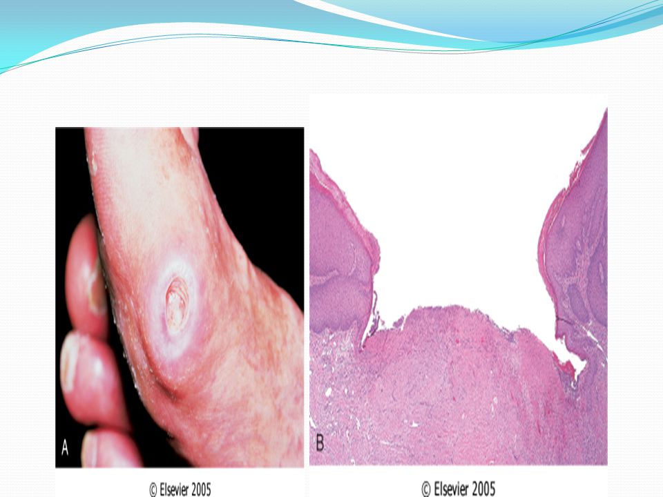


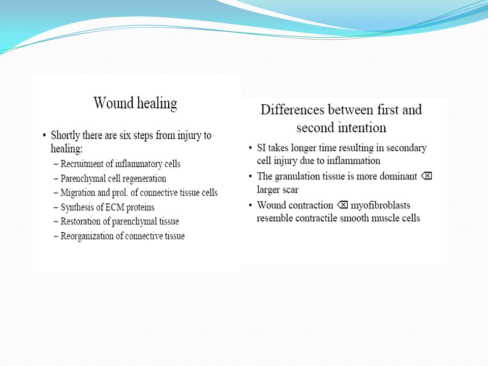


. LEARNING OBJECTIVES Review the normal physiology and concepts of cell proliferation, cell growth, cell “cycle”, and cell.>")


>")


