Download presentation
Presentation is loading. Please wait.

1
MOTOR PHYSIOLOGY © Wesner, M. F.

2
Many ways to study motor systems
Behavioral perspective - holistic approach to studying motor activity. Investigates the behavioral pattern of an animal. At the turn of the last century with the influence of Darwinian philosophy, naturalists were interested in studying behavior patterns of many species in the wild. The beginnings of Ethology. Why are behaviors elicited? Is there an evolutionary advantage to the modal action pattern (formally referred to as ‘Fixed Action Pattern’)-Species specific behaviors related to specific environmental situations.
-Species specific behaviors related to specific environmental situations.")
3
Behavioral perspective (cont.)
19th Century was very big in Reflexology..the study of reflexes. Reflex - a highly stereotyped, unlearned response to a particular stimulus.

4
DR. CHARLES SCOTT SHERRINGTON (1852-1957)
Nobel Prize in Medicine (1932) ..for his discoveries regarding the functions of neurons. Sherrington was born in London, studied in England and gravitated into physiology where he was strongly influenced by Michael Foster - "father of British Physiology”. His interest was in reflexes and he is best known for his lengthy studies in spinal reflexes. Also he will be remembered for his contributions to the physiology of perception, reaction and behavior.
 ..for his discoveries regarding the functions of neurons. Sherrington was born in London, studied in England and gravitated into physiology where he was strongly influenced by Michael Foster - father of British Physiology . His interest was in reflexes and he is best known for his lengthy studies in spinal reflexes. Also he will be remembered for his contributions to the physiology of perception, reaction and behavior.")
5
Sherrington understood that the brain made all necessary computations, but ultimately it was the reflexes that make up the spinal cord system that will be called upon to activate target muscles. He believed no matter how complex the motoric behavior, it was really nothing more than a series of unit reflexes chained together. CNS modulates the series. S R S R S R S R Note: Sherrington was Ivan Sechenov’s contemporary.

6
Ivan Sechenov ( ) Ivan Petrovich Pavlov ( ) Nobel Prize (1904) Sechenov, born in Russia, was known as the Father of Russian physiology. He introduced electrophysiology into laboratories and was also a notable teacher. His life work concentrated on neurophysiology. He wrote a major classic treatise "The Reflexes of the Brain."

7
Salivation is supposed to be an unlearned reflex governed by preprogrammed stimuli like food, not something that can be learned to a novel stimulus!

8
Ways to study motor systems
Behavioral perspective - holistic approach to studying motor activity. Investigates the overt behavioral patterns of an animal. Control systems perspective - an engineering view of systems (systems analysis). command Motor System detector Controller (CNS) input output Error Trans-ducer Closed-Loop System
. command. Motor System. detector. Controller (CNS) input. output. Error. Trans-ducer. Closed-Loop System.")
9
Ways to study motor systems
Behavioral perspective - holistic approach to studying motor activity. Investigates the behavioral pattern of an animal. Control systems perspective - an engineering view of systems (systems analysis). output Controller (CNS) command Input Motor System Open-Loop System - faster. Not relying on feedback signals Learned vs. innate behavior. Closed-loop while learning, then with experience develops into an open-loop efficient expression.
. output. Controller (CNS) command. Input. Motor System. Open-Loop System - faster. Not relying on feedback signals. Learned vs. innate behavior. Closed-loop while learning, then with experience develops into an open-loop efficient expression.")
10
Ways to study motor systems
Behavioral perspective - holistic approach to studying motor activity. Investigates the behavioral pattern of an animal. Control systems perspective - an engineering view of systems (systems analysis). Neurobiological view - interested in investigating the mechanical properties (i.e., the substrate) of motoric behavior. *
. Neurobiological view - interested in investigating the mechanical properties (i.e., the substrate) of motoric behavior. *")
11
The motor systems of the brain, spinal cord and peripheral nerves provide the means for us to move and thereby act upon our ever-changing environment. Afferent systems - transduce physical energies into neurological signals. Efferent systems - concerned with reverse process.. The transduction of neural signals into mechanical energy (or force).
.")
12
Descending Efferents:
Volitional movement Antigravity reflex inhibition Antigravity reflex excitation

13
Posterior median sulcus
Ventral median fissure

14
Descending tracts: Reticulospinal:
Automatic Antigravity & gravity reflexes. Uses sensory information about balance, body position & the visual environment to reflexively maintain posture and balance with movement (proximal / axial muscles).
.")
16
Somatic (striate) muscle
Fibers control flexors- striate muscles that pull limbs around a joint together (closing the knife). proximal Fibers control extensors- striate muscles that pull limbs around a joint apart (opening the knife).
. proximal. Fibers control extensors- striate muscles that pull limbs around a joint apart (opening the knife).")
17
flexion extension tendons antagonists synergists

19
each muscle fiber is a cell

20
Cholinergic neuromuscular endplate.
Amount of pool divergence or convergence defines motor resolution. More macroscopic..

21
Older, more radical treatments: plasmaphoresis; remove thymus
membrane immersed in liquid Nitrogen fractures the weak bonds along the hydrophobic layers Freeze fracture of a Neuromuscular Junction Myasthenia Gravis (grk. “severe muscle weakness”) - body produces antibodies that destroy nicotinic, cholinergic receptors. One remedy: Inhibit ACHE to prolong the presence of ACH in the cleft. Older, more radical treatments: plasmaphoresis; remove thymus
 - body produces antibodies that destroy nicotinic, cholinergic receptors. One remedy: Inhibit ACHE to prolong the presence of ACH in the cleft. Older, more radical treatments: plasmaphoresis; remove thymus.")
22
Multinucleated muscle fiber cell (cytoskeleton is primarily myofibrils).
- channels that allow connection to extracellular space. Shaped like a “T”. Contain extracell. fluid. I - high Ca++ like ER plasma in neurons. A - Openings allow membrane voltage change to travel into “deeper” intracellular myofibrils. - analogous to axolemma.

23
myofibril Sarcoplasmic reticulum

25
..during excitation,the I band shrinks in size. I A
sarcomere Z-line Sarcoplasmic reticulum H-band

26
actin 5-nm diameter A-band myosin 10-nm diameter

28
Transduction process: How does the neural signal translate into contraction?

29
The release of Ca++ from the sarcoplasmic reticulum
The release of Ca++ from the sarcoplasmic reticulum. Depolarization of the T-tubule membrane causes conformational changes in proteins that are linked to calcium channels in the sarcoplasmic reticulum, releasing stored Ca++ into the cytosol of the muscle fiber.

30
The molecular basis of muscle contraction
The molecular basis of muscle contraction. The binding of Ca++ to troponin allows the myosin heads to bind to the actin filament. Then the myosin heads rotate, causing the filaments to slide with respect to one another.

31
Excitation: AP arrives at a-motor neuron endplate. ACh release into cleft. ACh binds to nicotinic ACH receptors: EPSP. Na+ channels open (inc. gNa+) at sarcolemma. T-tubules continue to convey the membrane depolarization toward the myofibril membranes. Depolarization of the T-tubules causes conformational change of Ca++ SR channels yielding cytosolic Ca++ release. Contraction: Ca++ binds to troponin protein. Myosin binding sites exposed. Myosin heads bind to actin & myosin heads rotate (ratchet). Myosin heads disengage (ATP required) Cycle continues until ATP and Ca++ levels are depleted.
 at sarcolemma. T-tubules continue to convey the membrane depolarization toward the myofibril membranes. Depolarization of the T-tubules causes conformational change of Ca++ SR channels yielding cytosolic Ca++ release. Contraction: Ca++ binds to troponin protein. Myosin binding sites exposed. Myosin heads bind to actin & myosin heads rotate (ratchet). Myosin heads disengage (ATP required) Cycle continues until ATP and Ca++ levels are depleted.")
32
Relaxation: Ca++ is sequestered by the SR by an ATP-driven pump. Myosin binding sites on actin are once again covered by troponin. A lot of ATP required to run muscles. Here and during contraction, not to mention the usual ATP demands associated with synaptic transmission. Muscles are very metabolically demanding.

33
Proprioception is an integral part of kinesthesis-the perception or knowing of the position of the body as muscles, tendons and joints move, in addition to the visual, auditory and vestibular systems responsible for our perception of body position. Proprioception is usually a reference to unconscious perception of body position.

34
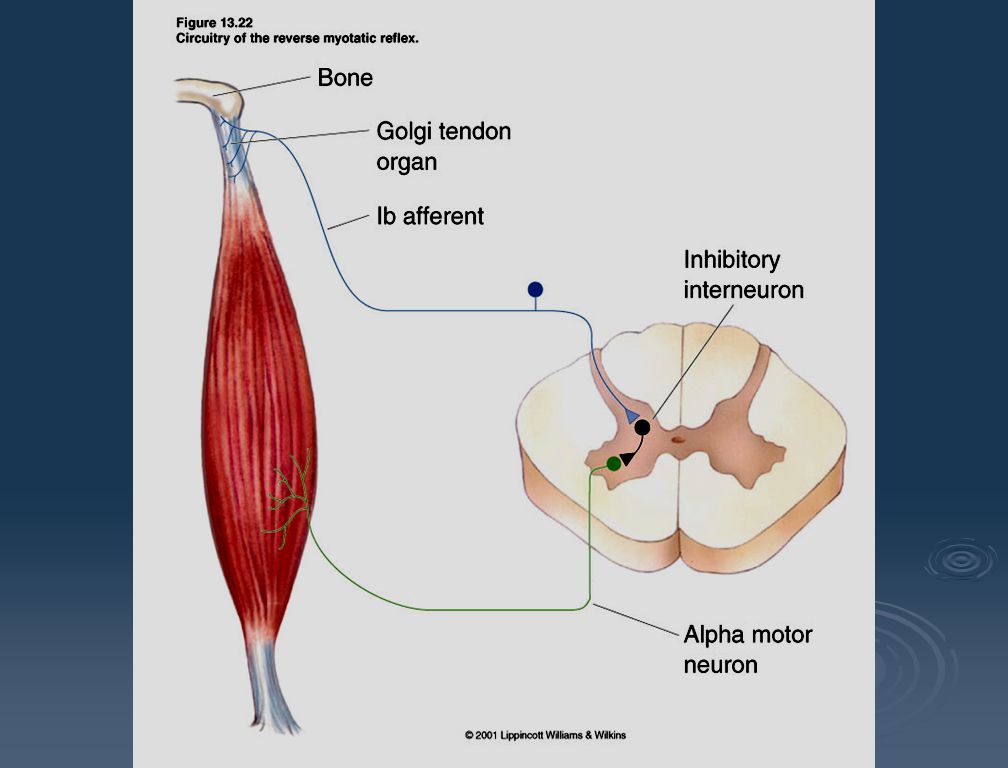
Proprioception - Motor sensory system designed to signal changes in muscle length. Two main types of proprioceptors: Muscle Spindles - found within the muscle, parallel to the muscle fibers (stretch or length). Golgi tendon apparatus - found in the tendons, in series to the muscle fibers (contraction-’pulls’ at the tendons, tension).
. Golgi tendon apparatus - found in the tendons, in series to the muscle fibers (contraction-’pulls’ at the tendons, tension).")
36
The stretch (or myotatic) reflex..
- Group 1a

38
There is a secondary motor neuron (the g-motor neuron which has smaller axons & less myelin. It is part of a proprioceptive gain system designed to avoid sensory saturation.

40
Resets the spindle stretch receptors so that 1a can pick up subsequent stretch responses.
a motor neuron -- myotatic correction and subsequent g-induced reset. start with a passive stretch

41
Muscle Spindles - found within the muscle, parallel to the muscle fibers (stretch or length).
Golgi tendon apparatus - found in the tendons, in series to the muscle fibers (contraction-’pulls’ at the tendons, tension).
.")
44
Knee Jerk reflex is also monosynaptic (intrasegmental).
Tap “pulls” on the tendon. This ‘interprets’ as a stretch of the quadricepts which in turn activates the muscle spindles (& thus 1a sensory neuron). Neural signals carry through dorsal root ganglion and excites synapsing a motor neuron. This results in contraction of extrafusal fibers of the quadriceps (extension).. ..& corresponding inhibition of antagonistic hamstrings. Patellar
. Neural signals carry through dorsal root ganglion and excites synapsing a motor neuron. This results in contraction of extrafusal fibers of the quadriceps (extension).. ..& corresponding inhibition of antagonistic hamstrings. Patellar.")
46
Pain reflex is polysynaptic.
Synergistic flexion- Note: intersegmental reflex.

47
Reciprocal inhibition: the complexities of antagonism
Reciprocal inhibition: the complexities of antagonism. Want to avoid being in a catatonic (rigid) state.
 state.")
49
Motor functional distinctions:

50
Descending tracts: Voluntary movements Pyramidal tracts
A more primitive tract originating from the red nuclei. Function subsumed by corticospinal projections. Pyramidal tracts - Higher end motor activity. Decussate at the enlarged ventral region of the medulla (cross-section looks triangular; thus the name “pyramidal”).
.")
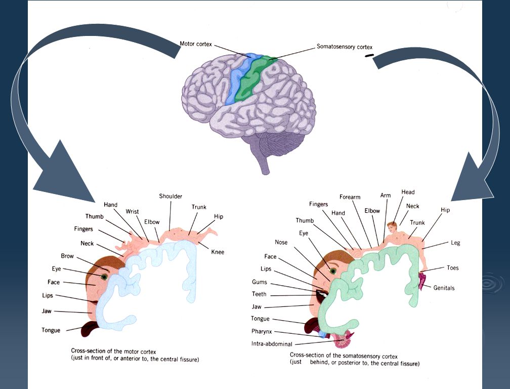
51
“rt. hand” M1, Area #4 ..in the tegmentum Terminates in the dorsolateral portion of the ventral horn.

52
Rubrospinal tract is an alternative route for the mediation of voluntary movement. It is responsible for LARGE muscle movement such as the arms and the legs as well as for fine motor control in non-primate species. It facilitates the flexion and inhibits the extension in the upper extremities. It is small and rudimentary in humans. NOTE: In some other primates, however, experiments have shown that over time, the rubrospinal tract can assume almost all the duties of the corticospinal when the corticospinal tract is lesioned.

53
Descending tracts: Originates from colliculus..Coordination of eye and head position (e.g., vestibular ocular reflex [VOR] used for perceived image stabilization.) Originates from vestibular nuclei, near the cochlear nuclei (part of VIII). Movement of the head activates vestibular system. Reflex circuitry controls neck and back muscles to ensure head stability (proprioceptive control as our body moves). Balance and body posture. Makes use of spinal reflexes, and sensory information from proprioceptors, visual system & vestibular system.
 Originates from vestibular nuclei, near the cochlear nuclei (part of VIII). Movement of the head activates vestibular system. Reflex circuitry controls neck and back muscles to ensure head stability (proprioceptive control as our body moves). Balance and body posture. Makes use of spinal reflexes, and sensory information from proprioceptors, visual system & vestibular system.")
55
Descending tracts: Activates or inhibits antigravity muscles. lateral
reticulospinal tracts lateral medial

56
+ - Terminates in the dorsomedial portion of the ventral horn.

57
The Reticulospinal projections
Pontine Reticulospinal - Enhances antigravity reflexes. Medullar Reticulospinal - Inhibits antigravity reflexes. Allows for voluntary override!

59
V1 V2 V3 S1 M1 SMA V4 MT( V5) A1 PMA Posterior Parietal
 A1 PMA Posterior Parietal")
63
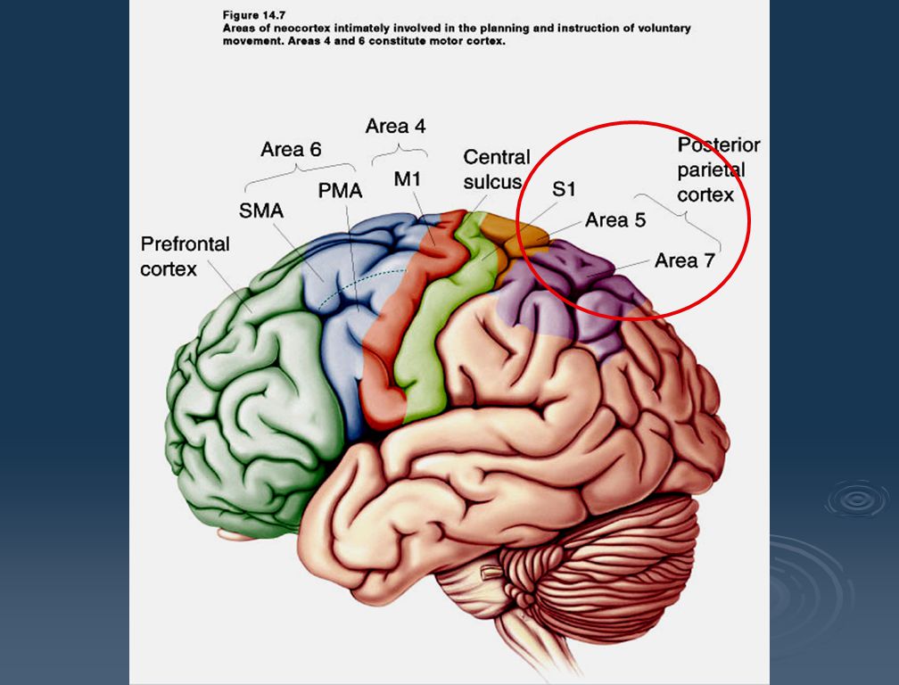
Brodmann’s Classification

64
Premotor area - involves reticulospinal tracts (proximal) muscle areas.
Supplementary motor area - involves distal muscle areas.

65
Psychomotor Cortex A lot of this through subcortical basal ganglia (i.e., lenticular to thalamus to #6)
")
66
From “What” the actions should be to “How” they will be carried out..
“Get Ready” Posterior Parietal (#5,7) Prefrontal Cortex (#8) “Set” PMA & SMA (#6) - lesions here produce apraxia = lack of complex motor skills. “Go” M1 (#4) - due to release of subcortical inhibition on SMA (#6). Spinal Cord = sends signals to motor pools.
 Prefrontal Cortex (#8) Set PMA & SMA (#6) - lesions here produce apraxia = lack of complex motor skills. Go M1 (#4) - due to release of subcortical inhibition on SMA (#6). Spinal Cord = sends signals to motor pools.")
67
Edward Evarts (NIH) “Behavioral Neurophysiology” Motoric activity in awake animals and measuring single cell activity--reminiscent of vision science research with RFs decades earlier.

68
Basal Ganglia Motor Loop: Part of the extrapyramidal system and key to setting up motor commands for execution through M1 to spinal cord..

69
Basal ganglia VLo

71
Receives input from globus pallidus
lenticular Internal capsule

72
#6 Striatum (putamen) external internal
 external internal")
73
#6 (proximal muscles) VLo Nigrastriatal tracts
 VLo Nigrastriatal tracts")
75
Basal ganglia VLo Disinhibition- globus pallidus is “turned off”, allowing VLo to activate SMA (distal motor pools).
.")
76
Cortical cells excite putamen neurons..
2. Activated putamen cells are inhibitory. They inhibit globus pallidus (GPe) cells. 5. Result: Excitation of SMA (a release from active supression). 3. GPe cells are inhibitory. They inhibit VL cells.. 4. but when they are inhibited by putamen cells, they in turn “turn off” their inhibition on VLo cells (disinhibition).
 cells. 5. Result: Excitation of SMA (a release from active supression). 3. GPe cells are inhibitory. They inhibit VL cells.. 4. but when they are inhibited by putamen cells, they in turn turn off their inhibition on VLo cells (disinhibition).")
77
Basal Ganglia facilitates or inhibits the initiation of volitional movement..
1. At rest, Globus pallidus always inhibits VLo. 2. Striatum inhibits globus pallidus. 3. This, in turn, activates VLo (disinhibition). 4. This activates SMA.. 5. which in turn activates M1..
. 4. This activates SMA.. 5. which in turn activates M1..")
78
An imbalance of the motor loop can lead to dysfunction:
Hypokinesia: Paucity of movement. e.g., Parkinson’s Disease (s. nigra defect) No striatal excitation. (bradykinesia, akinesia) Hyperkinesia: Excess of movement. e.g., Huntington’s Disease (subthalamic defect) No controlled inhibition of VLo. (chorea, dementia). No disinhibition
 No striatal excitation. (bradykinesia, akinesia) Hyperkinesia: Excess of movement. e.g., Huntington’s Disease (subthalamic defect) No controlled inhibition of VLo. (chorea, dementia). No disinhibition.")
79
Corticospinal fibers are primarily emerging from layer V and VI of the cortex (M1 is agranular). Largest descending cells are Betz cells in the corticospinal tract. Majority are the pyramidal cells.

80
A comparison being made between “What was intended” to “What has happened?”
An important loop for motor skills learning. The VLc loop

81
Corticospinal cells in M1 code for movement “force” and direction (Georgopolus study suggests a motoric receptive field?)..
..")
82
“Motor cell tuning”. This particular cell really “likes” leftward arm movement (180°):
:")
83
Population vectors. Combining gaussian- distributed response properties for multiples of M1 cells can lead to many different directional characteristics. (i.e., the ratio of M1-cell broadband outputs can provide a large gamut of directional movements. ANALOGY: Color vision-the ratio of 3 cone outputs in the retina can provide a large gamut of perceived colors. 45°

Similar presentations