Download presentation
Presentation is loading. Please wait.

1
Mediastinal Staging Samer Kanaan, M.D.

2
Overview Importance of accurate nodal staging
Accuracy of radiographic staging Mediastinoscopy EUS EBUS

3
Staging

4
TNM Definitions T Stage Size of the Primary Tumor
Adjacent structures invaded into by Tumor N Stage Nodal disease involvement M Stage Metastatic disease involvement

5
Stage TNM Classifcation IA T1N0M0 IB T2N0M0 IIA T1N1M0 IIB T2N1M0 or T3N0M0 IIIA T1-3N2M0 or T3N1M0 IIIB T4NanyM0 or TanyN3M0 IV TanyNanyM1

7
Stage IA, cancer is in the lung only, less than 3cm in size.
Stage IB, the cancer is: (a) greater than 3cm in size (b) involve the main bronchus (c) invade visceral pleura (d) associated with obstructive pneumonitis.
 greater than 3cm in size (b) involve the main bronchus (c) invade visceral pleura (d) associated with obstructive pneumonitis.")
8
Stage IIA, cancer is less than 3cm in size and involves ipsilateral hilar lymph nodes.
Stage IIB, cancer is either the same as in stage IB and has also spread to ipsilateral hilar lymph nodes or Cancer has not spread to lymph nodes but has spread to one or more of the following: (a) the chest wall, (b) the diaphragm, (c) mediastinal pleura, (d) pericardium, (e) the main bronchus less than 2cm from the carina, and/or (f) associated obstructive pneumonitis of the entire lung.
 the chest wall, (b) the diaphragm, (c) mediastinal pleura, (d) pericardium, (e) the main bronchus less than 2cm from the carina, and/or (f) associated obstructive pneumonitis of the entire lung.")
9
Stage IIIA The cancer has spread to ipsilateral mediastinal or subcarinal lymph nodes (N2). Similar to Stage IIB, It may also spread to one or more of the following: (a) the chest wall, (b) the diaphragm, (c) mediastinal pleura, (d) pericardium, (e) the main bronchus less than 2cm from the carina, and/or (f) associated obstructive pneumonitis of the entire lung.
 the chest wall, (b) the diaphragm, (c) mediastinal pleura, (d) pericardium, (e) the main bronchus less than 2cm from the carina, and/or (f) associated obstructive pneumonitis of the entire lung.")
10
Cancer may also spread to the pleural fluid (T4).
Stage IIIB The cancer has spread to (a) contralateral mediastinal or hilar nodes or ipsilateral supraclavicular nodes. The cancer may also spread to one or more of the following: (b) the heart, (c) the inferior vena cava and the aorta, (f) the trachea, and (g) the esophagus. Cancer may also spread to the pleural fluid (T4). Separate nodules in the same lobe is also (T4)*
 contralateral mediastinal or hilar nodes or ipsilateral supraclavicular nodes. The cancer may also spread to one or more of the following: (b) the heart, (c) the inferior vena cava and the aorta, (f) the trachea, and (g) the esophagus. Cancer may also spread to the pleural fluid (T4). Separate nodules in the same lobe is also (T4)*")
12
Staging

13
Stage TNM Classifcation 5 Year Survival IA T1N0M0 67
IB T2N0M IIA T1N1M IIB T2N1M0 or T3N0M0 39 IIIA T1-3N2M0 or T3N1M0 23 IIIB T4NanyM0 or TanyN3M0 5 IV TanyNanyM Mountain, Chest 1997

14
Why is accurate nodal staging essential?
N1 disease N2 disease N3 disease

15
Treatment of Lung Cancer According to Stage
Primary treatment Adjuvant therapy Five-year survival rate (%) Non-small cell carcinoma I Resection Chemotherapy 60 to 70 II Chemotherapy with or without radiotherapy 40 to 50 IIIA (resectable) Resection with or without preoperative chemotherapy 15 to 30 IIIA (unresectable) or IIIB (involvement of contralateral or supraclavicular lymph nodes) Chemotherapy with concurrent or subsequent radiotherapy None 10 to 20 IIIB (pleural effusion) or IV Chemotherapy or resection of primary brain metastasis and primary T1 tumor 10 to 15 (two-year survival) Small cell carcinoma Limited disease Chemotherapy with concurrent radiotherapy 15 to 25 Extensive disease < 5 Adapted with permission from Spira A, Ettinger DS. Multidisciplinary management of lung cancer. N Engl J Med 2004;350:388.
 Non-small cell carcinoma. I. Resection. Chemotherapy. 60 to 70. II. Chemotherapy with or without radiotherapy. 40 to 50. IIIA (resectable) Resection with or without preoperative chemotherapy. 15 to 30. IIIA (unresectable) or IIIB (involvement of contralateral or supraclavicular lymph nodes) Chemotherapy with concurrent or subsequent radiotherapy. None. 10 to 20. IIIB (pleural effusion) or IV. Chemotherapy or resection of primary brain metastasis and primary T1 tumor. 10 to 15 (two-year survival) Small cell carcinoma. Limited disease. Chemotherapy with concurrent radiotherapy. 15 to 25. Extensive disease. < 5. Adapted with permission from Spira A, Ettinger DS. Multidisciplinary management of lung cancer. N Engl J Med 2004;350:388.")
16
Treatment – Stage IIIA Stage IIIA N2 disease 5 year survival is 10-15% overall Stage IIIA bulky mediastinal involvement (visible on CXR) have 5 year survival of 2-5% Radiation: Treatment with 60 Gy can achieve long term survival benefit in 5-10% of patients Chemotherapy and Radiation: Meta analysis from 11 randomized studies showed cisplatin based chemotherapy with radiation resulted in 10% reduction in the risk of death compared to radiation therapy alone. Combined SurgicalTherapy: Neoadjuvant chemotherapy plus surgery had median survival > 3X versus surgery alone Neoadjuvant chemotherapy and radiation allowed 65-75% patients to undergo surgical resection these patients had 27% 3 year survival.
 have 5 year survival of 2-5% Radiation: Treatment with 60 Gy can achieve long term survival benefit in 5-10% of patients. Chemotherapy and Radiation: Meta analysis from 11 randomized studies showed cisplatin based chemotherapy with radiation resulted in 10% reduction in the risk of death compared to radiation therapy alone. Combined SurgicalTherapy: Neoadjuvant chemotherapy plus surgery had median survival > 3X versus surgery alone. Neoadjuvant chemotherapy and radiation allowed 65-75% patients to undergo surgical resection these patients had 27% 3 year survival.")
17
N2 Disease Patients benefit from neoadjuvant therapy and surgery versus resection followed by adjuvant therapy. Patients are more likely to complete chemotherapy regimen pre operatively than post operatively. Awaiting definitive results of the NATCH (Neoadjuvant Taxol Carboplatin Hope) trial available 2009 Alam N, et al. Lung Cancer 2005;47: Depierre A, et al. J Clin Oncol 2002;20:
 trial available Alam N, et al. Lung Cancer 2005;47: Depierre A, et al. J Clin Oncol 2002;20:")
18
What is the accuracy of radiographic staging?

19
CT Scan

20
Information gained by CT
Tumor size Tumor number Central tumor or Peripheral Lymph node enlargement (>1 cm) Extent Discrete lymph nodes versus mediastinal infiltration Metastatic disease
 Extent. Discrete lymph nodes versus mediastinal infiltration. Metastatic disease.")
21
Accuracy of CT in Staging
CT scan Tumor Sensitivity = 63% Specificity = 84% Mediastinum Sensitivity = 51-75% Specificity = 66-86% Positive predictive value = 60% Negative predictive value = 80% Toloza E, et al. Chest 2003(suppl):137s–146s Gould MK, et al. Ann Intern Med 2003; 139:879–892 Dwamena et al. Radiology 1999; 213:530–536
:137s–146s. Gould MK, et al. Ann Intern Med 2003; 139:879–892. Dwamena et al. Radiology 1999; 213:530–536.")
22
Accuracy of CT in Staging the Mediastinum
CT scanning alone is not sufficient to determine nodal staging However, certain characteristics can guide further staging

23
CT Staging of the Mediastinum
Group A: mediastinal infiltration Group B: discrete mediastinal lymph node enlargement Group C: central tumor or suspected N1 disease Group D: peripheral tumor, no mediastinal involvement Invasive biopsy N2, N3 involvement 20-25% Invasive biopsy ???

24
Prevalence of N2 disease in clinical stage I
Location? Central 9-11% Peripheral 6-19% Cell Type? Adenocarcinoma 14% Squamous 8.9% Tumor Stage? T1 8.4% T2 10.4% Suzuki K et al, JTCVS; 1999;117:593-8 Daly BD, et al. JTCVS; 1993;105:904-10 Uy KFL et al, Difficult Decisions in Thoracic Surgery; 2007:68-74.

25
CT Staging of the Mediastinum
Group A: mediastinal infiltration Group B: discrete mediastinal lymph node enlargement Group C: central tumor or suspected N1 disease Group D: peripheral tumor, no mediastinal involvement Invasive biopsy N2, N3 involvement 20-25% Invasive biopsy ??? But must assume at least 10% chance of N2 disease Invasive biopsy

26
PET Scan

27
MacManus MP, et al. Int J Radiat Oncol Biol Phys 2001; 50:287–293
PET in Staging Detecting both size and activity of tumor Detecting size and activity of lymph nodes Provides whole-body information M1 disease found in 1-8% of patients thought to be stage I by CT M1 disease found in 7-18% of patients thought to be stage II by CT Reed CE, et al. JTCVS 2003; 126:1943–1951 MacManus MP, et al. Int J Radiat Oncol Biol Phys 2001; 50:287–293

28
Accuracy of PET in Staging the Mediastinum
PET Scan Tumor Sensitivity = 83-96% Specificity = 73-78% Mediastinum Sensitivity = 64-91% Specificity = 77-93% Distant Metastasis Sensitivity = 95% Specificity = 83%

29
Recommendations of PET in Staging the Mediastinum
Stage IA consider Stage IB-IIIB should undergo PET Any abnormal result in the mediastinum should prompt lymph node sampling

30
PET/CT combined

31
Sensitivity Specificity
CT 86% 67% PET 94% 59% PET/CT 97% 44%

32
Is the combination of PET/CT good enough to obviate mediastinoscopy?
Radiographic N2, N3 = need for tissue biopsy prior to neoadjuvant therapy NO Radiographic N1 = 20-25% occult N2 disease Radiographic N0 CONTROVERSIAL

33
What is the prevalence of undetected N2 disease after PET/CT
PET/CT staging was node negative At mediastinoscopy found to have N2 disease 11.7% of the time (n=137) Gonzalez-Stawinski et al. JTCVS 2003;126:
 Gonzalez-Stawinski et al. JTCVS 2003;126:")
34
What is the prevalence of undetected N2 disease after PET/CT
After CT 19.2% (n=2224) After CT + PET 6.7% (n=906) After CT + mediastinoscopy 8.3% (n=869) After CT + PET + mediastinoscopy 4.5% (n=178) Uy KFL et al, Difficult Decisions in Thoracic Surgery; 2007:68-74.
 After CT + PET 6.7% (n=906) After CT + mediastinoscopy 8.3% (n=869) After CT + PET + mediastinoscopy 4.5% (n=178) Uy KFL et al, Difficult Decisions in Thoracic Surgery; 2007:")
35
What is the prevalence of undetected N2 disease after PET/CT
PET/CT staging was node negative but at thoracotomy found to have N2 disease 5.6% PET/CT/mediastinoscopy staging was node negative but at thoracotomy found to have N2 disease 4.5% Meyers JTCVS 2006;131:

36
Choice of lymph node sampling

37
Emergency Sternotomy = 0.12%
Mediastinoscopy Sensitivity = 70-95% Specificity = 100% Negative Predictive value = 88-93% Positive Predictive value = 100% Complication rate = 0.6% Mortality rate = 0.08% Emergency Sternotomy = 0.12%

38
Staging with Mediastinoscopy
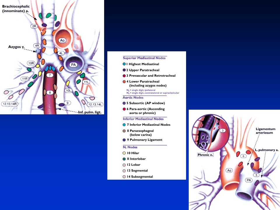
Define N1, N2, N3 disease 1, 3, 2L, 2R, 4L, 4R, 7 + enlarged nodes Not 5,6,8,9 Luke WP, Pearson FG, et al. JTCVS; 1986: 91(1) Kiser AC, Detterbeck FC. Diagnosis and treatment of lung cancer: an evidencebased guide for the practicing clinician. Philadelphia, PA: WB Saunders, 2001; 133–147
 Kiser AC, Detterbeck FC. Diagnosis and treatment of lung cancer: an evidencebased guide for the practicing clinician. Philadelphia, PA: WB Saunders, 2001; 133–147.")
40
What’s the real problem with mediastinoscopy?

41
ACS survey in 2001 of 729 hospitals including 40,090 patients
Mediastinoscopy performed in 27.1% of patients going to curative resection Of these mediastinoscopies, only 46.6% had documented node biopsy

42
Perhaps they are utilizing PET/CT?
26.5% received PET Perhaps they are sampling at the time of thoracotomy? Only 42.2% of surgical resections had mediastinal lymph nodes 59.5% Stage I, 17.5% Stage II, 17.0% Stage III, 6.0% Stage IV

43
Endoscopic Ultrasound Endobronchial Ultrasound
Bronchoscopy Endoscopic Ultrasound Endobronchial Ultrasound

44
EUS Can be done with conscious sedation
Can detect and biopsy adrenal, celiac, liver metastasis Can detect T4 invasion into the mediastinum EUS for T staging Sensitivity 88% Specificity 98% False Negative 1% False Positive 30% EUS for N staging Sensitivity 84% Specificity 100% False Negative 19% False Positive 8% Varadarajulu S, et al. Gastrointest Endosc 2004; 59:345–348 Detterbeck et al. Chest;2007:132 (3 Supplement): 202S. Annema JT, et al. JAMA 2005; 294:931–936, only study to investigate positive lymph with surgical incision
: 202S. Annema JT, et al. JAMA 2005; 294:931–936, only study to investigate positive lymph with surgical incision.")
45
Detterbeck et al. Chest;2007:132 (3 Supplement): 202S.
EUS Pre-selected patient population CT evidence of N2, N3 nodal disease Detterbeck et al. Chest;2007:132 (3 Supplement): 202S.
: 202S.")
46
EUS in the Setting of a Negative CT
Saved an inappropriate thoracotomy in 9/67 = 13% LeBlanc et al. Am J Resp and Crit Care Med. 171 (2): 177. (2005)
: 177. (2005)")
47
EUS in the Setting of a Negative CT
Failed to detect N2 disease in 17/67 = 25% Would never be able to detect N2 disease in 5/67 = 7% LeBlanc et al. Am J Resp and Crit Care Med. 171 (2): 177. (2005)
: 177. (2005)")
48
Endobronchial Ultrasound-Guided Needle Aspiration (EBUS)
Stations 1, 2, 3, 4, 7, 10 Not 5, 6, 8, 9 Only EBUS-positive nodes sampled

49
EBUS Sensitivity 90% Specificity 100% False Positive 0%
False Negative 20% Few complications Up to 25% suspected malignancy can be benign Minimize mediastinal scarring for future biopsy or resection Can be done with conscious sedation, no need for intubation Detterbeck et al. Chest;2007:132 (3 Supplement): 202S. Doelken P, Reed C, Silvestri G, et al. Ann Thorac Surg 2008;85:224 –30
: 202S. Doelken P, Reed C, Silvestri G, et al. Ann Thorac Surg 2008;85:224 –30.")
50
Detterbeck et al. Chest;2007:132 (3 Supplement): 202S.
EBUS Detterbeck et al. Chest;2007:132 (3 Supplement): 202S.
: 202S.")
51
EBUS in Patients with Normal CT
Biopsies taken from any identifiable lymph node 2, 4, 7, 10, 11 199 lymph nodes Average size 8mm Measured against pathologic specimen Sensitivity 92.3% Specificity 100% Negative predictive value 96.3%

52
EBUS in Patients with Normal CT
Upstaged to N1 disease in 3% patients Upstaged to N2 disease in 13% patients Upstaged to N3 disease in 3% patients Saved an inappropriate thoracotomy in 16% Failed to identify N2 or N3 disease in 2% of patients

53
EUS + EBUS EBUS: stations 1-4, 7 EUS: stations 8, 9 Conscious sedation
52 minutes

55
EUS + EBUS = Medical Mediastinoscopy
Vilmann P, Puri R. The complete ''medical'' mediastinoscopy (EUS-FNA + EBUS-TBNA).Minerva Med Aug;98(4):331-8. Sensitivity 100% Specificity 100%
.Minerva Med Aug;98(4): Sensitivity 100% Specificity 100%")
56
Med EUS EBUS EUS+ Sensitivity Specificity False Neg False Pos Station
Limitations Med 78-95 100 11 1, 3, 2, 4, 7 Utilization EUS 87 19 8 5, 6, 8, 9 Limited location EBUS 90 20 1, 3, 2, 4, 7, 10 No standard protocol EUS+ 95 1, 2, 3, 4, 8, 9, 10 No data

57
* * Sensitivity in the setting of radiographic stage 1 disease Med EUS
Specificity False Neg False Pos Station Limitations Med 42 78-95 100 11 1, 3, 2, 4, 7 Utilization EUS 66 87 19 8 5, 6, 8, 9 Limited location EBUS 94 90 20 1, 3, 2, 4, 7, 10 No standard protocol EUS+ 97 95 1, 2, 3, 4, 8, 9, 10 No data * Sensitivity in the setting of radiographic stage 1 disease

58
Summary Mediastinum should be staged invasively utilizing mediastinoscopy, EUS, EBUS or EUS+EBUS. PET/CT alone will miss N2 disease (5-12%) Perhaps future lies with medical mediastinoscopy of EUS+EBUS

Similar presentations


 Geometry (29%)>")
,>")
 Motion Controller Design for A Class of Second-order Systems Center for Self-Organizing Intelligent.>")
