Download presentation
Presentation is loading. Please wait.

1
Adult Respiratory Distress Syndrome

2
Case presentation A 45-year-old man develops ARDS after sustaining multiple broken bones in an automobile accident. The man weighs 70 kg. Mechanical ventilation is initiated in the AC mode with the following settings: (PEEP), 10 cm H2O; (FiO2), 70%; respiration rate, 12/min. The most appropriate Tidal volume at this point: 1000 ml 420 ml 500 ml 560 ml 700 ml
, 10 cm H2O; (FiO2), 70%; respiration rate, 12/min. The most appropriate Tidal volume at this point: 1000 ml. 420 ml. 500 ml. 560 ml. 700 ml.")
3
Bernard GR et al., Am J Respir Crit Care Med 1994
ARDS Definition The 1994 North American-European Consensus Conference (NAECC) criteria: Onset - Acute and persistent Radiographic criteria - Bilateral pulmonary infiltrates consistent with the presence of edema Oxygenation criteria - Impaired oxygenation regardless of the PEEP concentration, with a Pao2/Fio2 ratio 300 torr (40 kPa) for ALI and 200 torr (27 kPa) for ARDS Exclusion criteria - Clinical evidence of left atrial hypertension or a pulmonary-artery catheter occlusion pressure of 18 mm Hg. Bernard GR et al., Am J Respir Crit Care Med 1994
 criteria: Onset - Acute and persistent. Radiographic criteria - Bilateral pulmonary infiltrates consistent with the presence of edema. Oxygenation criteria - Impaired oxygenation regardless of the PEEP concentration, with a Pao2/Fio2 ratio 300 torr (40 kPa) for ALI and 200 torr (27 kPa) for ARDS. Exclusion criteria - Clinical evidence of left atrial hypertension or a pulmonary-artery catheter occlusion pressure of 18 mm Hg. Bernard GR et al., Am J Respir Crit Care Med")
4
Stratification System of Acute Lung Injury GOCA
Letter Meaning Scale Definition G Gas exchange (to be combined with the numeric descriptor) 1 2 3 A B C D Pao2/Fio2 301 Pao2/Fio Pao2/Fio – 200 Pao2/Fio2 100 Spontaneous breathing, no PEEP Assisted breathing, PEEP 0-5 cmH2O Assisted breathing, PEEP 6-10 cmH2O Assisted breathing, PEEP 10 cmH2O O Organ failure Lung only Lung + 1 organ Lung + 2 organs Lung + 3 organs Cause Unknown Direct lung injury Indirect lung injury Associated diseases No coexisting disease that will cause death within 5 yr Coexisting disease that will cause death within 5 yr but not within 6 mo Coexisting disease that will cause death within 6 mo Artigas A, et al. Am J Respir Crit Care Med
 A. B. C. D. Pao2/Fio2 301. Pao2/Fio Pao2/Fio2 101 – 200. Pao2/Fio2 100. Spontaneous breathing, no PEEP. Assisted breathing, PEEP 0-5 cmH2O. Assisted breathing, PEEP 6-10 cmH2O. Assisted breathing, PEEP 10 cmH2O. O. Organ failure. Lung only. Lung + 1 organ. Lung + 2 organs. Lung + 3 organs. Cause. Unknown. Direct lung injury. Indirect lung injury. Associated diseases. No coexisting disease that will cause death within 5 yr. Coexisting disease that will cause death within 5 yr but not within 6 mo. Coexisting disease that will cause death within 6 mo. Artigas A, et al. Am J Respir Crit Care Med")
5
The ARDS Lung ARDS Focal Patchy Diffuse Chest x-ray (zero PEEP)
Focal heterogeneous loss of aeration in caudal and dependent lung region Bilateral and diffuse x-ray densities respecting lung apices Bilateral and diffuse hyperdensities “White lungs” Chest CT scan Loss of aeration Upper lobes normally aerated despite a regional excess of lung tissue – Lower lobes poorly or non aerated Lower lobes massively nonaerated – The loss of aeration involves partially the upper lobes Massive, diffuse and bilateral non- or poorly aerated lung regions – No normally aerated lung region Response to PEEP PEEP <10-12 cmH2O ++++ Lung recruitment curve Open lung concept Risk of overinflation of the aerated lung regions Recruitment of non aerated lung unit Low potential for recruitment High potential for recruitment Rouby JJ, et al. Eur Respir J Rouby JJ, et al. Anesthesiology

6
The ARDS Lung Early phases of ARDS Direct insult of the lung
Primary pulmonary ARDS “Indirect” insult of the lung Secondary extrapulmonary ARDS Pathologic changes Lung tissue consolidation Severe intra-alveolar damage (Edema, fibrin, collagen neutrophil aggregates, red cells) Microvascular congestion Interstitial edema Alveolar collapse Less severe alveolar damage End-expiratory lung volume EELV Static elastance of the total respiratory system Est,rs Static elastance of the chest wall Est,w / Static lung elastance Est,L / / Intra-abdominal pressure Response to PEEP Est,rs [Est,L >> Est,w] Stretching phenomena Est,rs [Est,L Est,w] Recruitment of previously closed alveolar spaces Lung recruitment ++++ Gattinoni L, et al. Am J Respir Crit Care Med
 Microvascular congestion. Interstitial edema. Alveolar collapse. Less severe alveolar damage. End-expiratory lung volume EELV. Static elastance of the total respiratory system Est,rs. Static elastance of the chest wall Est,w / Static lung elastance Est,L. / / Intra-abdominal pressure. Response to PEEP. Est,rs [Est,L >> Est,w] Stretching phenomena. Est,rs [Est,L Est,w] Recruitment of previously closed alveolar spaces. Lung recruitment Gattinoni L, et al. Am J Respir Crit Care Med")
7
ARDS Mortality Trend 28% 2006

8
Crit Care Med 2009 Vol. 37, No. 5

9
Crude 60-day mortality among Acute Respiratory Distress Syndrome (ARDS) Network patients, 1996–2005.
24%

10
Baby Lung Concept In acute lung injury/acute respiratory distress syndrome, the normally aerated tissue has the dimensions of the lung of a 5- to 6- year-old child (300–500 g aerated tissue) What appears dangerous is not the VT/kg ratio but instead the VT/”baby lung” ratio. The practical message is straightforward: the smaller the “baby lung,” the greater is the potential for unsafe mechanical ventilation.
 What appears dangerous is not the VT/kg ratio but instead the VT/ baby lung ratio. The practical message is straightforward: the smaller the baby lung, the greater is the potential for unsafe mechanical ventilation.")
11
ARDS: Baby lungs

12
The amount of normally aerated tissue, measured at end-expiration, was in the order of 200–500 g in severe ARDS, i.e., roughly equivalent to the normally aerated tissue of a healthy boy of 5/6 years.

14
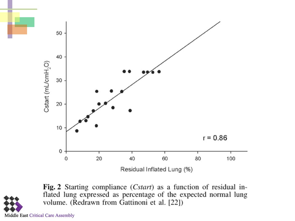
Stiff or Small? ARDS lung is not “stiff” at all, but small
The elasticity of the residual inflated lung is nearly normal, as indicated by: The specific tissue compliance: (compliance/normally aerated tissue) “baby lung” was a healthy anatomical structure, located in the nondependent regions of the original lungs
 baby lung was a healthy anatomical structure, located in the nondependent regions of the original lungs.")
15
Ventilating ARDS with Normal VT
Straining of the “baby lung”

16
Supine Prone Supine

17
Sponge Lung Concept The densities in the dependent lung regions are in fact due not to an increase in the amount of edema but to a loss of alveolar gases, as the result of the compressive gravitational forces, including the heart weight

18
Baby lung at end-inspiration Spectrum of Regional Opening Pressures
Inflated Superimposed Pressure Consolidation Small Airway Collapse 10-20 cmH2O Alveolar Collapse (Reabsorption) 20-60 cmH2O = Lung Units at Risk for Tidal Opening & Closure (from Gattinoni)
 cmH2O. = Lung Units at Risk for Tidal. Opening & Closure. (from Gattinoni)")
19
Baby Lung and VILI Elastic fibers (spring) Collagen fibers (string).
 Collagen fibers (string).")
20
Transpulmonary Pressure

22
Ventilator Induced Lung Injury

23
Recognized Mechanisms of Airspace Injury
Airway Trauma “Stretch” “Shear”

24
Pathways to VILI Moderate Stress/Strain Extreme Stress/Strain Rupture
End-Expiration Extreme Stress/Strain Tidal Forces (Transpulmonary and Microvascular Pressures) Moderate Stress/Strain Rupture Signaling Mechano signaling via integrins, cytoskeleton, ion channels inflammatory cascade Cellular Infiltration and Inflammation Marini / Gattinoni CCM 2004
 Moderate Stress/Strain. Rupture. Signaling. Mechano signaling via. integrins, cytoskeleton, ion channels. inflammatory cascade. Cellular Infiltration and Inflammation. Marini / Gattinoni CCM")
25
Stress distribution homogeneous system
FT min max L. Gattinoni, 2003 Mead J et al. J. Appl. Physiol. 28(5):
:")
26
Stress distribution High Stiffness Zone
min max L. Gattinoni, 2003 Mead J et al. J. Appl. Physiol. 28(5):
:")
27
Ventilator-induced lung injury is initiated by the application of excessive stress
Gattinoni, L. et al. CMAJ 2008;178: Copyright ©2008 Canadian Medical Association or its licensors

28
NEJM 2000;342:

29
NEJM 2000;342:

30
ARDS

31
Cytokines, complement, prostanoids, leukotrienes, O2- Proteases
Pinsp = 40 mbar Volutrauma Atelectrauma PEEP = 5 mbar Cytokines, complement, prostanoids, leukotrienes, O2- Proteases Biotrauma

32
Barotrauma

33
Distal Organ Dysfunction
MV and MODS: A Possible Link Biophysical Injury shear overdistention cyclic stretch D intrathoracic pressure Biochemical Injury (Biotrauma) mf cytokines, complement, PGs, LTs, ROS, proteases bacteria Epithelium/ interstitium neutrophils Distal Organ Dysfunction alveolar-capillary permeability cardiac output organ perfusion ? sFasL DEATH Slutsky, Tremblay Am J Resp Crit Care Med. 1998;157:1721-5
 mf. cytokines, complement, PGs, LTs, ROS, proteases. bacteria. Epithelium/ interstitium. neutrophils. Distal Organ Dysfunction. alveolar-capillary. permeability. cardiac output. organ perfusion. sFasL. DEATH. Slutsky, Tremblay Am J Resp Crit Care Med. 1998;157:")
34
Principles and goals of mechanical ventilation in ards

35
Respiratory Pressure/Volume (P/V) Curve
Healthy subject In normal healthy volunteers, the P/V curve explore the mechanical properties of the respiratory system (lung + chest wall) ARDS RV, Residual volume; FRC, Functional residual capacity; TLC, Total lung capacity; UIP, Upper inflection point; LIP, Lower inflection point. The critical opening pressure above which most of the collapsed units open up and may be recruited - CLIN Compliance of the intermediate, linear segment of the P/V curve Maggiore SS, et al. Eur Respir J Rouby JJ, et al. Eur Respir J
 ARDS. RV, Residual volume; FRC, Functional residual capacity; TLC, Total lung capacity; UIP, Upper inflection point; LIP, Lower inflection point. The critical opening pressure above which most of the collapsed units open up and may be recruited - CLIN Compliance of the intermediate, linear segment of the P/V curve. Maggiore SS, et al. Eur Respir J Rouby JJ, et al. Eur Respir J")
36
Ventilator-induced Lung Injury (VILI)
Upper Deflection point Lower Inflection point

37
Principles and Goals of MV in ARDS
Appropriate oxygenation (PO2 = 55-60) Accept hypercapnea and mild acidosis (pH~ 7.3) Limit distending pressure=limit transpulmonary pressure: Pplateau <28 cm H2O Limit tidal volume: 4-6 ml/Kg Best PEEP: cm H2O
 Accept hypercapnea and mild acidosis (pH~ 7.3) Limit distending pressure=limit transpulmonary pressure: Pplateau <28 cm H2O. Limit tidal volume: 4-6 ml/Kg. Best PEEP: cm H2O.")
38
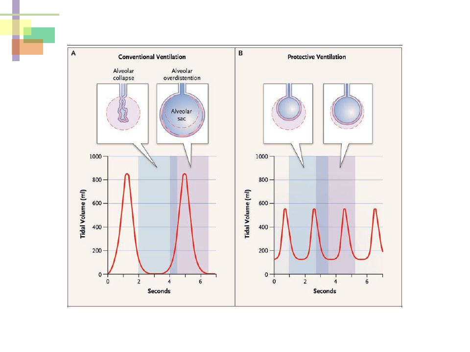
Preventing Overdistention and Under-Recruitment Injury
“Lung Protective” Ventilation 10-16 cm H2O < 28 cm H2O 4-6 mL/kg Add PEEP V O L U M E Limit Distending Pressure Limit VT Transpulmonary Pressure= Airway Pressure-Pleural Pressure Pressure

40
Lung protective ventilatory strategy
CT at end-expiration Pelosi P et al, AJRCCM 2001;164:

41
Lung Protective Ventilator Strategies
volutrauma zone of overdistension V UIP DON’T EVEN THINK OF PARKING HERE zone of derecruitment and atelectasis "safe" window LIP atelectrauma P

42
Intervention Control TV <6 ml/Kg PEEP >PFlex TV (10-12 ml/Kg)
1998 53 patients Intervention Control TV <6 ml/Kg PEEP >PFlex TV (10-12 ml/Kg) Lowest PEEP 28 day mortality Intervention Control 38% 71%
 Lowest PEEP. 28 day mortality. Intervention. Control. 38% 71%")
43
ARMA Trial Intervention Control 31% 40% Intervention Control
28 day mortality Intervention Control 31% 40% 861 patients Intervention Control TV (4-6 ml/Kg) PEEP 8.5 TV (10-12 ml/Kg) PEEP 8.6 861 patients Intervention Control Pplateau <30 Pplateau <50
 PEEP 8.5. TV (10-12 ml/Kg) PEEP patients. Intervention. Control. Pplateau <30. Pplateau <50.")
44
NIH ARDS Network trial NEJM 2000;342:1301
ARDS net mortality Reducing from 12 to 6 ml/kg VT saved lives

45
NIH ARDS Network trial NEJM 2000;342:1301
Low TV High TV P = Mortality 31 40 0.007 Days of free MV 12 10 Days free of organ failure 15 0.006 Reducing from 12 to 6 ml/kg VT saved lives

46
Tradeoffs with 6 ml/kg Crs also better in the HIGH Vt group
Better Oxygenation in the high TV but more death Crs also better in the HIGH Vt group

47
ARDS Network: Improved Survival with Low VT
1.0 0.9 0.8 0.7 0.6 0.5 0.4 0.3 0.2 0.1 0.0 180 160 140 120 100 80 60 40 20 Proportion of Patients Lower tidal volumes Survival Discharge Traditional tidal values The Acute Respiratory Distress Syndrome Network (ARDS Net) conducted a multicenter, randomized trial comparing traditional ventilation treatment (VT = 12 mL/kg, PPlat 50 cm H2O) vs. lower tidal volumes (VT = 6 mL/kg, PPlat 30 cm H2O) in patients with ARDS. The trial was stopped early when the lower tidal volume group experienced significantly lower mortality (P=0.007). As shown on this slide, the proportion of patients who were breathing without assistance by day 28 (65.7% vs 55.0%) was statistically significant (P<0.001) in favor of ventilation with lower tidal volumes. The Acute Respiratory Distress Syndrome Network. Ventilation with lower tidal volumes as compared with traditional tidal volumes for acute lung injury and the acute respiratory distress syndrome. N Engl J Med 2000;342: Survival Discharge Days after Randomization ARDS Network. N Engl J Med
 conducted a multicenter, randomized trial comparing traditional ventilation treatment (VT = 12 mL/kg, PPlat 50 cm H2O) vs. lower tidal volumes (VT = 6 mL/kg, PPlat 30 cm H2O) in patients with ARDS. The trial was stopped early when the lower tidal volume group experienced significantly lower mortality (P=0.007). As shown on this slide, the proportion of patients who were breathing without assistance by day 28 (65.7% vs 55.0%) was statistically significant (P<0.001) in favor of ventilation with lower tidal volumes. The Acute Respiratory Distress Syndrome Network. Ventilation with lower tidal volumes as compared with traditional tidal volumes for acute lung injury and the acute respiratory distress syndrome. N Engl J Med 2000;342: Survival. Discharge. Days after Randomization. ARDS Network. N Engl J Med")
48
Randomized Trials of MV in ARDS
1996-9 1990s

49
10-16 20-26 25-32 29-38

51
Tidal hyperinflation during Low TV ventilation in ARDS
30 patients with ARDS Ventilatory strategy (ARMA protocol) 6 ml/Kg IBW BAL ► cytokine measurements CT scan on mechanical ventilation Hyperinflated Normally aerated Poorly aerated Non-aerated
 6 ml/Kg IBW. BAL ► cytokine measurements. CT scan on mechanical ventilation. Hyperinflated. Normally aerated. Poorly aerated. Non-aerated.")
52
Tidal hyperinflation during low TV ventilation in ARDS
Hyperinflated Normally aerated Poorly aerated Non-aerated Less protected More protected

53
Despite the use of protective ventilatory strategy (6 ml/Kg) …..
30 % of patients hyperinflated Plateau Pressure: Protected (25.5 0.5) vs. unprotected (28.9 0.9) Higher inflammatory cytokines in unprotected Number of ventilator-free days: Protected (7 8) vs. unprotected (1 2) Mortality: Protected (30%) vs. unprotected (40%) Limit plateau pressure to < 28 “Small baby lung”: the ARDS network protective ventilatory strategy does not fully protect the lungs from VILI because hyperinflation of the small normal lung may occur despite lowering TV to 6 ml/Kg and limiting plateau pressure to < 30
 vs. unprotected (28.9 0.9) Higher inflammatory cytokines in unprotected. Number of ventilator-free days: Protected (7 8) vs. unprotected (1 2) Mortality: Protected (30%) vs. unprotected (40%) Limit plateau pressure to < 28. Small baby lung : the ARDS network protective ventilatory strategy does not fully protect the lungs from VILI because hyperinflation of the small normal lung may occur despite lowering TV to 6 ml/Kg and limiting plateau pressure to < 30.")
54
Neuromuscular Blockers in Early Acute Respiratory Distress Syndrome
340 patients Cisatracurium besylate Placebo # of Patients 178 162 TV 6-8 ml/Kg PEEP > 5 90 day mortality 31.6% 40.7% p= 0.08 Figure 2 Probability of Survival through Day 90, According to Study Group. Papazian L et al. N Engl J Med 2010;363:

55
Neuromuscular Blockers in Early Acute Respiratory Distress Syndrome
Figure 2 Probability of Survival through Day 90, According to Study Group. Hazard Ratio: 0.68 (95% confidence interval [CI], 0.48 to 0.98; P = 0.04) Papazian L et al. N Engl J Med 2010;363:
 Papazian L et al. N Engl J Med 2010;363:")
56
Possible Mechanisms by Which Neuromuscular Blocking Agents Might Lead to improved Survival
Figure 1. Possible Mechanisms by Which Neuromuscular Blocking Agents (NMBAs) Might Lead to Improved Survival in Patients with the Acute Respiratory Distress Syndrome (ARDS). Respiratory physiological features of a patient with ARDS are illustrated before (top) and after (bottom) paralysis induced with the use of NMBAs. Before paralysis, increased respiratory drive from multiple causes can lead to increased tidal volumes, active exhalation, and patient–ventilator asynchrony, all of which can potentially worsen various forms of ventilator-induced lung injury. In addition, muscle activation may divert blood flow away from vital organs and lead to a lower mixed venous partial pressure of oxygen (PO2). These mechanisms may lead to increased organ dysfunction and ultimately death. After paralysis, the administered NMBAs prevent patient-initiated generation of high and low lung volumes and also prevent active expiration, allowing for better lung-protective ventilation and less ventilator-induced lung injury. Ventilator-induced lung injury may also be lessened by less pulmonary blood flow due to decreased oxygen consumption. NMBAs may also indirectly improve arterial oxygenation by decreasing blood flow to active muscle groups (because of decreased oxygen requirements) and by improving the distribution of ventilation relative to perfusion (V̇/̇Q). (Arterial PO 2 may also be decreased through this mechanism if V̇/̇Q is worsened.) Finally, NMBAs may have a direct antiinflammatory effect. The relative effect of NMBAs on many of these mechanisms depends on the state of muscle activation before paralysis, which is dependent on a number of factors, including the patient's level of sedation.
 Might Lead to Improved Survival in Patients with the Acute Respiratory Distress Syndrome (ARDS). Respiratory physiological features of a patient with ARDS are illustrated before (top) and after (bottom) paralysis induced with the use of NMBAs. Before paralysis, increased respiratory drive from multiple causes can lead to increased tidal volumes, active exhalation, and patient–ventilator asynchrony, all of which can potentially worsen various forms of ventilator-induced lung injury. In addition, muscle activation may divert blood flow away from vital organs and lead to a lower mixed venous partial pressure of oxygen (PO2). These mechanisms may lead to increased organ dysfunction and ultimately death. After paralysis, the administered NMBAs prevent patient-initiated generation of high and low lung volumes and also prevent active expiration, allowing for better lung-protective ventilation and less ventilator-induced lung injury. Ventilator-induced lung injury may also be lessened by less pulmonary blood flow due to decreased oxygen consumption. NMBAs may also indirectly improve arterial oxygenation by decreasing blood flow to active muscle groups (because of decreased oxygen requirements) and by improving the distribution of ventilation relative to perfusion (V̇/̇Q). (Arterial PO 2 may also be decreased through this mechanism if V̇/̇Q is worsened.) Finally, NMBAs may have a direct antiinflammatory effect. The relative effect of NMBAs on many of these mechanisms depends on the state of muscle activation before paralysis, which is dependent on a number of factors, including the patient s level of sedation.")
57
Possible Mechanisms by Which Neuromuscular Blocking Agents Might Lead to improved Survival
Figure 1. Possible Mechanisms by Which Neuromuscular Blocking Agents (NMBAs) Might Lead to Improved Survival in Patients with the Acute Respiratory Distress Syndrome (ARDS). Respiratory physiological features of a patient with ARDS are illustrated before (top) and after (bottom) paralysis induced with the use of NMBAs. Before paralysis, increased respiratory drive from multiple causes can lead to increased tidal volumes, active exhalation, and patient–ventilator asynchrony, all of which can potentially worsen various forms of ventilator-induced lung injury. In addition, muscle activation may divert blood flow away from vital organs and lead to a lower mixed venous partial pressure of oxygen (PO2). These mechanisms may lead to increased organ dysfunction and ultimately death. After paralysis, the administered NMBAs prevent patient-initiated generation of high and low lung volumes and also prevent active expiration, allowing for better lung-protective ventilation and less ventilator-induced lung injury. Ventilator-induced lung injury may also be lessened by less pulmonary blood flow due to decreased oxygen consumption. NMBAs may also indirectly improve arterial oxygenation by decreasing blood flow to active muscle groups (because of decreased oxygen requirements) and by improving the distribution of ventilation relative to perfusion (V̇/̇Q). (Arterial PO 2 may also be decreased through this mechanism if V̇/̇Q is worsened.) Finally, NMBAs may have a direct antiinflammatory effect. The relative effect of NMBAs on many of these mechanisms depends on the state of muscle activation before paralysis, which is dependent on a number of factors, including the patient's level of sedation.
 Might Lead to Improved Survival in Patients with the Acute Respiratory Distress Syndrome (ARDS). Respiratory physiological features of a patient with ARDS are illustrated before (top) and after (bottom) paralysis induced with the use of NMBAs. Before paralysis, increased respiratory drive from multiple causes can lead to increased tidal volumes, active exhalation, and patient–ventilator asynchrony, all of which can potentially worsen various forms of ventilator-induced lung injury. In addition, muscle activation may divert blood flow away from vital organs and lead to a lower mixed venous partial pressure of oxygen (PO2). These mechanisms may lead to increased organ dysfunction and ultimately death. After paralysis, the administered NMBAs prevent patient-initiated generation of high and low lung volumes and also prevent active expiration, allowing for better lung-protective ventilation and less ventilator-induced lung injury. Ventilator-induced lung injury may also be lessened by less pulmonary blood flow due to decreased oxygen consumption. NMBAs may also indirectly improve arterial oxygenation by decreasing blood flow to active muscle groups (because of decreased oxygen requirements) and by improving the distribution of ventilation relative to perfusion (V̇/̇Q). (Arterial PO 2 may also be decreased through this mechanism if V̇/̇Q is worsened.) Finally, NMBAs may have a direct antiinflammatory effect. The relative effect of NMBAs on many of these mechanisms depends on the state of muscle activation before paralysis, which is dependent on a number of factors, including the patient s level of sedation.")
58
volutrauma V atelectrauma P UIP "safe" window LIP Optimal PEEP
zone of derecruitment and atelectasis volutrauma zone of overdistension UIP "safe" window V Optimal PEEP atelectrauma LIP P

59
The PEEP Effect NEJM 2006;354:1839-1841
taken at the end of inspiration. A=0, B=15, C=15 x3, D= 15*5 F=0*1, G = 0*3, H =0*5 NEJM 2006;354:

60
Higher vs. Lower PEEP Overinflated Recruitment

61
Positive End-Expiratory Pressure Setting in Adults With Acute Lung Injury and Acute Respiratory Distress Syndrome (EXPRESS) Ventilation Strategy Using Low Tidal Volumes, Recruitment Maneuvers, and High Positive End-Expiratory Pressure for Acute Lung Injury and Acute Respiratory Distress Syndrome “LOVS” ALVEOLI

62
Intervention Control TV <6 ml/Kg PEEP >PFlex TV (10-12 ml/Kg)
1998 53 patients Intervention Control TV <6 ml/Kg PEEP >PFlex TV (10-12 ml/Kg) Lowest PEEP 28 day mortality Intervention Control 38% 71%
 Lowest PEEP. 28 day mortality. Intervention. Control. 38% 71%")
63
Intervention Control TV 5-8 ml/Kg 9-11 ml/Kg PEEP Pflex + 2 cm H2O
50 patients 53 Patients Intervention Control TV 5-8 ml/Kg 9-11 ml/Kg PEEP Pflex + 2 cm H2O >5 cm H2O ICU Mortality 32% 53% P= 0.04 Crit Care Med l 1311

64
Positive End-Expiratory Pressure Setting in Adults With Acute Lung Injury and Acute Respiratory Distress Syndrome (EXPRESS) 385 patients 382 Patients Intervention Control TV 6 ml/Kg PEEP Plateau 28-30 16±3 cm H2O 5-9 cm H2O ICU Mortality NS Mercat A, Richard JM, Vielle B, et al. (EXPRESS). JAMA. 2008;299:
. JAMA. 2008;299:")
65
ALVEOLI 549 patients Low PEEP High PEEP TV 6 ml/Kg PEEP 8.3 ± 3.2
13.2 ± 3.5 ICU Mortality 24.9% 27.5% NS N E J Med : 327

66
Ventilation Strategy Using Low Tidal Volumes, Recruitment Maneuvers, and High Positive End-Expiratory Pressure for Acute Lung Injury and Acute Respiratory Distress Syndrome LOVS 475 patients 508 Patients Intervention Control TV 6 ml/Kg PEEP Pplat < 40 PEEP 20 cm H2O RM Pplat < 30 Low PEEP ICU Mortality 36.4% 40.4% NS Meade et al JAMA. 2008;299(6):
:")
67
PEEP in ARDS Bad Good 67

68
JAMA, March 3, 2010—Vol 303, No. 9

69
Time to Unassisted Breathing for Higher and Lower Positive End-Expiratory Pressure (PEEP) Stratified by Presence of Acute Respiratory Distress Syndrome (ARDS) at Baseline
 Stratified by Presence of Acute Respiratory Distress Syndrome (ARDS) at Baseline")
70
Time to Death in Hospital for Higher and Lower Positive End-Expiratory Pressure (PEEP) Stratified by Presence of Acute Respiratory Distress Syndrome (ARDS) at Baseline
 Stratified by Presence of Acute Respiratory Distress Syndrome (ARDS) at Baseline")
71
Optimal PEEP

72
Principle for FiO2 and PEEP Adjustment
PEEP Table by ARDSNet ARDS Network, 2000: Multicenter, randomized 861 patients Principle for FiO2 and PEEP Adjustment FiO2 0.3 0.4 0.5 0.6 0.7 0.8 0.9 1.0 PEEP 5 5-8 8-10 10 10-14 14 14-18 18-24 用的是預期體重 NEJM 2000; 342:

73
PV Curve Best PEEP Rotta, J Pediatr (Rio J0 2003:79(Suppl 2):S149
:S149")
74
Issues with PV Curves Require sedation/paralysis
Difficult to identify “inflection points” May require esophageal pressure to separate lung from chest wall effects Pressure volume curves of individual lung units are not known

75
Optimal PEEP by Compliance
15 normovolemic patients requiring MV for ARF PEEP resulting in maximum oxygen transport and the lowest dead space fraction resulted in highest compliance Optimal PEEP varied from 0- to 15 cm H2O Mixed venous PO2 increased from 0 PEEP to the PEEP resulting in maximum oxygen transport, but then decreased at higher PEEP Conclusion: compliance may be used to indicated the PEEP likely to result in optimum cardiopulmonary function Suter, N Eng J Med 1975:292:284

76
Concerns when using lung-protective strategy…
Heterogeneous distribution Hypercapnia Auto-PEEP Sedation and paralysis Patient-ventilator dyssynchrony Increased intrathoracic pressure Maintenance of PEEP

77
Permissive Hypercapnia
Low Vt (6ml/kg) to prevent over-distention Increase respiratory rate to avoid very high level of hypercapnia If Respiratory rate can’t be increased further then the PaCO2 allowed to rise Accept PH > 7.25 Usually well tolerated May be beneficial (shift oxygen dissociation curve to the right) May use bicarb infusion till the kidney is able to retain bicarb
 to prevent over-distention. Increase respiratory rate to avoid very high level of hypercapnia. If Respiratory rate can’t be increased further then the PaCO2 allowed to rise. Accept PH > Usually well tolerated. May be beneficial (shift oxygen dissociation curve to the right) May use bicarb infusion till the kidney is able to retain bicarb.")
78
Permissive Hypercapnia – When would you NOT do it?
Renal failure High intracranial pressures Cardiovascular problems

79
“Lung Protective” Ventilation
Conclusion “Lung Protective” Ventilation 10-16 cm H2O < 28 cm H2O 4-6 mL/kg Add PEEP V O L U M E Limit Distending Pressure Limit VT Pressure

80
Management of refractory hypoxemia
PEEP Pee (diuresis) Prone Paralysis Pleural evacuation (pleural effusion drainage) Prostacyclin (or iNO) More PEEP/recruitment maneuvers
 Prone. Paralysis. Pleural evacuation (pleural effusion drainage) Prostacyclin (or iNO) More PEEP/recruitment maneuvers.")
81
What next? Prone position Inhaled nitric oxide
High-frequency oscillation ECMO

82
Other Ventilator Strategies
Lung recruitment maneuvers Prone positioning High-frequency oscillatory ventilation (HFOV) ECMO
 ECMO.")
83
Recruitment Maneuvers
Application of high airway pressure (35-40cmH2O) for approximately 40 seconds. Most common methodology 40 cm H2O CPAP 40 seconds Employed to open atelectatic alveolar units that occur with ARDS and particularly with any disconnection from ventilator If successful, PaO2 will increase by 20% or more. Must use PEEP after procedure to keep recruited alveoli open.
 for approximately 40 seconds. Most common methodology. 40 cm H2O CPAP. 40 seconds. Employed to open atelectatic alveolar units that occur with ARDS and particularly with any disconnection from ventilator. If successful, PaO2 will increase by 20% or more. Must use PEEP after procedure to keep recruited alveoli open.")
84
Effects of Recruitment Maneuvers to Promote Homogeneity within the Lung
Malhotra A. N Engl J Med 2007;357:

85
Lung Recruitment To open the collapsed alveoli
A sustained inflation of the lungs to higher airway pressure and volumes Ex.: PCV, Pi = 45 cmH2O, PEEP = 5 cmH2O, RR = 10 /min, I : E = 1:1, for 2 minutes 高低的分界是9% NEJM 2006; 354:

86
ARDSnet protocol Vs open lung protocol
Tv 6 ml/kg Plateau pressure <30 Conventional PEEP (titrate for FIO2 <0.6) Experimental protocol Tv 6 ml/kg Plateau pressure <40 Recruitment maneuvers High PEEP (10-15) JAMA, February 2008
 Experimental protocol. Tv 6 ml/kg. Plateau pressure <40. Recruitment maneuvers. High PEEP (10-15) JAMA, February")
87
ARDSnet protocol Vs open lung protocol
JAMA, February 2008

88
Lung Recruitment 不論哪一組,都大約有24%的肺無法打開 NEJM 2006; 354:

89
Lung Recruitment NEJM 2006; 354:

90
Lung Recruitment Potentially recruitable (PEEP 5 15 cmH2O)
Increase in PaO2:FiO2 Decrease in PaCO2 Increase in compliance The effect of PEEP correlates with the percentage of potentially recruitalbe lung The percentage of recruitable lung correlates with the overall severity of lung injury Sensitivity : 71% Specificity : 59% NEJM 2007; 354:

91
Lung Recruitment The percentage of potentially recruitable lung:
Extremely variable, Strongly associated with the response to PEEP Not routinely recommended

92
Prone Position

93
Prone Position Mechanisms to improve oxygenation:
Increase in end-expiratory lung volume Better ventilation-perfusion matching More efficient drainage of secretions

94
Prone Position Improved gas exchange More uniform alveolar ventilation
Recruitment of atelectasis in dorsal regions Improved postural drainage Redistribution of perfusion away from edematous, dependent regions

95
Prone Positioning

96
Prone Position NEJM 2001;345:

97
Prone Position NEJM 2001;345:

98
Prone Position Improve oxygenation in about 2/3 of all treated patients No improvement on survival, time on ventilation, or time in ICU Might be useful to treat refractory hypoxemia Optimum timing or duration ? Routine use is not recommended

99
High-Frequency Oscillatory Ventilation (HFOV)
")
100
HFV - the “ultimate” lung protective strategy?
Over-distended Protected Under-recruit

101
Frequency: 180-600 breaths/min (3-10Hz)
HFOV Frequency: breaths/min (3-10Hz)
")
102
Effect of HFOV on gas exchange in ARDS patients
AJRCCM 2002; 166:801-8

103
Survival difference of ARDS patients treated with HFOV or CMV
30-day: P=0.057 90-day: P=0.078 AJRCCM 2002; 166:801-8

104
HFOV Complications: Failed to show a mortality benefit
Recognition of a pneumothorax Desiccation of secretions Sedation and paralysis Lack of expiratory filter Failed to show a mortality benefit Combination with other interventions ? Chest 2007; 131:

105
Acute Lung injury Decreased lung compliance results in high airway pressures Low tidal volume 6-8 ml/kg ideal body weight Maintain IPP 30 cm H2O PEEP to improve oxygenation

106
Conclusions The only treatment that shows mortality benefit:
lung-protective ventilation strategy Low tidal volume (6ml/Kg), high PEEP, adequate Pplat (<30 cmH2O) Modalities to improve oxygenation: Prone position, steroid, fluid treatment, steroid, HFOV, NO Combining other treatments: Antibiotics, EGDT…etc
, high PEEP, adequate Pplat (<30 cmH2O) Modalities to improve oxygenation: Prone position, steroid, fluid treatment, steroid, HFOV, NO. Combining other treatments: Antibiotics, EGDT…etc.")
107
Be Inspired The University of Michigan

Similar presentations