Download presentation
Presentation is loading. Please wait.

1
Cardiac CT Shiva Roy FRACP 2008

2
Why change current practise?
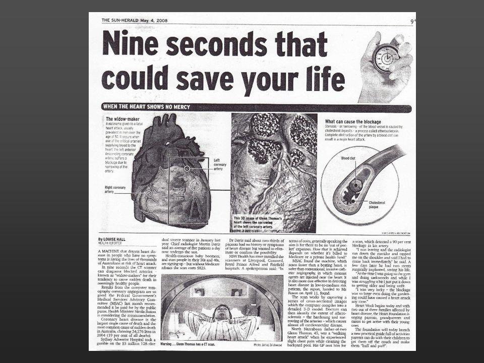
Poor at predicting cardiac events 50% of first cardiac events are MI. 50% events occur in low to mod risk patients >50% patients with MI “average” lipids Functional testing inaccurate “So I’m going to live?” Expensive business! Hospital admission / angio $ Perfusion scan $1000 Cardiologist / stress echo $500 Functional testing: 70-80% sensitivity and specificity in best hands. Up to 2% annual incidence of events in trial patients with negative stress echo’s – 20% 10 yr incidence i.e. high risk (we reassure!) Expensive: 1 Cardiac death in Eastern Suburbs - Millions of lost revenue in taxes, millions in insurance payouts
 Expensive: 1 Cardiac death in Eastern Suburbs - Millions of lost revenue in taxes, millions in insurance payouts.")
3
Lipid Management Frequently uncertain who to treat
NCEP supports statins in high risk (>20% 10 yr) Moderate risk (10-20% 10yr) group challenging Akosah et al: Young pts mean age 50 presenting MI 70% in lower risk category and statin ineligible Early plaque detection / lipid lowering therapy What about the asymptomatic 30 yr old smoker with a family history, LDL of 3.6 and HDL of1.2? PBS guidelines do not take total risk into account NCEP Surely the early detection of plaque and commencement of lipid lowering therapy would have prevented some events. Clearly we need more sensitive screening to detect early plaque formation facilitating early introduction of lipid lowering therapy in at risk people. Coronary calcium has been identified as a way of defining a CAD equivalent.
 Moderate risk (10-20% 10yr) group challenging. Akosah et al: Young pts mean age 50 presenting MI. 70% in lower risk category and statin ineligible. Early plaque detection / lipid lowering therapy. What about the asymptomatic 30 yr old smoker with a family history, LDL of 3.6 and HDL of1.2 PBS guidelines do not take total risk into account. NCEP. Surely the early detection of plaque and commencement of lipid lowering therapy would have prevented some events. Clearly we need more sensitive screening to detect early plaque formation facilitating early introduction of lipid lowering therapy in at risk people. Coronary calcium has been identified as a way of defining a CAD equivalent.")
4
Coronary Calcification
Misguided bias against technique Proven robust technique in identifying at risk population Coronary Calcium Score >100 or >75th pecentile identifies a CAD equivalent Technique discredited early on, levelled at the entrepreneurial approach of it’s introduction rather than on any scientific basis and in fact it has proven to be a very robust technique in identifying at risk population but not an at risk individual. N.B. – CTA not discredited for the same reason. Coronary Calcium scores are given in all patients undergoing CTA except graft patients and some stent patients (not necessary, have diagnosed obstructive disease)
")
6
New Guidelines From AHA

7
AHA – Circulation 2005 Given the evolving literature since the last ACC/AHA Expert Consensus statement (2000), current data indicate that CAD risk stratification is possible with CAC measures. Specifically, low CAC scores are associated with a low adverse event risk, and high CAC scores are associated with a worse event-free survival. This recommendation to measure atherosclerosis burden, in clinically selected intermediate–CAD risk patients (eg, those with a 10% to 20% Framingham 10-year risk estimate) to refine clinical risk prediction and to select patients for altered targets for lipid-lowering therapies.
, current data indicate that CAD risk stratification is possible with CAC measures. Specifically, low CAC scores are associated with a low adverse event risk, and high CAC scores are associated with a worse event-free survival. This recommendation to measure atherosclerosis burden, in clinically selected intermediate–CAD risk patients (eg, those with a 10% to 20% Framingham 10-year risk estimate) to refine clinical risk prediction and to select patients for altered targets for lipid-lowering therapies.")
8
Original Article Coronary Calcium as a Predictor of Coronary Events in Four Racial or Ethnic Groups
Robert Detrano, M.D., Ph.D., Alan D. Guerci, M.D., J. Jeffrey Carr, M.D., M.S.C.E., Diane E. Bild, M.D., M.P.H., Gregory Burke, M.D., Ph.D., Aaron R. Folsom, M.D., Kiang Liu, Ph.D., Steven Shea, M.D., Moyses Szklo, M.D., Dr.P.H., David A. Bluemke, M.D., Ph.D., Daniel H. O'Leary, M.D., Russell Tracy, Ph.D., Karol Watson, M.D., Ph.D., Nathan D. Wong, Ph.D., and Richard A. Kronmal, Ph.D. N Engl J Med Volume 358(13): March 27, 2008
: March 27,")
9
Conclusion The coronary calcium score is a strong predictor of incident coronary heart disease and provides predictive information beyond that provided by standard risk factors in four major racial and ethnic groups in the United States. No major differences among racial and ethnic groups in the predictive value of calcium scores were detected

10
Introduction to Coronary CTA
Imaging technique accounting for cardiac motion through ECG gating Early 1980’s conventional CT 1987 EBCT 1999 Multi Detector CT Accelerated progression in imaging capability over past decade will continue into forseeable future Diagnostic capability has at times preceded the critical evaluation of clinical application Successive generations of CT scanners have been applied to Cardiac imaging This has resulted in Accelerated…. As a result the Diagnostic…..

11
Coronary Motion

12
Technology Cardiac motion – Translational, Rotational and Accordian-type movements Selective coronary angiography gold standard Whole heart covered, real time imaging Temporal resolution of 10msec Spatial resolution 100um But Lumen only Limited angles No cross sectional reconstruction The problem with the heart is that it moves through multiple planes and we need to account for translational, rotational and accordian-type movements in attemting to create a still picture we can analyse. Presently, selective coronary angiography is the gold standard in assessing coronary artery patency. The whole heart is covered throughout the imaging cycle, the Temporal resolution is 10msec which means a still image is created every .01sec. Spatial resolution is extremely high with flat plane imaging and can discriminate .01 of a mm. We need to benchmark CT coronary angiography against this. To obtain a still frame we need a Detector array covering the whole heart – around 14cm, spatial resolution of 0.3mm which is about the threshold of clinical relevance and temporal resolution of msec which is sufficient to suppress all cardiac motion. We’re not quite there yet but we’re not far off

13
Technology 64 slice CT pivotal technology “Can Do”
Spatial resolution 0.35 mm – “isotropic” Slice, dice, any angle, cross sectional analysis Temporal resolution msec Detector array 4 cm wide Infinite Grey scale, image vessel wall, characterise plaque “Can Do” Sensitivity and Specificity ~95% c.f. invasive angiography, ~5% segments unevaluable 64 slice CT is the pivotal technology that has brought CT coronary angiography to clinical relevance ..Spatial resolution of 0.35mm is probably at the threshold of clinical relevance and allows for isotropic”imaging. This means that the cube of information has identical X, Y and Z axes and therefor appears the same from any projection ..Temporal resolution of 65 to 200 msec depending on whether half segment or multisegment reconstruction ..Detector array 4 cm wide so the whole heart is covered in four rotations which takes 1.4cm. Practically each segment is scanned 2, 3 or 4 times so scan time on the GE machine is 6 seconds. Siemens detector array is about half the width so the scan time is twice as long….. Don’t believe everything you here.

14
Helical Scanning Helical scanning involves continuous x-ray exposure and table movement to acquire the most image data in the shortest time.

15
Snapshot Pulse is most dose efficient
At Z location, waiting for desired heart phase NO XRAYS Z location Time

16
METHODS - CTA 0.5-0.625 mm slices Single Breath-hold Imaging
80 cc Non-ionic (IODINATED) contrast Aggressive B blockade
 contrast. Aggressive B blockade.")
17
Normal Study

18
Accuracy of Noninvasive CT Angiography: Trial exclusions
Technically inadequate scans not included in analysis Patient exclusion criteria Rapid heart rates Irregular heart beat/arrhythmias Renal dysfunction Contrast Allergies Beta-blocker intolerant Obesity limits interpretation

19
Diagnostic accuracy of CTA
Analysis Sensitivity (%) Specificity (%) PPV (%) NPV (%) Stenoses > 50%, per patient 93 82 62 97 Stenoses > 50%, per vessel 84 91 51 98 Stenoses > 70%, per patient 49 Stenoses > 70%, per vessel 85 92 33 99 ACCURACY study. 232 pts. Chest pain syndromes. Low ppv due to low prevalence of disease in this gp. PPV=positive predictive value NPV=negative predictive value Min J. Radiological Society of North America 2007; November 25-30, 2007; Chicago, IL.
 Specificity (%) PPV (%) NPV (%) Stenoses > 50%, per patient Stenoses > 50%, per vessel Stenoses > 70%, per patient. 49. Stenoses > 70%, per vessel ACCURACY study. 232 pts. Chest pain syndromes. Low ppv due to low prevalence of disease in this gp. PPV=positive predictive value. NPV=negative predictive value. Min J. Radiological Society of North America 2007; November 25-30, 2007; Chicago, IL.")
20
Radiation Dose with CT EBCT – calcium scan – 0.7 mSv
MSCT – Calcium scan – 1.2 mSv MSCT – Angiogram – 9-18 mSv Dose Modulation – up to 47% savings Coronary Angiogram – mSv Nuclear Imaging – 6-15 mSv

21
Case 1 - 43 male 43 year old man commenced a new exercise program
Left side chest discomfort on exertion Cholesterol 6.0, LDL 3.6, HDL 1.3 No smoking, diabetes, HT or family history of IHD BMI 26 kg/m2 Medications – nil Resting ECG – normal What next ? CIA Mar 08

22
Functional Test Objectively negative stress echocardiogram – 13 minutes However, vague left sided chest pain at peak exercise “Is my heart OK ?” CIA Mar 08

23
LAD CIA Mar 08

24
Case 2 48 yr old man Consistent exertional bilateral arm tightness
“like the compression of a blood pressure cuff” Chol 7.8, LDL 5.1. Father and brother IHD in their 50s. On no medical therapy at time of presentation Negative Stress Echo after 12 minutes of Bruce protocol. No symptoms with stress test Worrying symptoms and CV risk factors, but negative functional test

25
Volume rendered image of Coronary CT
Severe LAD and Diagonal branch stenosis

26
Outcome This patient had a concerning history and risk factor profile. He declined the offer of an invasive angiogram given his negative stress test. He agreed to have a CT coronary angiogram which detected severe proximal LAD disease which required revascularisation.

27
Case 3 37 yr old pastry chef- referred Aug 05
Sudden death of 40 yr old first cousin Background SVT 3 episodes over last 15 years Ex smoker 1year (since age 20) FH of IHD father CABG at 63 Chol 5.4, LDL 3.4, HDL 1.2, TG 1.9 Chol/HDL ratio 4.5 Overweight at 100kg (BMI 33)
 FH of IHD father CABG at 63. Chol 5.4, LDL 3.4, HDL 1.2, TG 1.9. Chol/HDL ratio 4.5. Overweight at 100kg (BMI 33)")
28
Assessment SR 86bpm BP 130/90 ECG ECHO
Exercise stress test (echo assisted) 11.5 mins of Bruce protocol Normal haemodynamic response Limited by fatigue no CP No ECG or ECHO evidence of ischaemia
 11.5 mins of Bruce protocol. Normal haemodynamic response. Limited by fatigue no CP. No ECG or ECHO evidence of ischaemia.")
29
Management Lifestyle changes Stay off cigarettes weight loss
dietary and exercise advice Further investigation ? (asymptomatic) novel risk factors Hs CRP, Lp(a), homocysteine GTT ambulatory BP monitor ? Sleep study Pharmacological intervention ?
 novel risk factors Hs CRP, Lp(a), homocysteine. GTT. ambulatory BP monitor. Sleep study. Pharmacological intervention")
30
Cardiovascular Risk Assessment
Assessment of 5-10 year risk of coronary event Framingham risk score (10 year risk) NZ risk calculator ( 5 year risk) Pharmacological intervention if risk >2%/yr or 20%/10yrs
 NZ risk calculator ( 5 year risk) Pharmacological intervention if risk. >2%/yr or 20%/10yrs.")
31
- 4 +7 +8 (assume still smoking) +1 13 (12%/10yrs)
 (12%/10yrs)")
32
Management Aspirin…recommended when 10yr risk> 10%
per 1000 people treated per year prevent 14 AMI at expense of 4 bleeds Statins.. NHF Risk > 20%/10yrs PBS subsidy ineligible

33
NHF Guidelines Known CHD, PVD or CVD AAA DM CRF
Familial Hyperlipidaemia Absolute risk of >2%/yr

34
NHF Guidelines Increased absolute risk: LDL > 4.0 or Chol > 6.0
+ any 2 or more risk factors HDL< 1.0 FH HT Overweight Smoking IGT Microalbuminuria Age> 45

36
64 slice coronary CT angiography
Referred by GP June Calcium score 360

37
CT + Catheter Angiogram correlation

38
CT – Cardiac Applications
Coronary Calcification (CAS) Non-invasive Coronary Angiography Aortic Assessment (anuerysm, dissection) Pulmonary Embolism Pericardial disease Congenital heart disease Cardiac thrombi & tumor Quantification cardiac anatomy & volumes, global & regional function Venous Anatomy – Pulmonary and Coronary veins pre-procedure
 Non-invasive Coronary Angiography. Aortic Assessment (anuerysm, dissection) Pulmonary Embolism. Pericardial disease. Congenital heart disease. Cardiac thrombi & tumor. Quantification cardiac anatomy & volumes, global & regional function. Venous Anatomy – Pulmonary and Coronary veins pre-procedure.")
40
Open Bypass Grafts 40

41
Coronary aneurysms

42
Apical Thrombus and Infarction

43
Left Atrial Appendage Thrombus
Another interesting finding: Filling defect/Thrombus in the left atrial appendage. (low flow is the cause of thrombis- AF patients) ? The function of the Appendages (vestigial- left over – evolution) Concern is that a part may break off and travel north and cause the patient to stroke
 The function of the Appendages. (vestigial- left over – evolution) Concern is that a part may break off and travel north and cause the patient to stroke.")
44
ASD

45
Patent Ductus Arteriosus
Ao

46
Pericardial Thickening

47
Pulmonary Veins

48
Placement of LV Lead

49
Appropriateness criteria for Cardiac CT CCT/CMR writing group JACC 2006:48(7):1-21
Appropriate/uncertain/inappropriate indications for cardiac CT based on symptom status/ECG/biomarkers/ability to exercise/pre-test risk profile Pre PV isolation/BiV pacing Anomalous coronary anatomy/pericardial disease/cardiac masses/cardiomyopathy/non-coronary cardiac surgery Possible Pulmonary embolus/aortic dissection

50
Triple Rule In

51
In the ER? High negative predictive value, therefore CT may help avoid unnecessary hospital admissions, however… Patient preparation 24hr scan workup usually not logistically feasible Scanner availability Coronary physiology and other investiagations(ECG and biomarkers) well validated for prognosis All coronary segments may not be visible Apparent non-flow limiting lesion potentially unstable “Triple rule out” –high radiation and contrast doses High volume centre usually provides high quality service
 well validated for prognosis. All coronary segments may not be visible. Apparent non-flow limiting lesion potentially unstable. Triple rule out –high radiation and contrast doses. High volume centre usually provides high quality service.")
53
Potential use of CT Coronary Angiography
Intermediate Risk Low risk High Risk Asymptomatic?/atypical pain No Investigation or Functional testing (Stress ECG/Echo/Nuclear) Suspicious pain/Neg FT Atypical pain/Pos FT Poorly compliant with Lifestyle or med Rx angina, ECG +ve Troponin +ve, functional test +ve MSCT Mild atheroma Moderate atheroma Severe atheroma Functional test Medical therapy No ischaemia Ischaemia Medical therapy Angio / revascularisation Risk as calculated by conventional vascular risk factors: Low<10%, intermediate 10-20%, High>20%
 Suspicious pain/Neg FT. Atypical pain/Pos FT. Poorly compliant with. Lifestyle or med Rx. angina, ECG +ve. Troponin +ve, functional test +ve. MSCT. Mild atheroma. Moderate atheroma. Severe atheroma. Functional test. Medical therapy. No ischaemia. Ischaemia. Medical therapy. Angio / revascularisation. Risk as calculated by conventional vascular risk factors: Low<10%, intermediate 10-20%, High>20%")
Similar presentations


 Detection and Treatment of Asymptomatic Atherosclerosis for Primary Prevention.>")


 SHAPE Guidelines Prevention of Fatal Cardiovascular Events (Heart.>")
