Download presentation
Presentation is loading. Please wait.

1
Coronary MDCTA Applications
Thomas H. Hauser MD, MMSc, MPH, FACC Director of Nuclear Cardiology Beth Israel Deaconess Medical Center Assistant Professor of Medicine Harvard Medical School Boston, MA Good afternoon. I thank the organizers and sponsors of this meeting for kindly inviting me to give this presentation.

2
Outline Possible indications for coronary MDCTA
How to approach a coronary MDCTA study

3
Outline Possible indications for coronary MDCTA
How to approach a coronary MDCTA study

4
Possible Indications for Cardiac CT
Coronary artery CAD/Plaque Stents Grafts Anomalous coronaries Ventricular size and function Valve imaging Myocardial perfusion Infarct imaging Cardiac vein imaging Congenital heart disease Cardiac masses Cardiomyopathy Pulmonary vein imaging

5
Accuracy investigators, RSNA 2007
Detection of CAD Accuracy investigators, RSNA 2007

6
Clinical Evaluation of Coronary CTA

7
Multi-Center Trial: 16-Slice MDCT
Garcia, M. J. et al. JAMA 2006;296:

8
Multi-Center Trials: CORE-64, Accuracy
CORE-64 reported at AHA 2007 (Toshiba) 291 patients at 9 institutions Sensitivity 85% Specificity 90% Excluded patients with calcium score >600 ACCURACY reported at RSNA (GE) 229 patients at 16 institutions Sensitivity 93% Specificity 82%
 291 patients at 9 institutions. Sensitivity 85% Specificity 90% Excluded patients with calcium score >600. ACCURACY reported at RSNA 2007 (GE) 229 patients at 16 institutions. Sensitivity 93% Specificity 82%")
9
J Am Coll Cardiol Budoff et al. online only
ACCURACY Trial J Am Coll Cardiol Budoff et al. online only

10
Limitations of Coronary CTA
Coronary Motion Slab artifacts Ventricular Ectopy Ventilatory Motion Calcium Stents Radiation Dose

11
Hoffmann et al, J Nucl Med 2006; 47:797–806
Coronary Motion Hoffmann et al, J Nucl Med 2006; 47:797–806

12
Higher Heart Rate = More Motion
Hoffmann, M. H. K. et al. Radiology 2005;234:86-97

13
Hoffmann et al, J Nucl Med 2006; 47:797–806
Slab Artifact Hoffmann et al, J Nucl Med 2006; 47:797–806

14
Hoffmann et al, J Nucl Med 2006; 47:797–806
Calcium Hoffmann et al, J Nucl Med 2006; 47:797–806

15
Raff et al, J Am Coll Cardiol 2005;46:552–7
Calcium Raff et al, J Am Coll Cardiol 2005;46:552–7

16
Gaspar, T. et al. J Am Coll Cardiol 2005;46:1573-1579
Stents Gaspar, T. et al. J Am Coll Cardiol 2005;46:

17
Stents

18
Grafts

19
Grafts

20
Grafts

21
Grafts

22
Malagutti et al. Eur Heart J 2006 epub
Grafts Vessels Segments Sens Spec Grafts % 96% Run-off % 93% Non-BP % 86% Malagutti et al. Eur Heart J 2006 epub

23
Radiation Dose: High Einstein et al, JAMA. 2007;298:

24
J Am Coll Cardiol Maruyama et al. 52 (18): 1450
Radiation Dose J Am Coll Cardiol Maruyama et al. 52 (18): 1450
:")
25
J Am Coll Cardiol Maruyama et al. 52 (18): 1450
Radiation Dose J Am Coll Cardiol Maruyama et al. 52 (18): 1450
:")
26
Problems Correlating with Angiography
Angiographic stenosis is not perfectly correlated with functional significance Potential advantages for combining with functional imaging Identification of non-obstructive plaque may identify patients at increased risk for adverse events Ongoing prospective studies of prognosis

27
Angiographic vs. Functional Stenosis
Meijboom et al, J Am Coll Cardiol, 2008; 52:

28
Ostrom et al, J Am Coll Cardiol, 2008; 52:1335-1343
Outcomes after CTA Ostrom et al, J Am Coll Cardiol, 2008; 52:

29
Plaque Characterization
Leber et al, J Am Coll Cardiol, 2005; 46:

30
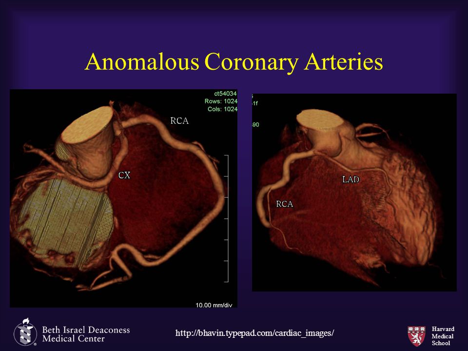
Anomalous Coronary Arteries
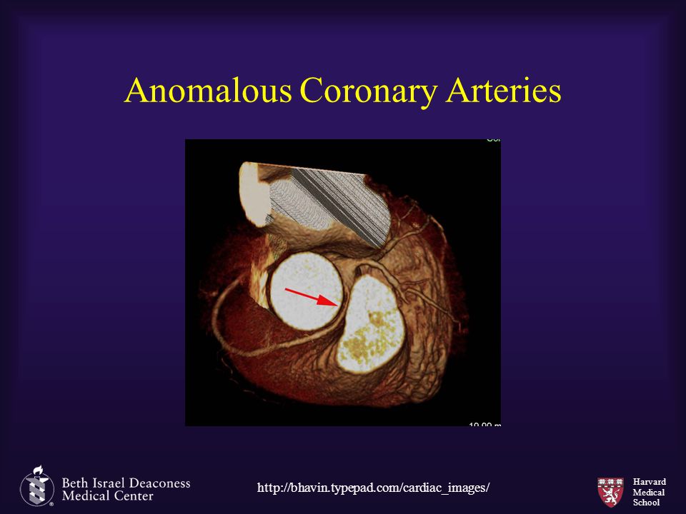
31
Anomalous Coronary Arteries
32
Ventricular Function

33
Ventricular Function

34
Ventricular Function: Compared to CMR
Segung et al, Circulation 2006;114: ; 31 patients

35
Ventricular Function: Compared to CMR
Segung et al, Circulation 2006;114: ; 31 patients, radial method

36
Valvular Function

37
Valvular Function

38
Pouleur et al, Radiology 2007;244:745-754
Aortic Stenosis Pouleur et al, Radiology 2007;244:

39
Pouleur et al, Radiology 2007;244:745-754
Aortic Stenosis Pouleur et al, Radiology 2007;244:

40
Pouleur et al, Radiology 2007;244:745-754
Aortic Stenosis Agreement between multidetector CT and TTE in the detection of normal (AVA 2 cm2), mildly stenotic (AVA 1.2 cm2 and < 2.0 cm2), moderately stenotic (AVA 0.8 cm2 and < 1.2 cm2), or severely stenotic (AVA < 0.8 cm2) aortic valve opening was excellent ( = 0.88, P < .001) Pouleur et al, Radiology 2007;244:
, mildly stenotic (AVA 1.2 cm2 and < 2.0 cm2), moderately stenotic (AVA 0.8 cm2 and < 1.2 cm2), or severely stenotic (AVA < 0.8 cm2) aortic valve opening was excellent ( = 0.88, P < .001) Pouleur et al, Radiology 2007;244:")
41
Valvular Function

42
Valvular Dehiscence

43
Valvular Dehiscence

44
Perfusion and Late Enhancement
Nieman et al. Radiology.2008; 247: 49-56

45
Perfusion and Late Enhancement
Nieman et al. Radiology.2008; 247: 49-56

46
Perfusion and Late Enhancement
Nieman et al. Radiology.2008; 247: 49-56

47
Cardiac CT Possible indications for coronary MDCTA
How to approach a coronary MDCTA study

48
How to Review a Coronary CTA Study
Review the axial images Interrogate multiple reconstructions at different points in the cardiac cycle to determine which has the least amount of artifact If any abnormalities, further investigate them with MIPs MPRs, and curved MPRs. Volume rendered images can be helpful to communicate your findings Generally not diagnostic Especially helpful in graft cases The entire dataset beyond the heart needs to be reviewed to ensure that there are no other significant findings.

49
Axial Stack

50
Axial Slice

51
MIP

52
Volume Rendered Image

53
cMPR with SAX and VA

54
cMPR with SAX and VA, Orthogonal

55
Importance of Interactive Reconstructions
Ferencik et al, Radiology: Volume 243: Number 3—June 2007

56
Outline Possible indications for coronary MDCTA
Coronary artery imaging is becoming established Stenosis Plaque characterization Stents Grafts Ventricular function Aortic Stenosis How to approach a coronary MDCTA study Axial images contain all of the primary data Use interactive reconstructions to aid in assessing problem areas

Similar presentations


 Inclusion criteria.>")


