Download presentation
Presentation is loading. Please wait.

1
Xanthelasma A Case Series
Victoria Smith BSc (Hons) ITEC VTCT Director of Aesthetics
 ITEC VTCT. Director of Aesthetics.")
2
Portrait of Mona Lisa painted in 1506
Portrait of Mona Lisa painted in 1506 Suggests Xanthelasma prevalent in 16th Century

3
What is Xanthelasma? Lipoprotein Yellow plaques Eyelids Harmless
flat or slightly raised Eyelids lower and upper Harmless cosmetically unattractive, irritating Can grow 3mm – 6cm diameter Sharp distinct border

4
Causes Cause unknown High cholesterol High level of plasma lipids
High BP Heart disease Genetic factors

5
Who gets Xanthelasma? Rare More prevalent in women
Hampton R. Oct 2011, Medscape More prevalent in women F:M ratio = 2:1 (Christoffersen et al. BMJ 2011\0 Age of onset = yrs Peak age = 40 – 50 yrs

6
Existing Treatments for Xanthelasma
Surgery - skin grafts, flaps Cryotherapy- Liquid Nitrogen Laser e.g. Nd:YAG Trichloroacetic acid Camouflage Make Up

7
History of Electrolysis
Invented by Dr Charles E Michel 1875 Developed for Trichiasis Uses galvanic/short wave diathermy current Insertion of needle - emitting current FDA approved for permanent hair removal

8
Advanced Electrolysis
Lumps, Bumps and Skin lesions Skin tags Milia Telangiectasia Seborrhoeic Keratosis Dermatosis Papulosa Nigra “Age spots”

9
The Procedure Clean environment Short wave diathermy
17-19 MHz Sterex (UK) One piece stainless steel needle 2 techniques used
 One piece stainless steel needle. 2 techniques used.")
10
Technique 1 “Scratching” removal of epithelial layer
“scratching” with needle to lift plaques

11
Technique 2 used if plaques don’t raise repeated “tapping” of plaque
“Dotting” used if plaques don’t raise repeated “tapping” of plaque heat accumulates plaque darkens and crusts

12
Sometimes bleeds - before scab forms
After Procedure No discomfort Area resembles a graze Sometimes bleeds - before scab forms Scab comes away days

13
Results 10 patients (M:F 4:6 Age: 39-53years) 7 completed treatment
1 failed to complete 2 part way through treatment 7 completed treatment all xanthelasma removed 100% patient satisfaction 4wks - 1year post treatment variable healing, blemish remaining all: would have the treatment repeated would recommend

14
Results at 1 year 2 patients at 1 year No re-growth in treated area
Slight pink scarring easy to cover Little re-growth in new areas Increased self esteem

15
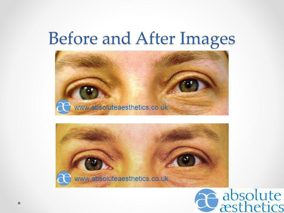
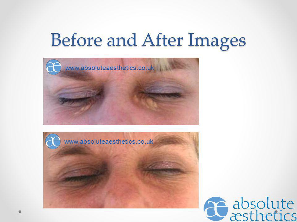
Before and After Images
16
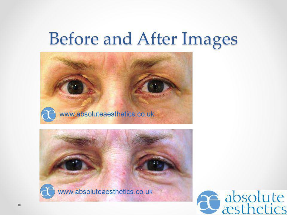
Before and After Images
17
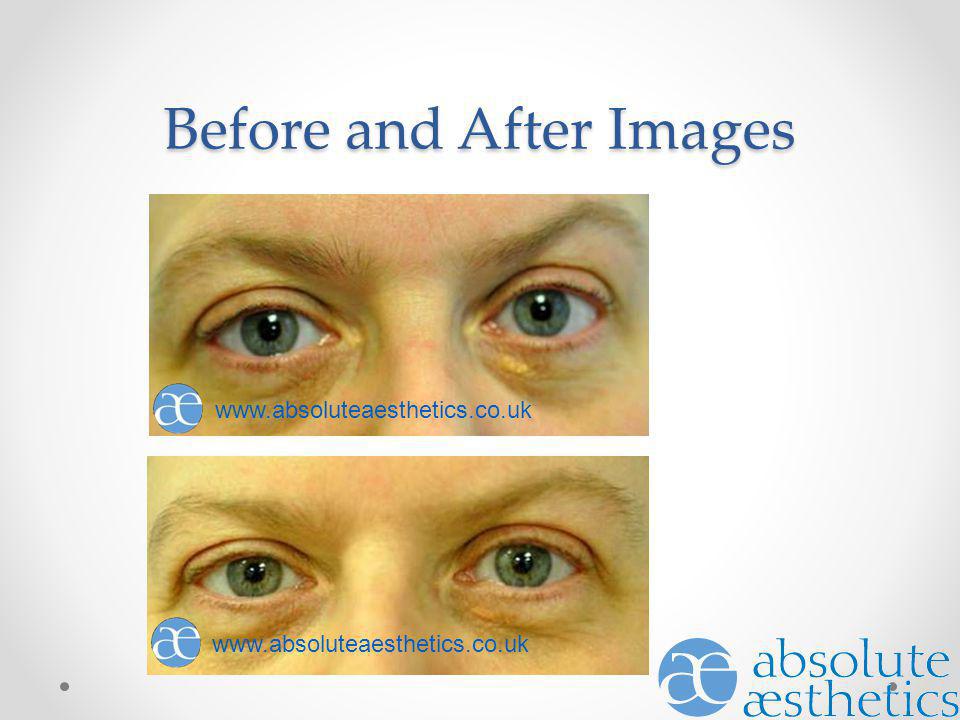
Before and After Images
18
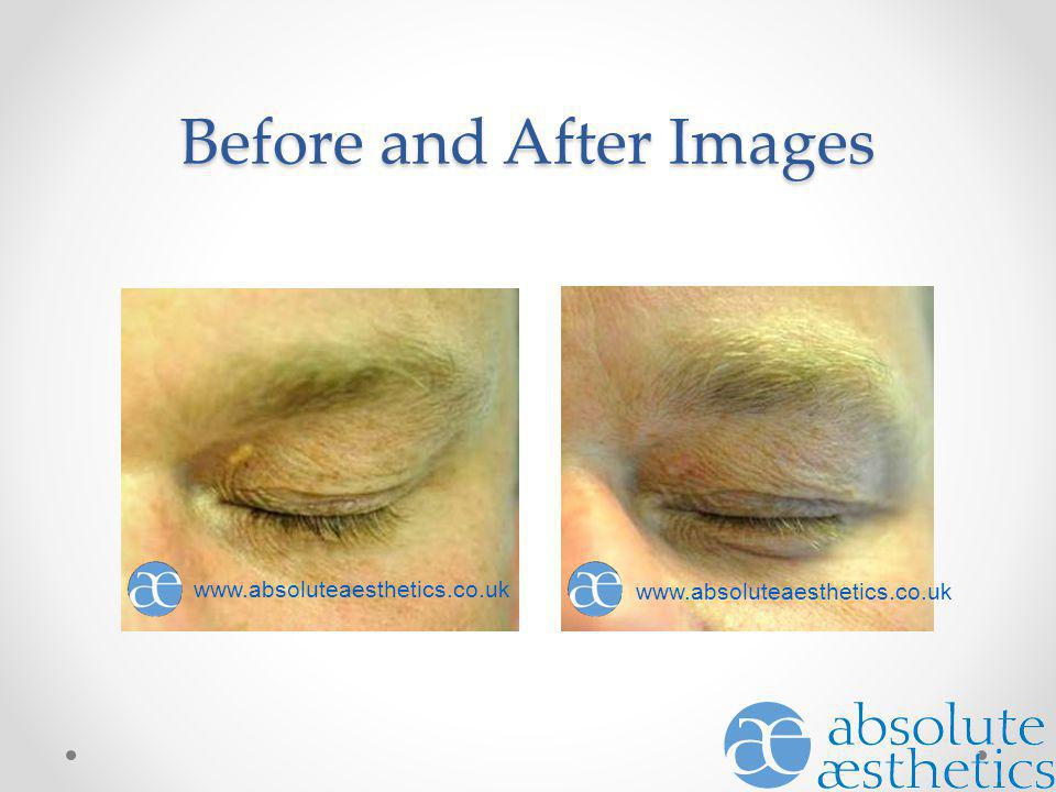
Before and After Images
19
Before and After Images
20
Conclusion Successful removal of xanthelasma Scrapping technique
more exposed wound - longer to heal Dotting technique less of an open wound - quicker to heal Significant increase in patient self esteem Low cost Low downtime

Similar presentations


 For Treating Cutaneous Leishmaniasis Dr. Eshtyag Abdeen Ali Dermatologist.>")

 ITEC VTCT Director of Aesthetics.>")

 A National Public Awareness Campaign from the P.A.D. Coalition and the National Heart,>")