Download presentation
Presentation is loading. Please wait.

1
Congenital Heart Disease (CHD) By Alireza Pourtalebi
 By Alireza Pourtalebi")
2
Blood circulation after birth: The transformation from fetal to neonatal circulation involves two major changes: 1. A marked increase in systemic resistance. caused by loss of the low-resistance placenta. 2. A marked decrease in pulmonary resistance. caused by pulmonary artery dilation with the neonate’s first breaths.

3
Fetal Circulation 8 Hours old24-72 hrs

4
With the first breaths of air the baby takes at birth, the fetal circulation changes. A larger amount of blood is sent to the lungs to pick up oxygen. Because the ductus arteriosus (the normal connection between the aorta and the pulmonary valve) is no longer needed, it begins to wither and close off. (72 hrs.) The circulation in the lungs increases and more blood flows into the left atrium of the heart pressure causes the foramen ovale to close and blood circulates normally Blood circulation after birth:
 is no longer needed, it begins to wither and close off. (72 hrs.) The circulation in the lungs increases and more blood flows into the left atrium of the heart pressure causes the foramen ovale to close and blood circulates normally Blood circulation after birth:.")
5
Epidemiology of CHD Incidence - 8/1000 live births - 3-4/100 still born - 2/100 premature infants excluding PDA -10-25/100 abortuses Most congenital defects are well tolerated during fetal life. Etiology - Unknown in most cases - Genetic factors - single gene defect - Chromosomal abnormality - Environmental factors - Gender differences in type of CHD - occur during the 1st 8 wks. of fetal development

6
Factors Contributing to CHD 85 to 90 % of cases, there is no identifiable cause for the heart defect generally considered to be caused by multifactorial inheritance. factors are usually both genetic and environmental, where a combination of genes from both parents, in addition to unknown environmental factors, produce the trait or condition. Maternal Factors: seizure disorders w/ intake of anti-seizure medications intake of lithium for depression uncontrolled IDDM lupus german measles (rubella) – 1 st trimester of pregnancy
 – 1 st trimester of pregnancy.")
7
Family History: risk increases when either parent has CHD, or when another sibling was born w/ CHD If you have had one child with CHD, the chance that another child will be born with CHD ranges from 1.5 to 5 %, depending on the type of CHD in the first child. If you have had two children with CHD, then the risk to 5 to 10 %, to have another child with CHD. If the mother has CHD, the risk for a child to be born with CHD ranges from 2.5 to 18 percent, with an average risk of 6.7 percent. Chromosome abnormalities: 5 to 8 % of all babies with CHD have a chromosome abnormality includes Down syndrome, trisomy 18 and trisomy 13, Turner’s syndrome, Cri-du-chat syndrome Factors Contributing to CHD

8
Relative Frequency of Congenital Heart Lesions Lesions% of all Lesions - Ventricular septal defect25-30 - Atrial septal defect (Secundum)6-8 - Patent ductus arteriosus6-8 - Coarctation of aorta5-7 - Tetralogy of Fallot5-7 - Pulomnary Valve Sterosis5-7 - Aortic Valve Stenosis4-7 - d-Transposition of great arteries3-5 - Hypoplastic left ventricle1-3 8
6-8 - Patent ductus arteriosus6-8 - Coarctation of aorta5-7 - Tetralogy of Fallot5-7 - Pulomnary Valve Sterosis5-7 - Aortic Valve Stenosis4-7 - d-Transposition of great arteries3-5 - Hypoplastic left ventricle1-3 8")
9
Relative Frequency Lesions% of all Lesions - Hypoplastic right ventricle1-3 - Truncus arteriosus1-2 - Total anomalous PVR1-2 - Tricuspid atresia1-2 - Single ventricle1-2 - Double-outlet right ventricle1-2 - Others5-10 9

10
Clues for Evaluation of an Infant with suspected CHD 1. On History and Physical Examination color) Acyanotic Cyanotic 2. Chest roentgenogram Normal Increased/Plethora pulmonary blood flow Decreased/Oligemia 3. Electrocardiogram - Right - Left hypertrophy - Biventricular Final diagnosis - Precordial examinant - Echocardiography
 Acyanotic Cyanotic 2. Chest roentgenogram Normal Increased/Plethora pulmonary blood flow Decreased/Oligemia 3. Electrocardiogram - Right - Left hypertrophy - Biventricular Final diagnosis - Precordial examinant - Echocardiography.")
11
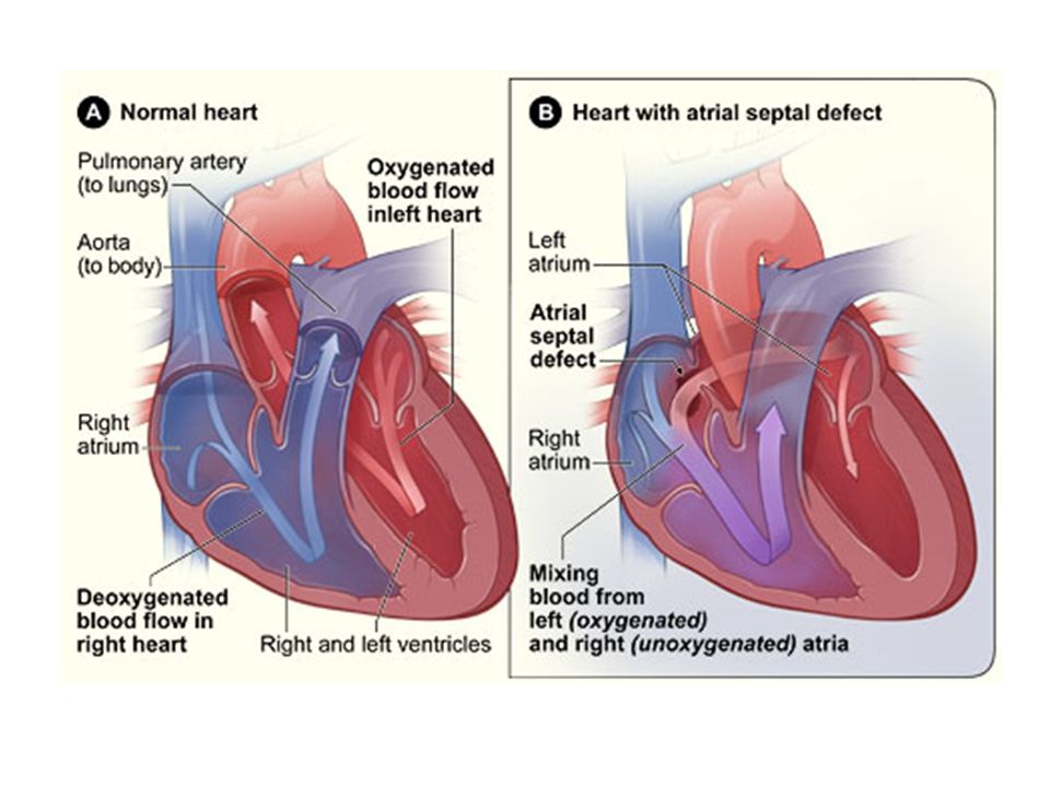
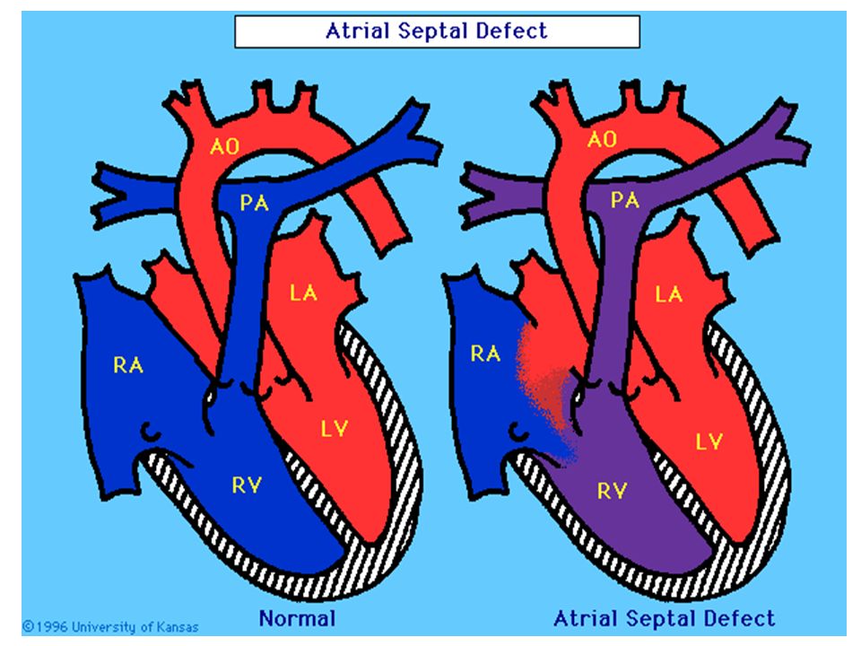
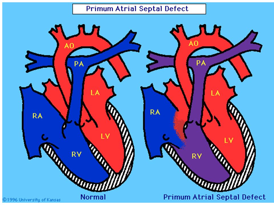
Acyanotic Congenital Heart Diseases 1. Left to Right Shunt Lesions Pink Baby (L R shunt) L R shunts cause CHF and pulmonary hypertension. This leads to RV enlargement, RV failure These babies present with CHF and respiratory distress. 1.1. Atrial Septal Defect Defect occur in any portion of the atrium - Ostium secundum (at fossa ovalis) - Ostium primum (ECD) (lower atrial septum) - Sinus venosus (upper atrial septum) Pathophysiology Left to right shunt - Transatrial in OS & SV - Transatrial & transventricular in OP
 L R shunts cause CHF and pulmonary hypertension. This leads to RV enlargement, RV failure These babies present with CHF and respiratory distress Atrial Septal Defect Defect occur in any portion of the atrium - Ostium secundum (at fossa ovalis) - Ostium primum (ECD) (lower atrial septum) - Sinus venosus (upper atrial septum) Pathophysiology Left to right shunt - Transatrial in OS & SV - Transatrial & transventricular in OP.")
13
Acyanotic CHD Clinical Manifestations Most are asymptomatic Right ventricular lift Wide & fixed split of 2nd heart sound Systolic ejection murmur Mid-diastolic murmur at tricuspid area Holosystolic murmur at mitral area in OP

14
Acyanotic CHD Diagnosis Clinical CXR - Right. V & A enlargement - Large pulm. artery - ↑ed pulm. vascularity ECG - volume overload, - right axis deviation - minor right ventricular conduction delay Echocardiography Catheterization Prognosis - Well tolerated Complications - pulm. Hypertension, Eismenger syndrome Treatment Surgery - For all symptomatic ASD - Asymptomatic patients with shunt ratio > 2:1

17
17

18
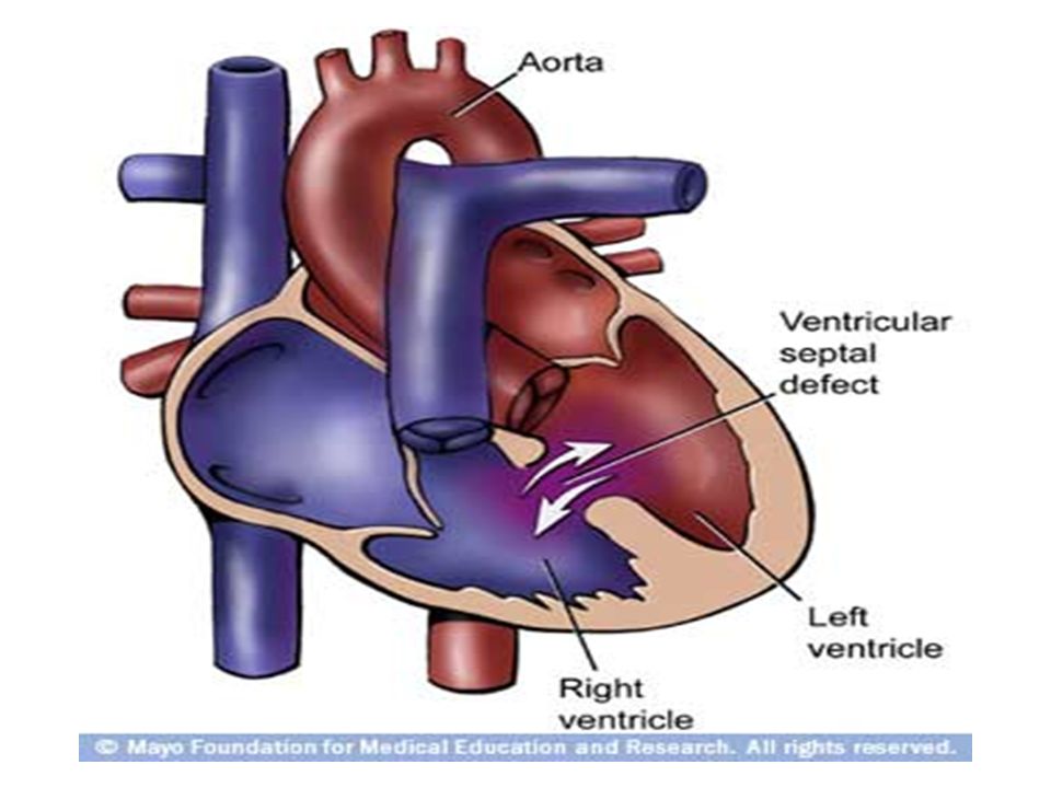
1.2 Ventricular Septal Defect An opening in the ventricular septum allows oxygenated blood to pass from the left ventricle, through the opening in the septum, and then mix with unoxygenated blood in the right ventricle. VSDs are the most commonly occurring type of congenital heart defect, occurring in 14-17 % of babies born each year. occur when the partitioning process does not occur completely, leaving an opening in the ventricular septum. The most common cardiac malformation Defect occur in any portion of the septum - Majority membranous - Muscular – Single or Swiss-cheese defect Pathophysiology Lt to Rt shunt Restrictive if defect is small (0.5cm2) Non-restrictive - large defect (> 1cm2) - Right and left vent. Pressure equalized EFFECTS: When blood passes through the VSD from the left ventricle to the right ventricle a larger volume of blood than normal must be handled by the right side of the heart extra blood then passes through the pulmonary artery into the lungs pulmonary hypertension and pulmonary congestion pulmonary arteries become thickened and obstructed due to increased pressure If VSD is not repaired, and lung disease begins to occur pressure in the right side of the heart will eventually exceed pressure in the left R to L shunt cyanosis Due to high pressure --- tissue damage may eventually occur in the right ventricle bacteria in the bloodstream can easily infect this injured area bacterial endocarditis.
 Non-restrictive - large defect (> 1cm2) - Right and left vent. Pressure equalized EFFECTS: When blood passes through the VSD from the left ventricle to the right ventricle a larger volume of blood than normal must be handled by the right side of the heart extra blood then passes through the pulmonary artery into the lungs pulmonary hypertension and pulmonary congestion pulmonary arteries become thickened and obstructed due to increased pressure If VSD is not repaired, and lung disease begins to occur pressure in the right side of the heart will eventually exceed pressure in the left R to L shunt cyanosis Due to high pressure --- tissue damage may eventually occur in the right ventricle bacteria in the bloodstream can easily infect this injured area bacterial endocarditis..")
20
Signs and Symptoms fatigue sweating tachypnea murmur heavy breathing congested breathing disinterest in feeding, or tiring while feeding poor weight gain The larger the opening, the greater the amount of blood that passes through and overloads the right ventricle and lungs. Diagnosis - Clinical - CXR - Cardiomegaly - Plethoric lung - ECG - Echocardiography Prognosis - 30-50% small defects close by 2 yr of age - Rarely moderate to large defects close

21
Treatment Medical management - digoxin - diuretics Adequate nutrition - high-calorie formula or breast milk - supplemental tube feedings Prophylactic antibiotics to prevent bacterial endocarditis Surgical repair – VSD will be closed w/ stitches or special patch Interventional cardiac catheterization – Septal occluder Complications - Infective endocarditis - Recurrent lung infection - Heart failure - Pulmonary HTN - Acquired pulmonary stenosis -- aortic valve regurgitation

22
22

23
In many children, there is no known reason for the ductus arteriosus remaining open. However, PDA is seen more often in the following: premature infants infants born to a mother who had rubella during the first trimester of pregnancy EFFECTS: PDA oxygenated blood passes from the aorta to the pulmonary artery & mixes w/ the unoxygenated blood w/c goes to the lungs blood volume to the lungs pulmonary hypertension & congestion Further, because blood is pumped at high pressure through the PDA, the lining of the pulmonary artery will become irritated and inflamed. Bacteria in the bloodstream can easily infect this injured area bacterial endocarditis. Patent Ductus Arteriosus (PDA) Functional closure soon after birth Aortic end of the ductus distal to the origin of left subclavian artery and the other end at bifurcation of pulmonary artery. Male to female ratio 1:2 Pathology Deficiency of mucoid endothelial layer & muscular media in term infant. Lt to Rt shunt - size- ratio of pulm. to systemic resistance Reversal of shunt
 Functional closure soon after birth Aortic end of the ductus distal to the origin of left subclavian artery and the other end at bifurcation of pulmonary artery. Male to female ratio 1:2 Pathology Deficiency of mucoid endothelial layer & muscular media in term infant. Lt to Rt shunt - size- ratio of pulm. to systemic resistance Reversal of shunt.")
24
Signs and Symptoms fatigue sweating tachypnea shortness of breath congested breathing disinterest in feeding, or tiring while feeding poor weight gain murmur increase systolic BP bounding pulse Diagnosis - Clinical - Chest X-ray - ECG - Echocardiography Prognosis - Small PDA - normal life - Large PDA - CHF Complications - Infective Endocarditis/Endarteritis - CHF - Embolization - Pulmonary HTN Treatment - Medical - Surgical closure

25
Medical Management Indomethacin IV (prostaglandin inhibitor) may help close a PDA. - works by stimulating the muscles inside the PDA to constrict, thereby closing the connection Digoxin Diuretics adequate nutrition (premature infants or those infants with a large PDA may become tired when feeding, and are not able to eat enough to gain weight) high-calorie formula or breast milk Special nutritional supplements may be added to formula or pumped breast milk that increase the number of calories in each ounce, thereby allowing your baby to drink less and still consume enough calories to grow properly. supplemental tube feedings - infants who can drink part of their bottle, but not all, may be fed the remainder through the feeding tube - infants who are too tired to bottle-feed may receive their formula or breast milk through the feeding tube alone. PDA surgical repair or closure - Repair is usually indicated in infants younger than 6 months of age who have large defects that are causing symptoms, such as poor weight gain and rapid breathing - Transcatheter coil closure of the PDA - PDA ligation - involves closing the open PDA with stitches or the vessel connecting the aorta and pulmonary artery may be cut and cauterized
 high-calorie formula or breast milk Special nutritional supplements may be added to formula or pumped breast milk that increase the number of calories in each ounce, thereby allowing your baby to drink less and still consume enough calories to grow properly. supplemental tube feedings - infants who can drink part of their bottle, but not all, may be fed the remainder through the feeding tube - infants who are too tired to bottle-feed may receive their formula or breast milk through the feeding tube alone. PDA surgical repair or closure - Repair is usually indicated in infants younger than 6 months of age who have large defects that are causing symptoms, such as poor weight gain and rapid breathing - Transcatheter coil closure of the PDA - PDA ligation - involves closing the open PDA with stitches or the vessel connecting the aorta and pulmonary artery may be cut and cauterized.")
26
26

27
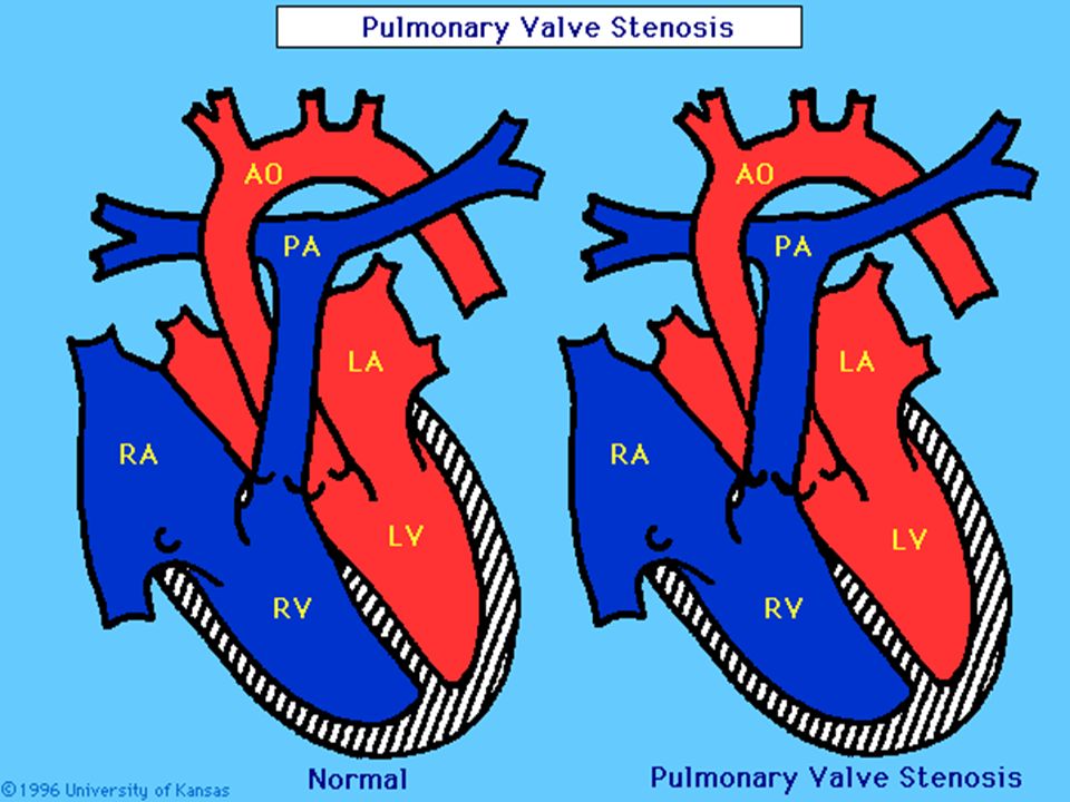
2. Obstructive Lesions 2.1. Pulmonic Stenosis - 4 types - Valvular - Infundibular - Supra valvular - Peripheral Pathophysiology - Rt outlet obstruction → Pressure work ↓ Rt vent. hyperthropy Clinical Manifestation - Mild to moderate - asymptomatic - Critical stenosis - Systolic ejection murmur - Heart failure in neonates & infants - Rarely cyanosis Diagnosis - Clinical - CXR - Rt vent. hypertrophy - reduced pulm. blood flow - ECG - Echocardiography Prognosis - good in mild to moderate Complications - CHF in severe Ps - rarely IE Treatment - vavular PS - ballon valvoplasty - surgery

29
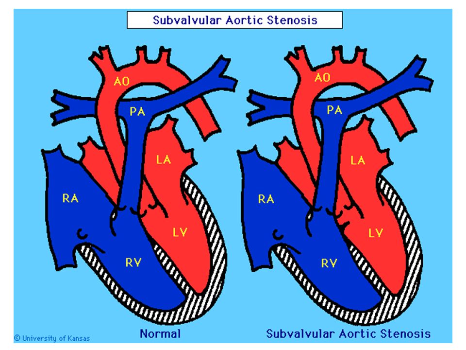
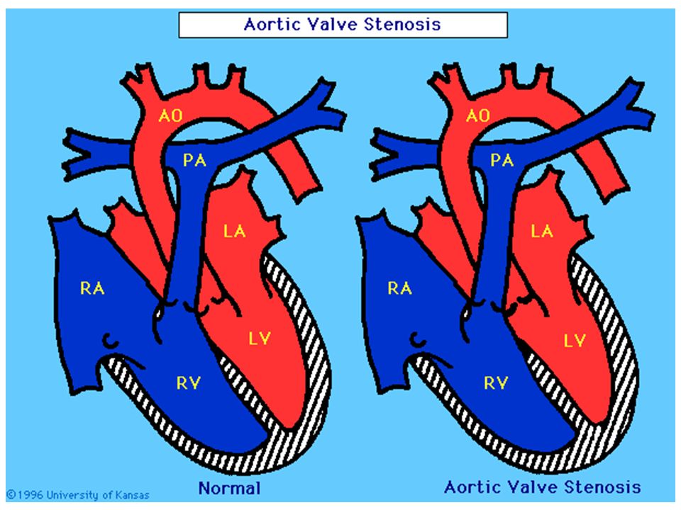
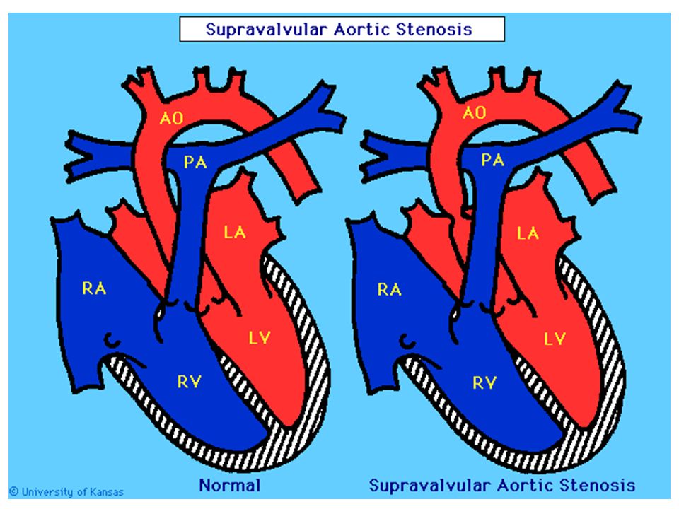
2.2 Aortic Stenosis -Valvular - the commonest -Supra valvular -Subvalvular (subaortic) 29 Clinical Manifestation Mild stenosis - Normal pulse & apical impulse - Systolic ejection M - Normal to enlarged heart size Critical stenosis - Left ventricular failure - pulm. edema, cardiomegaly - Weak peripheral pulses - Weak systolic M - Paradoxical split 2nd heart sound Diagnosis - Clinical - CXR - ECG - Echocardiography - Graded exercise testing Prognosis is good for mild to moderate Treatment - Balloon valvoplasty - Surgical

33
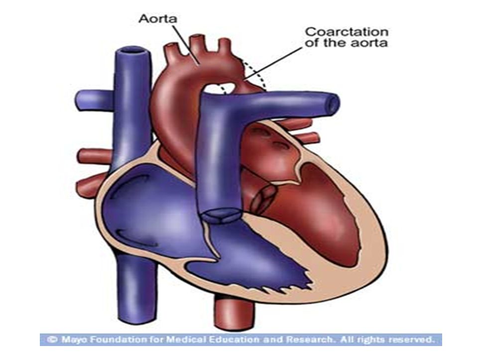
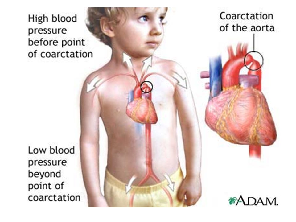
Coarctation of the Aorta Narrowing of the aorta can occur anywhere, but is most likely to happen in the segment just after the aortic arch. This narrowing restricts the amount of blood to the lower part of the body occurs in about 8-11 % of all children with CHD EFFECTS: The left ventricle has to work harder to try to move blood through the narrowing in the aorta left-sided heart failure BP is higher above the narrowing, and lower below the narrowing. Older children may have headaches from too much pressure in the vessels in the head, or cramps in the legs or abdomen from too little blood flow in that region. The walls of the arteries may become weakened by high pressure spontaneous tears cause a stroke or uncontrollable bleeding. risk for bacterial endocarditis. -Occur at any site from the arch of aorta to iliac bifurcation -98% juxta ductal Pathogenesis In utero arch hypoplasia Rt heart output passes through the ductus

36
irritability pale skin sweating heavy and/or rapid breathing poor feeding poor weight gain cold feet and/or legs diminished or absent pulses in the feet BP in the arms significantly greater than the BP in the legs Mild narrowing may not cause symptoms at all. Often, a school-aged child or adolescent is simply noted to have high BP or a heart murmur on a physical examination. Some may complain of headaches or cramps in the lower sections of the body. Signs and Symptoms Clinical Manifestation Hypertension → mechanical obstruction→ renal Differential cyanosis → pink upper extr. → cyanosed lower extr. Classic signs - Disparity in pulse & BP - Radio-femoral delay - Systolic M at LMSB & inter-scapular area

37
Diagnosis - Clinical - CXR - cardiomegaly & pulm. congestion - Notching of ribs - ECG - Echocardiography Prognosis – Untreated cases succumb by 20-40 years Complications - CVA - I/E - Aneurysms Treatment - Medical - IV PGE 1 in neonatal age - Surgery interventional cardiac catheterization - During the procedure, the child is sedated and a small, thin, flexible tube (catheter) is inserted into a blood vessel in the groin and guided to the inside of the heart - once the catheter is in the heart, the cardiologist will pass an inflated balloon through the narrowed section of the aorta to stretch the area open. - A small device, called a stent, may also be placed in the narrowed area after the balloon dilation to keep the aorta open. surgical repair Your child's coarctation of the aorta may be repaired surgically in an operating room. The surgical repair is performed under GA. The narrowed area is either surgically removed, or made larger with the help of surrounding structures or a patch.
 is inserted into a blood vessel in the groin and guided to the inside of the heart - once the catheter is in the heart, the cardiologist will pass an inflated balloon through the narrowed section of the aorta to stretch the area open. - A small device, called a stent, may also be placed in the narrowed area after the balloon dilation to keep the aorta open. surgical repair Your child s coarctation of the aorta may be repaired surgically in an operating room. The surgical repair is performed under GA. The narrowed area is either surgically removed, or made larger with the help of surrounding structures or a patch..")
38
3. Pulmonary Vascular Disease (Eismenger syndrome) - Occur in shunt lesions VSD - mainly ASD PDA -Reversal of shunt due to pulm. HTN→ Cyanosis 4. Regurgitant Lessons - Pulmonary valvular insufficiency - Congenital mitral valve insufficiency - Mitral valve prolapse
 - Occur in shunt lesions VSD - mainly ASD PDA -Reversal of shunt due to pulm. HTN→ Cyanosis 4. Regurgitant Lessons - Pulmonary valvular insufficiency - Congenital mitral valve insufficiency - Mitral valve prolapse.")
39
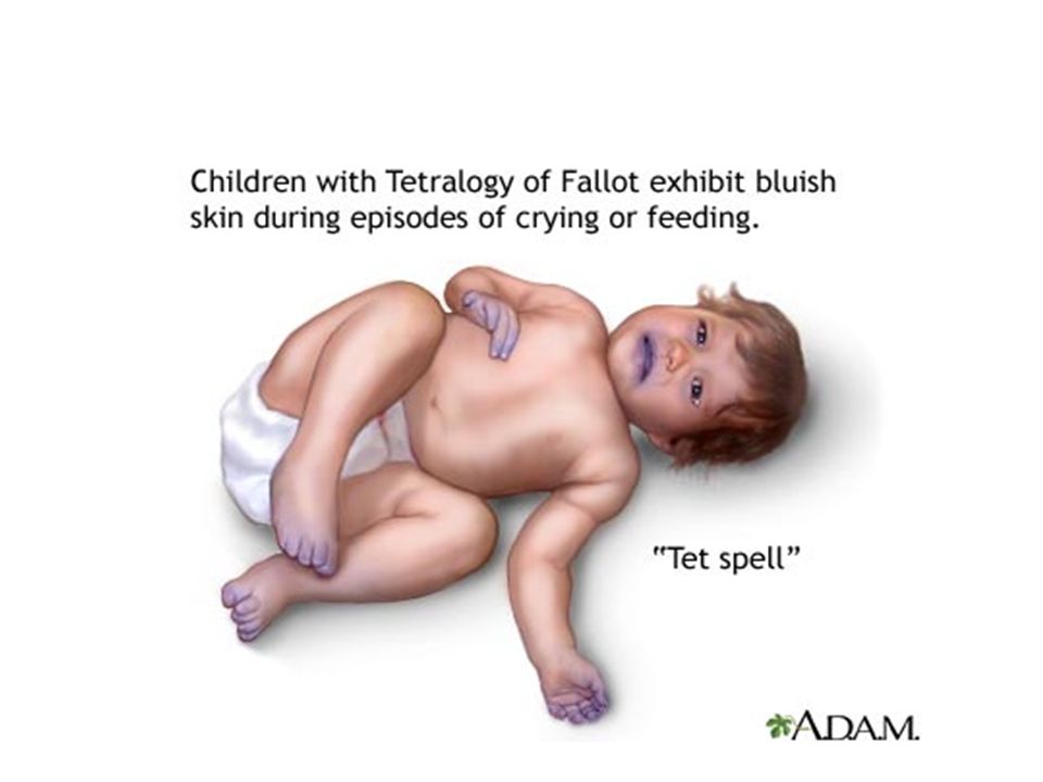
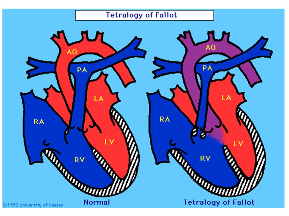
Cyanotic Congenital Heart Disease 1. Cyanotic lesions with decreased pulmonary blood flow 1.1. Tetralogy of Fallot Consists: 1. Rt ventricular outflow obst. 2. Ventricular septal defect 3. Dextroposition of the aorta 4. Right ventricular hypertrophy EFFECTS: If the right ventricle obstruction is severe, or if the pressure in the lungs is high a large amount of oxygen-poor (blue) blood passes through the VSD, mixes with the oxygen-rich (red) blood in the left ventricle, and is pumped to the body cyanosis The more blood that goes through the VSD, the less blood that goes through the pulmonary artery to the lungs oxygenated blood to the left side of the heart. Soon, nearly all the blood in the left ventricle is oxygen-poor (blue). This is an emergency situation, as the body will not have enough oxygen to meet its needs.
 blood passes through the VSD, mixes with the oxygen-rich (red) blood in the left ventricle, and is pumped to the body cyanosis The more blood that goes through the VSD, the less blood that goes through the pulmonary artery to the lungs oxygenated blood to the left side of the heart. Soon, nearly all the blood in the left ventricle is oxygen-poor (blue). This is an emergency situation, as the body will not have enough oxygen to meet its needs..")
40
Signs and Symptoms Cyanosis (blue color of the skin, lips, and nail beds) that occurs with such activity as crying or feeding Some babies do not have noticeable cyanosis, but may instead be very irritable or lethargic due to a decreasing amount of oxygen available in the bloodstream. Murmur Tachycardia Irritability Syncope Clubbing of fingers Pathophysiology - Outflow obstruction - Hypertrophy of subpulmonic muslce - Normal or small pulmonary valve annulus - Rarely pulmonary atresia - VSD - Non-restrictive, located just below aortic valve - Aortic arch is right side in 20% - Right ventricular output shunts to the aorta

41
Clinical Manifestation - Rarely pink TOF - in the absence of obstruction - Cyanosis - Clubbing - Squatting position in walking children - Paroxysmal hypercyanotic attacks occur during 1st 2 years - Systolic ejection M - Delayed growth & development - Single 2nd heart sound Diagnosis - CXR - Narrow base & uplifted apex - A boot or wooden shoe - decreased pulm. vascularity - Right side aortic arch in 20% - ECG - Echocardiography Complication - Cerebral thrombosis - in < 2 years - Brain abscess - Infective endocarditis - Polycythemia - CHF in pink TOF

43
Treatment Severe outflow obstruction - Medical Px - PGE 1 infusion - Prevent dehydration - Partial exchange transfusion - Oral propranolol for tet spells - Surgery - Blalock Taussig - Total correction

46
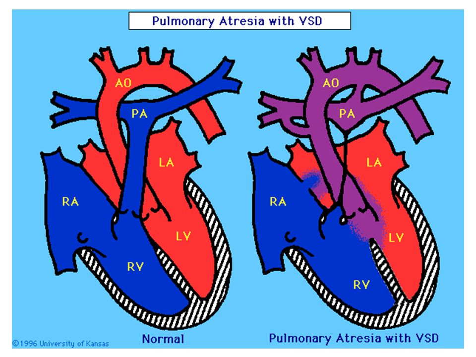
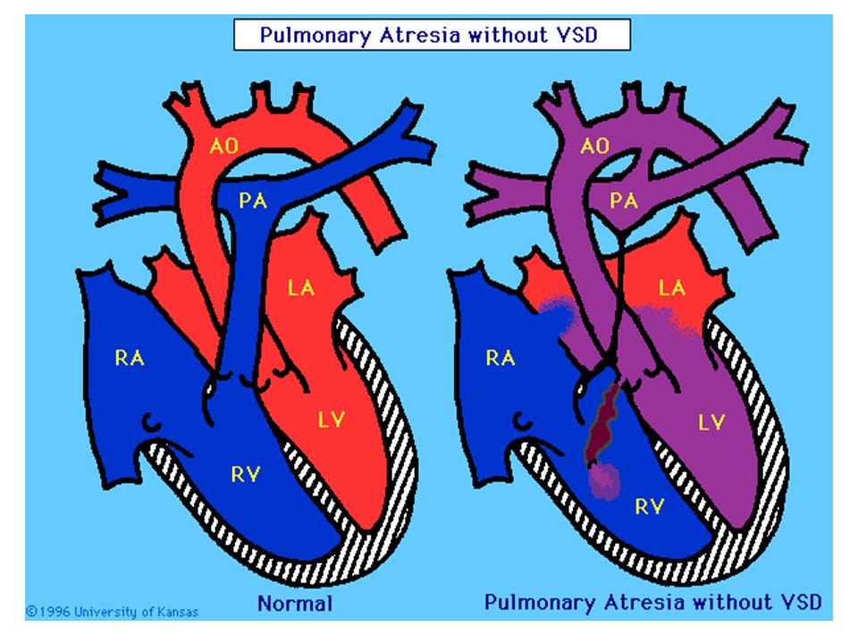
1.2. Pulmonary Atresia - With VSD - Extreme form of TOF - Without VSD - No egress of blood from Rt vent. - Shunt through foramen ovale to Lt atrium Left ventricle systemic circulation Aorta pulmonic circulation - Hypoplastic right ventricle (PDA) Clinical Manifestation - Cyanosis at birth - Respiratory distress - Single 2nd heart sound - No murmur Diagnosis - CXR - ECG - Echocardiography Treatment - PGE 1 - Surgery
 Clinical Manifestation - Cyanosis at birth - Respiratory distress - Single 2nd heart sound - No murmur Diagnosis - CXR - ECG - Echocardiography Treatment - PGE 1 - Surgery.")
48
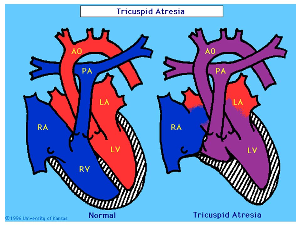
Tricuspid Atresia In this condition, there is no tricuspid valve, therefore, no blood flows from the right atrium to the right ventricle. Blood in right atrium foramen ovale left atrium and left ventricle aorta Tricuspid atresia defect is characterized by the following: – a small right ventricle – a large left ventricle – Small VSD and PDA – diminished pulmonary circulation – cyanosis - bluish color of the skin and mucous membranes caused from a lack of oxygen. A surgical shunting procedure is often necessary to increase the blood flow to the lungs. Clinical Manifestation - Cyanosis at birth - Polycythemia - Easily fatiguability - Exertional dyspnea Diagnosis - EXR -Pulm. Under circulation - ECG -Lt axis deviation & Lt vent. hypertrophy - Echocardiography Treatment - PGE1 - Surgery - Aortico - pulmonary Shunt - Bidirectional Glenn shunt - Modified Fontan operation

50
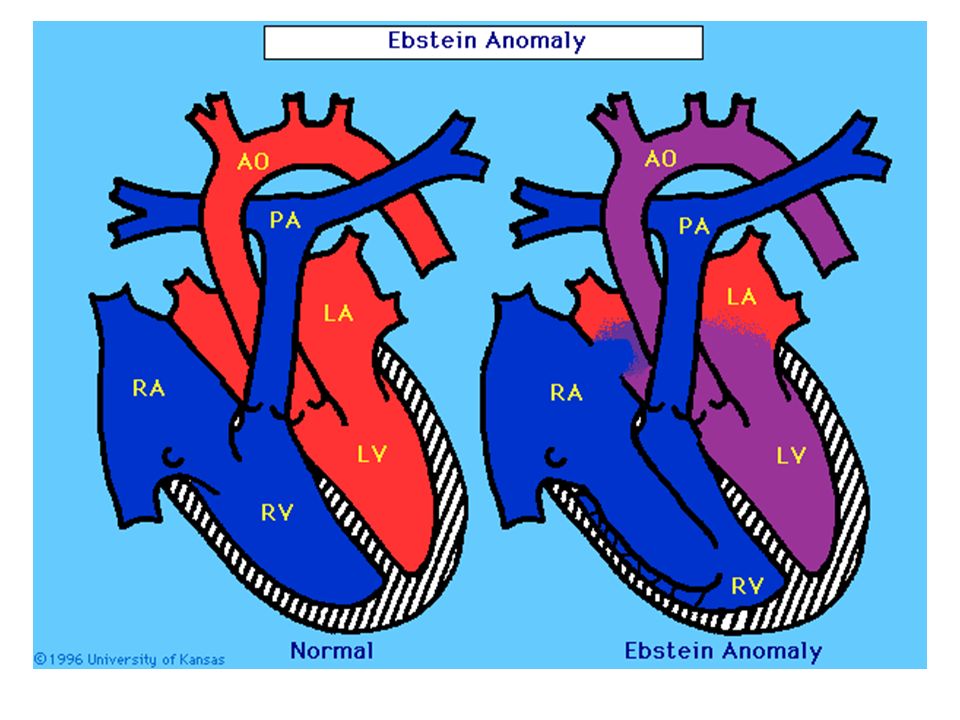
1.4. Ebstein Anomaly of the tricuspid valve - Down ward displacement of the tricuspid valve - Right ventricle with two parts - atrialized - normal ventricular myocardium - Abnormal tricuspid valve - Huge Rt atrium - Tricuspid regurgitation - Compromised Rt ventricular function Clinical Manifestations - Easly fatiguability - Cyanosis - Dysrhythmia - Rt to Lt shunt through formen ovale - Holosystolic M at tricuspid area - Heart failure Diagnosis - CXR - box shaped heart - ECG - Right BBB - Superior axis deviation Treatment - PGE 1 - Surgery

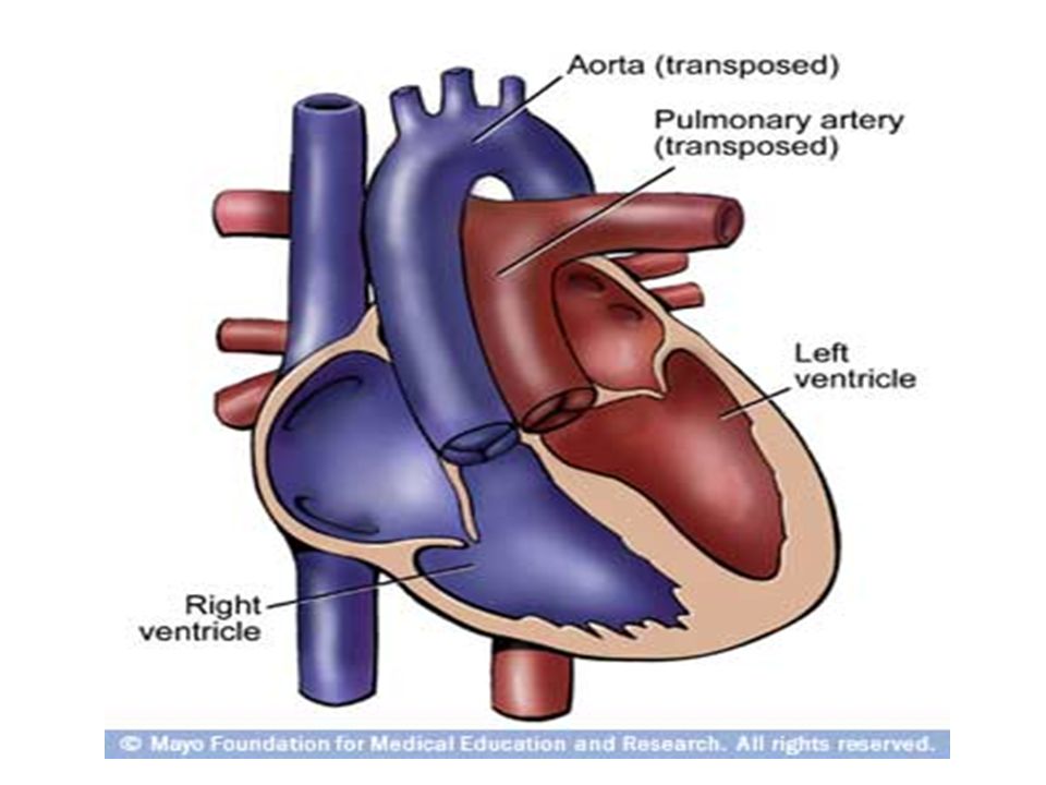
52
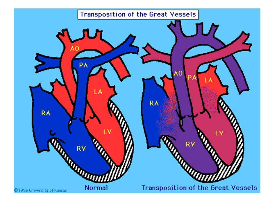
2. Cyanotic CHD With increased pulmonary blood flow 2.1 Transposition of the Great Arteries the aorta is connected to the right ventricle, and the pulmonary artery is connected to the left ventricle Oxygen-poor (blue) blood returns to the right atrium from the body passes through the right atrium and ventricle, into the misconnected aorta back to the body. Oxygen-rich (red) blood returns to the left atrium from the lungs passes through the left atrium and ventricle, into the pulmonary artery and back to the lungs. Other heart defects are often associated with TGA - atrial or ventricular septal defect -may be necessary in order for the infant with TGA to survive -Allow mixing of blood – providing at least smaller amounts of oxygen to the body a. D -TGA (uncorrected) - Systemic venous return to Rt atrium Normal - Pulmonary venous return to Lt atrium - Aorta arises from Right ventricle - Pulm. artery arises from Lt vent. Pathology
 blood returns to the right atrium from the body passes through the right atrium and ventricle, into the misconnected aorta back to the body. Oxygen-rich (red) blood returns to the left atrium from the lungs passes through the left atrium and ventricle, into the pulmonary artery and back to the lungs. Other heart defects are often associated with TGA - atrial or ventricular septal defect -may be necessary in order for the infant with TGA to survive -Allow mixing of blood – providing at least smaller amounts of oxygen to the body a. D -TGA (uncorrected) - Systemic venous return to Rt atrium Normal - Pulmonary venous return to Lt atrium - Aorta arises from Right ventricle - Pulm. artery arises from Lt vent. Pathology.")
54
Signs and Symptoms Cyanosis soon after delivery rapid breathing labored breathing rapid heart rate murmur cool, clammy skin *Systemic & Pulmonary Circulations Consists of two parallel circuits *Survival is with associated - patent foramen ovale or - VSD or – PDA Clinical Manifestations - Tachypnea & cyanosis at birth - Rarely congestive heart failure

55
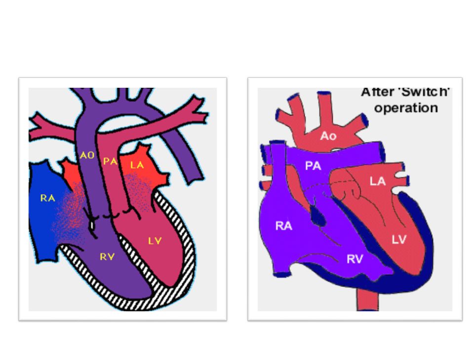
Treatment admitted to the NICU, placed on oxygen, and possibly even on a ventilator, IV medications to help the heart and lungs function more efficiently. a cardiac catheterization procedure will usually be performed to evaluate the defect(s) and the amount of blood that is mixing. as part of the cardiac catheterization, a balloon atrial septostomy may be performed to improve mixing of oxygen-rich (red) and oxygen-poor (blue) blood. – A catheter with a balloon in the tip is used to create an opening in the atrial septum – The catheter is guided through the foramen ovale (a small opening present in the atrial septum that closes shortly after birth) and into the left atrium. – The balloon is inflated. – The catheter is quickly pulled back through the hole, into the right atrium, enlarging the hole, allowing blood to mix between the atria. An IV prostaglandin E1 is given to keep the ductus arteriosus from closing. Within the first 1 to 2 weeks of age, TGA is surgically repaired. The “switch” operation is performed under GA, and involves the ff: The aorta is moved from the right ventricle to its normal position over the left ventricle. The pulmonary artery is moved from the left ventricle to its normal position over the right ventricle. The coronary arteries are moved so they will originate from the aorta and take oxygen-rich (red) blood to the heart muscle. Other defects, such as atrial or ventricular septal defects or a patent ductus arteriosus, are commonly closed.
 and the amount of blood that is mixing. as part of the cardiac catheterization, a balloon atrial septostomy may be performed to improve mixing of oxygen-rich (red) and oxygen-poor (blue) blood. – A catheter with a balloon in the tip is used to create an opening in the atrial septum – The catheter is guided through the foramen ovale (a small opening present in the atrial septum that closes shortly after birth) and into the left atrium. – The balloon is inflated. – The catheter is quickly pulled back through the hole, into the right atrium, enlarging the hole, allowing blood to mix between the atria. An IV prostaglandin E1 is given to keep the ductus arteriosus from closing. Within the first 1 to 2 weeks of age, TGA is surgically repaired. The switch operation is performed under GA, and involves the ff: The aorta is moved from the right ventricle to its normal position over the left ventricle. The pulmonary artery is moved from the left ventricle to its normal position over the right ventricle. The coronary arteries are moved so they will originate from the aorta and take oxygen-rich (red) blood to the heart muscle. Other defects, such as atrial or ventricular septal defects or a patent ductus arteriosus, are commonly closed..")
56
b. L. TGA (corrected transposition) Systemic VR to normally positioned Rt atrium Through bicuspid (Mitral) valve Right sided left ventricle Pulmo. artery pulm. venous return Normally positioned Lt atrium Through tricuspid valve Left sided Right ventricle Aorta Discordant atrio-ventricular relation (ventricular inversion) Transposition of great arteries Clinical Manifestation Depends on associated malformation Diagnosis - Clinical - CXR - Cardiomegaly - Narrow mediastinum - Increased pulmonary blood flow - ECG - Echocardiography Treatment - PGE 1 - emergency - Surgery
 Systemic VR to normally positioned Rt atrium Through bicuspid (Mitral) valve Right sided left ventricle Pulmo. artery pulm. venous return Normally positioned Lt atrium Through tricuspid valve Left sided Right ventricle Aorta Discordant atrio-ventricular relation (ventricular inversion) Transposition of great arteries Clinical Manifestation Depends on associated malformation Diagnosis - Clinical - CXR - Cardiomegaly - Narrow mediastinum - Increased pulmonary blood flow - ECG - Echocardiography Treatment - PGE 1 - emergency - Surgery.")
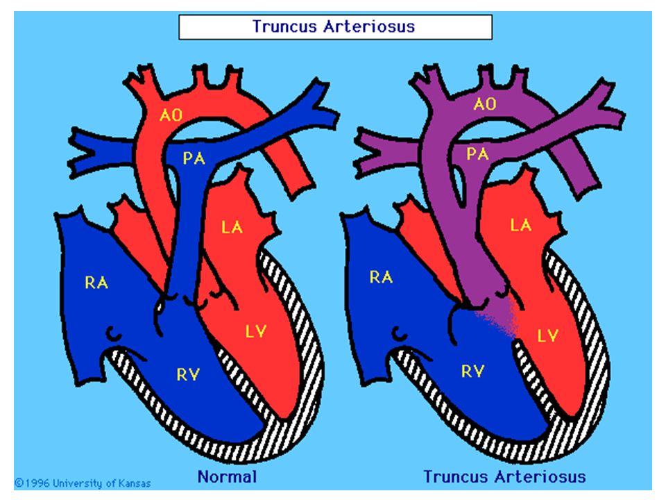
59
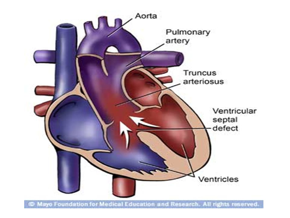
2.2 Truncus arteriosus The aorta and pulmonary artery start as a single blood vessel, which eventually divides and becomes two separate arteries. Truncus arteriosus occurs when the single great vessel fails to separate completely, leaving a connection between the aorta and pulmonary artery. Usually accompanied by a ventricular septal defect EFFECTS: oxygen-poor (blue) and oxygen-rich (red) blood mix back and forth through the ventricular septal defect. This mixed blood then flows through the common truncal vessel. Some of it will flow to pulmonary artery and on to the lungs, and some of the mixed blood will go into the aortic branch and to the body. The mixed blood that goes to the body does not have as much oxygen as normal, and will cause varying degrees of cyanosis - Single arterial trunk for both pulm. & systemic circ. - 4 types depending the origin of pulmonary artery Clinical Manifestation -Cyanosis -CHF at 2-3rd m -Systalic ejection m Treatment - surgery
 and oxygen-rich (red) blood mix back and forth through the ventricular septal defect. This mixed blood then flows through the common truncal vessel. Some of it will flow to pulmonary artery and on to the lungs, and some of the mixed blood will go into the aortic branch and to the body. The mixed blood that goes to the body does not have as much oxygen as normal, and will cause varying degrees of cyanosis - Single arterial trunk for both pulm. & systemic circ. - 4 types depending the origin of pulmonary artery Clinical Manifestation -Cyanosis -CHF at 2-3rd m -Systalic ejection m Treatment - surgery.")
61
Signs and Symptoms cyanosis fatigue sweating pale skin cool skin rapid breathing heavy breathing rapid heart rate congested breathing disinterest in feeding, or tiring while feeding poor weight gain Treatment Truncus arteriosus must be treated by surgical repair of the defects. However, medical support may be necessary until the best time for the operation to take place. medical management – Digoxin – Diuretics – ACE (angiotensin-converting enzyme) inhibitors - dilates the blood vessels, making it easier for the heart to pump blood forward into the body. adequate nutrition – high-calorie formula or breast milk – supplemental tube feedings surgical repair Surgery is usually performed after the infant is 2 weeks old, but before the blood vessels in the lungs are overwhelmed by extra blood flow and become diseased. The operation is performed under general anesthesia, and involves the following: The pulmonary arteries are detached from the common artery (truncus arteriosus) and connected to the right ventricle using a homograft (a section of pulmonary artery with its valves intact from a tissue donor). The ventricular septal defect is closed with a patch.
 inhibitors - dilates the blood vessels, making it easier for the heart to pump blood forward into the body. adequate nutrition – high-calorie formula or breast milk – supplemental tube feedings surgical repair Surgery is usually performed after the infant is 2 weeks old, but before the blood vessels in the lungs are overwhelmed by extra blood flow and become diseased. The operation is performed under general anesthesia, and involves the following: The pulmonary arteries are detached from the common artery (truncus arteriosus) and connected to the right ventricle using a homograft (a section of pulmonary artery with its valves intact from a tissue donor). The ventricular septal defect is closed with a patch..")
63
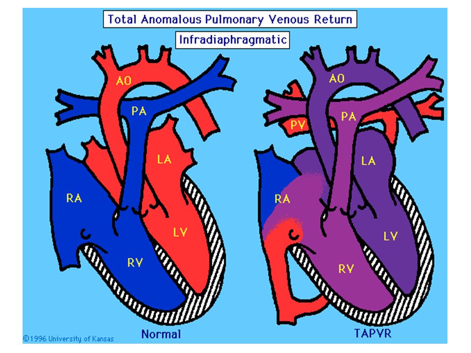
2.3 Total Anomalous Pulm. Venous return - Pulm. drainage into systemic vein

66
2.4Single Ventricle - No interventricular septum

67
2.5 Hypoplastic Left Heart Syndrome - Under development of Lt Side of the heart - Atretic aortic or mitral orifice - Non functional Lt ventricle - Hypoplasia of ascending aorta Right ventricle supplies both pulm. & systemic circulation 67

68
2.6 Persistent fetal circulation 2.6 Dextroposition of the heart 2.7 Dextrocardia

69
To be continue…

Similar presentations


 tricuspid valve 2. Hypoplastic right ventricle 3. Ventricular septal defect 4. Atrial.>")


