Download presentation
Presentation is loading. Please wait.

1
Amenorrhoea

2
GENERAL OBJECTIVE Students will understand amenorrhoea and types and causes as well as management.

3
Specific objectives After attending lecture, the student will can:
Know what is the amenorrhoea and it’s types. List causes of primary amenorrhoea. Given patient history ,examination and a set of laboratory data, correctly diagnose the disease. (problem-solving). Discuses treatment.
. Discuses treatment.")
4
Amenorrhea Amenorrhea is the absence of menstruation.
Amenorrhoea can be classified as: A. Physiological 1.Before puberty. 2. During pregnancy. 3. During lactation. 4. After the menopause.

5
Before puberty: Oestrogen level is not sufficient to promote adequate endometrial development to cause bleeding.

6
During pregnancy.

7
During lactation: The average time between delivery and the first subsequent period and associated Ovulation is: 10-14 weeks in those patients who do not breast-feed their infants. In those with breast feeding ,it depends on duration of breast feeding. During lactation, Prolactin is secreted in large amounts by anterior pituitary gland and there is suppression of LH secretion so the ovarian follicles may mature but fail to rupture.

8
After the menopause. Cessation of oestrogen production from the ovary due to no graffian follicles in ovary.

9
B. Pathological: can be classified as:
Primary amenorrhoea: No spontaneous uterine bleeding has occurred. Secondary amenorrhoea: cessation of the periods after menstruation has been established for more than six months in a normal female of reproductive age that is not due to pregnancy.

10
Primary amenorrhoea or
No spontaneous uterine bleeding has occurred by the age of: *14(13) years in the absence of secondary sexual characteristic or *16 years in presence of secondary sexual characteristic.
 years in the absence of secondary sexual characteristic. or. *16 years in presence of secondary sexual characteristic.")
11
Aetiology of primary amenorrhoea
classify the etiologies of primary amenorrhoea based on the presence or absence of secondary sexual characteristics.

12
A/ primary amenorrhoea with secondary sexual development:
1.Out flow obstruction: Imperforate hymen. Transverse vaginal septum. Cervical agenesis.

13
A/ primary amenorrhoea with secondary sexual development:
a. Mullerian agenesis (Absent uterus (Rokitansky syndrome). b. Surgical removal of the uterus. c. Damage after radiotherapy. d. Asherman's Syndrome (very rare in primary amenorrhoea)
. b. Surgical removal of the uterus. c. Damage after radiotherapy. d. Asherman s Syndrome (very rare in primary amenorrhoea)")
15
A/ primary amenorrhoea with secondary sexual development:
3. Complete androgen insensitivity syndrome (46XY) (testicular feminization).
 (testicular feminization).")
16
A/ primary amenorrhoea with secondary sexual development:
4. Ovarian cause: (unusual) Anovulation: poly cystic ovary syndrome (PCO) Hyperprolactinemia. Premature ovarian failure. Resistant ovary syndrome. Surgical removal of the ovaries. exposure to radiation.
 Anovulation: poly cystic ovary syndrome (PCO) Hyperprolactinemia. Premature ovarian failure. Resistant ovary syndrome. Surgical removal of the ovaries. exposure to radiation.")
17
A/ primary amenorrhoea with secondary sexual development:
5.Pituitary disorders: Pituitary adenomas. Sheehan's syndrome.

18
A/ primary amenorrhoea with secondary sexual development:
Hypothalamic disorders: Space occupying lesion: crainopharyngioma, tuberculosis, sarcoidosis. Congenital GnRH deficiency leads to low gonadotropin levels. When this occurs with anosmia, it is diagnosed as Kallman syndrome. Weight loss. Anorexia nervosa. Excessive exercise. Environment : Sudden change in the environment as in change of residence or in occupation can cause amenorrhoea. Emotional upsets and mental stress. Stress during examination can also cause loss of menses. major psychiatric disorders such as Psychoses, depression.

19
A/ primary amenorrhoea with secondary sexual development:
7.Pregnancy. 8. Idiopathic. Constitutional delay

20
A/ primary amenorrhoea with secondary sexual development:
9. Other causes : *Thyroid: (hypothyroidism and hyperthyroidism) * Diabetes mellitus. *Adrenal Tumours. *Chronic diseases.
 * Diabetes mellitus. *Adrenal Tumours. *Chronic diseases.")
21
B . primary ammenorrhoea without secondary sexual development (sexual infantilism):
Patients with primary amenorrhoea and no secondary sexual characteristics display the absence of gonadal hormone secreation. *Gonadal agenesis and dysgenesis (including Turner syndrome). *Delayed puberty. (Constitutional).
. *Delayed puberty. (Constitutional).")
22
B . primary ammenorrhoea without secondary sexual development (sexual infantilism):
*Anorexia nervosa. (if it occurred before development of secondary sexual characters) *17 alpha-hydroxylase deficiency. (Prevent synthesis of sex steroids).
 *17 alpha-hydroxylase deficiency. (Prevent synthesis of sex steroids).")
23
Diagnoses unique to primary amenorrhea include:
vaginal agenesis. androgen insensitivity syndrome. Turner syndrome (45,X). The remaining causes should be considered in patients with primary amenorrhea and in patients with secondary amenorrhea. Note: unique main only present in primary amenorrhoea.
. The remaining causes should be considered in patients with primary amenorrhea and in patients with secondary amenorrhea. Note: unique main only present in primary amenorrhoea.")
24
Management:

25
History An adequate history includes:
Childhood growth and development including height and weight charts and age at breast development if present. Presence or absence of cyclical symptoms mainly abdominal pain, difficulties in urination and even urinary retention, difficulties in defecation. (Outflow obstruction). Excessive weight loss/ presence of eating disorder.
. Excessive weight loss/ presence of eating disorder.")
26
History Excessive exercise. psychosocial condition should be known.
vasomotor symptoms, hot flushes, virilizing changes, galactorrhea, fatigue, palpitations, nervousness, headache, hearing loss, and visual changes. Family history of anosmia, androgen insensitivity in family as it may affect other females in the family and ascertaining the age at menarche of the patient's mother and sisters is advisable because the age at menarche in family members can occur within a year of the age in others. Any history of chronic illness, trauma, surgery, and medications is also important. A sexual history should be obtained if married.

27
Physical examination:
Pituitary infantilism due to lack of growth hormone. Turner’s syndrome. (Webbed neck, increased carrying angle, lack of breast development, and short stature). -The girl's stature may be tall which may be due to androgen insensitivity. * Girl weight and calculate her body mass index (BMI). *development of secondary sexual characteristic (breast development, axillary and pubic hair growth).
. -The girl s stature may be tall which may be due to androgen insensitivity. * Girl weight and calculate her body mass index (BMI). *development of secondary sexual characteristic (breast development, axillary and. pubic hair growth).")
28
Physical examination:
* Any evidence of abnormal virilization (as clitromegally) as seen in Adrenal or ovarian tumors. * hirsutism. *Fundoscopy examination, visual filed examination and neurological examination if pituitary tumour is suspected.
 as seen in Adrenal or ovarian. tumors. * hirsutism. *Fundoscopy examination, visual filed examination and neurological examination if pituitary tumour is suspected.")
29
Breast examination: Look for presence of breast and its stage of development. Presence of Galactorrhea.

30
Pelvic examination: Should be undertaken in the presence of the patient’s mother. Inspect vulva to see opening of introitus (opened, closed by bluish membrane, closed) A bimanual examination is inappropriate (contraindicated) in unmarried girl. Furthermore, it may be more appropriate to defer this from the first consultation to assure the patient’s confidence in future management and in most cases information taken from transabdominal ultrasound examination of the pelvis.
 A bimanual examination is inappropriate (contraindicated) in unmarried girl. Furthermore, it may be more appropriate to defer this from the first consultation to assure the patient’s confidence in future management and in most cases information taken from transabdominal ultrasound examination of the pelvis.")
31
Rectal examination shows the absence of uterus.

32
Investigations: Start investigation in cases with primary amenorrhoea
14 years in the absence of secondary sexual characteristic 16 years in presence of secondary sexual characteristic in some circumstances, it is reasonable to initiate an evaluation despite the absence of the above strict criteria as: in patient with the stigmata of Turner syndrome. in patient with obvious virilization. in a patient or her parents are concerned.

33
Investigations: The history and physical findings help in selecting tests in a female patient with amenorrhea. Chromosomal analysis: In uncertain diagnosis, full chromosomal analysis and Karyotype should be done. In Karyotype A buccal smear and examination of the polymorphnuclear leucocytes to determine if chromatin positive (XX) or chromatin negative (XO or XY) and some time full chromosomal analysis may be needed.
 or chromatin negative (XO or XY) and some time full chromosomal analysis may be needed.")
34
Investigation for chronic illness:
If the history or physical findings suggest a chronic disease process, the following may be indicated including measurement of the erythrocyte sedimentation rate (ESR), liver function tests, renal function test and urinalysis.
, liver function tests, renal function test and urinalysis.")
35
should include follicular stimulation hormon (FSH),lutinizing hormone ( LH), oestradiol, testosterone, Prolactin, thyroid function test, growth hormone , adrenocorticotropic hormone (ACTH).
,lutinizing hormone ( LH), oestradiol, testosterone, Prolactin, thyroid function test, growth hormone , adrenocorticotropic hormone (ACTH).")
36
The result in hypothalamic or pituitary cause is low FSH, LH, oestradiol.
The results in ovarian cause are high gonadotrophin and low estrogen.

37
Imaging studies: *Ultrasound: Determine the presence, state and size of ovaries and any follicular activity. Determine the presence and size of uterus. Congenital anatomic abnormalities of uterus or vagina or both are often associated with renal abnormalities such as unilateral solitary kidney so these patients should have intravenous pyelogram.

38
*a coned view of the sella turcica or MRI of the pituitary is indicated.
*Bone age X ray to determining bone age which is important in differentiating pubertal delays as a cause.

39
*Laproscopy Laproscopy rarely used to assess pelvic organ. It is useful in: * cases which there is doubt about the nature of the gonads. *cases where ovarian biopsy is needed to determine presence of primordial oocytes.

40
Most of the conditions are rare and constitutional delay without doubt is the most common diagnosis. However, as the rest of the diagnoses have serious implications this diagnosis of constitutional delay should only be made when all other syndromes have been excluded. Constitutional delayed puberty is a diagnosis of exclusion.

41
Investigative pathway for a patient with normal sexual characteristics.

42
Investigative pathway for a patient with no secondary sexual characteristics

43
Treatment: No attempt should ever be made to treat patients with primary amenorrhea until a firm diagnosis is reached.

44
Patient with sexual infantilism (without secondary sexual charcteristic)
Cases of Turner's syndrome Induce breast development by very gradually increasing oestrogen doses then change to definitive treatment of hormone replacement therapy (estrogen and progestrone). They have no hope to achieve pregnancy. In hypogonadotrophic hypogonadism who seek fertility will need therapy with either human menopausal gonadotrophin injection or gonadotrophin releasing hormone (GnRH). In 17-hydroxylase deficiency have no hope to achieve pregnancy.
. They have no hope to achieve pregnancy. In hypogonadotrophic hypogonadism who seek fertility will need therapy with either human menopausal gonadotrophin injection or gonadotrophin releasing hormone (GnRH). In 17-hydroxylase deficiency have no hope to achieve pregnancy.")
45
Patient with primary amenorrhoea with secondary sexual development:
androgen insensitivity * Excision of gonads as this gonad is a testis and there is a malignant potential in about 30% of cases *Creation of neovagina to permit sexual intercourse. *Treatment with oestrogen to augment breast development and prevent osteoporosis. * Psychological support. Those women are unable to be fertile at all.

46
Patient with primary amenorrhoea with secondary sexual development:
Imperforate hymen Treated by hymenectomy.

47
Patient with primary amenorrhoea with secondary sexual development:
Transverse vaginal septum Surgical correction is indicated but difficult technique is needed. Cervical agenesis: Hysterectomy is recommended.

48
Constitutional delay Girls in whom normal secondary sexual characteristics exist. There is no anatomical anomaly and endocrine investigations are all normal. It is reasonable to await events as these young women will eventually menstruate spontaneously as the maturation process proceeds. There is no need to suggest any treatment other than annual review. In some circumstances it may be useful to promote a menstruation using the oral contraceptive pill for one cycle to prove that menstruation can occur and this can be extremely reassuring. Other causes treated as it is (discussed in specific subject lectures)
")
49
Imperforate hymen The imperforate hymen may present at two ages of development. It may present in: Early childhood When the infant presents with a bulging hymen behind which is a mucocele, the vagina expanded by vaginal secretions of mucus. Treated by hymenectomy and does not subsequently cause any problems following hymenectomy.

50
At puberty The very distensible features of vagina allow quite large quantities of blood to collect in some cases. This situation is known as haematocolpos. When some blood does accumulate within the uterine cavity it is known as a haematometra.

51
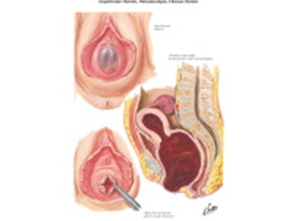
Diagram of hematometra and hematocolpos with imperforate distal transverse vaginal septum

52
History: A pubertal girl complains of intermittent cyclical abdominal pain. The pain is due to dysmenorrhoea associated with the accumulation of menstrual blood within the vagina. As the mass enlarges there may be associated difficulty with micturition and defaecation and even associated with retension of urine in some cases.

53
Examination: Normal stature and have normal secondary sexual characteristic. Abdominal examination will reveal on occasions an abdominal swelling Pelvic examination by inspection of external genitalia showed a tense bulging bluish membrane (which is the hymen) closing the introitus.
 closing the introitus.")
54
Abdominal mass with imperforate hymen

55
. observation of the introitus will display a tense bulging bluish membrane which is the hymen.

56
Vaginal agenesis. Not to be confused with imperforate hymen.

57
Investigation: Ultrasound showed blood collection in vagina and uterus.

58
Treatment: After explanation of the condition and obtaining parents consent,a cruciate incision (+) in the hymen allows drainage of the retained menstrual blood. From medico-legal point of view, the girl must be given a report confirm that the hymen was opened by surgical operation as treatment.
 in the hymen allows drainage of the retained menstrual blood. From medico-legal point of view, the girl must be given a report confirm that the hymen was opened by surgical operation as treatment.")
Similar presentations


>")


