Download presentation
Presentation is loading. Please wait.

1
2/25/08 Blood Vessels Chapter 19 – Day 1

2
2/25/08 Blood Vessel Structure Fig. 19.1

3
2/25/08 Blood Vessels - Intro Blood vessels are essentially a “pipeline” to: ♦Carry blood TO parts of the body = arteries ♦Carry blood BACK FROM parts of the body = veins Subdivisions ♦Pulmonary blood vessels: blood to the lungs and back ♦Systemic blood vessels: blood to and from the rest of the body

4
2/25/08 Blood Vessels - Intro Veins = mostly deoxygenated – except pulmonary vein & umbilical vein Arteries = mostly oxygenated – except pulmonary artery & umbilical artery Capillaries = interconnecting vessels ♦Enable gas exchange, etc. Blood vessel structure and comparison activity in lab

5
2/25/08 Blood Vessel Structure Fig. 19.1

6
2/25/08 Arteries vs. Veins - Similarities 3 Layers Tunica Externa ♦Connective Tissue Tunica Media ♦Smooth muscle cells ♦Elastic fibers (arteries) ♦Collagen fibers Tunica Interna ♦Elastic layer ♦Endothelial cells & connective tissue with elastic fibers (arteries)
 ♦Collagen fibers Tunica Interna ♦Elastic layer ♦Endothelial cells & connective tissue with elastic fibers (arteries).")
7
2/25/08 Arteries vs. Veins Arteries are thick walled Larger arteries have more elastic fibers ♦Tunica media – thicker, concentric & longitudinal ♦Tunica interna – internal elastic membrane ARTERIES ♦Elasticity Ability to stretch when full = high pressure Return to their original state when relaxed ♦Contractability More smooth muscle (than veins) Vasodilation, Vasoconstriction Both veins & large arteries need O2 – supplied by vaso vasorum
 Vasodilation, Vasoconstriction Both veins & large arteries need O2 – supplied by vaso vasorum.")
8
2/25/08 Arteries vs. Veins Hierarchy of organization ♦(learn examples from text) Largest vessels coming out of heart or to heart These carry the most volume ♦If arteries – they have the highest pressure Diagram on board and Fig. 19.2 Be able to work though these – know order and characteristics
 Largest vessels coming out of heart or to heart These carry the most volume ♦If arteries – they have the highest pressure Diagram on board and Fig Be able to work though these – know order and characteristics.")
9
2/25/08 Blood Vessel Hierarchy Fig. 19.2

10
2/25/08 Capillaries Fig. 19.5

11
2/25/08 Arteries vs. Veins Different jobs and different driving forces Arteries ♦Force of contraction pushes blood forward ♦Blood pressure = driving force ♦Moves downhill to lower extremities - gravity Veins ♦Lower extremities to heart = against gravity ♦Low pressure ♦Relies on other driving forces

12
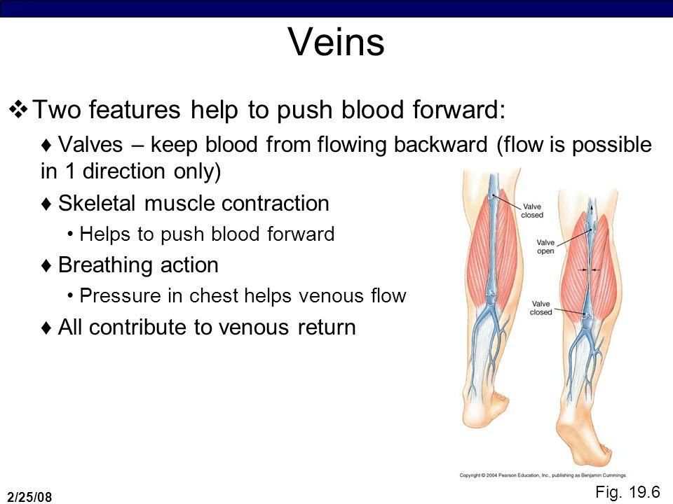
2/25/08 Veins Two features help to push blood forward: ♦Valves – keep blood from flowing backward (flow is possible in 1 direction only) ♦Skeletal muscle contraction Helps to push blood forward ♦Breathing action Pressure in chest helps venous flow ♦All contribute to venous return Fig. 19.6
13
2/25/08 Blood vessel function Ultimately blood delivers O 2 & nutrients to tissues as well as removing wastes How does this happen? Any organ: ♦Blood vessels (in & out): arteries – smallest branch = capillary ♦The organ is infused with capillaries ♦Nutrient exchange occurs at this level In the capillaries – only the endothelial layer is present Substances in the capillaries move to the Interstitial Fluid → then into cells
: arteries – smallest branch = capillary ♦The organ is infused with capillaries ♦Nutrient exchange occurs at this level In the capillaries – only the endothelial layer is present Substances in the capillaries move to the Interstitial Fluid → then into cells.")
14
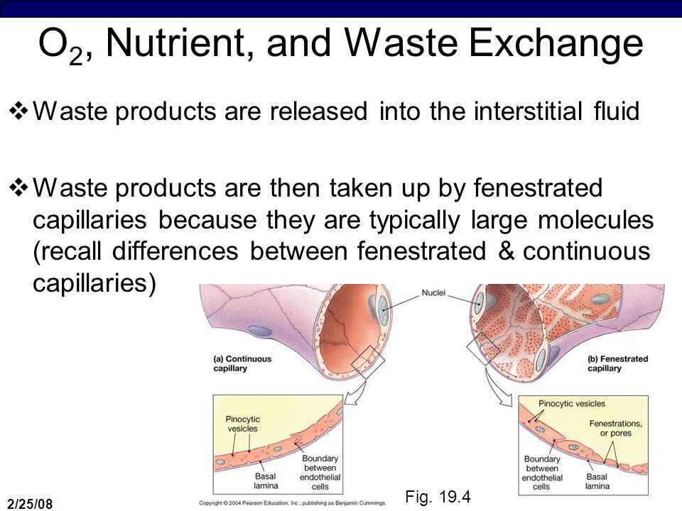
2/25/08 O 2, Nutrient, and Waste Exchange Waste products are released into the interstitial fluid Waste products are then taken up by fenestrated capillaries because they are typically large molecules (recall differences between fenestrated & continuous capillaries) Fig. 19.4
15
2/25/08 O 2, Nutrient, and Waste Exchange Exchange between blood and interstitial fluid 1.Simple (& Facilitated) Diffusion: ♦In response to a concentration gradient 2.Filtration ♦Force pushes out substances – based on pressure 3.Osmosis ♦Reabsorption of water
 Diffusion: ♦In response to a concentration gradient 2.Filtration ♦Force pushes out substances – based on pressure 3.Osmosis ♦Reabsorption of water")
16
2/25/08 Diffusion Ions and small organic molecules (glucose, amino acids, urea – move through pores in fenestrated capillaries or move via diffusion between endothelial cells of adjacent capillaries Ions (Na+, K, etc.) diffuse across endothelial cells by passing through channels in cell membranes Large water-soluble compounds can only work enter or leave blood stream via fenestrated capillaries Lipids (FAs, steroids) and lipid-soluble compounds (esp. CO2, O2) cross capillary walls by diffusion through endothelial cell membranes Plasma proteins can only diffuse through in sinusoids (such as those in the liver)
 cross capillary walls by diffusion through endothelial cell membranes Plasma proteins can only diffuse through in sinusoids (such as those in the liver).")
17
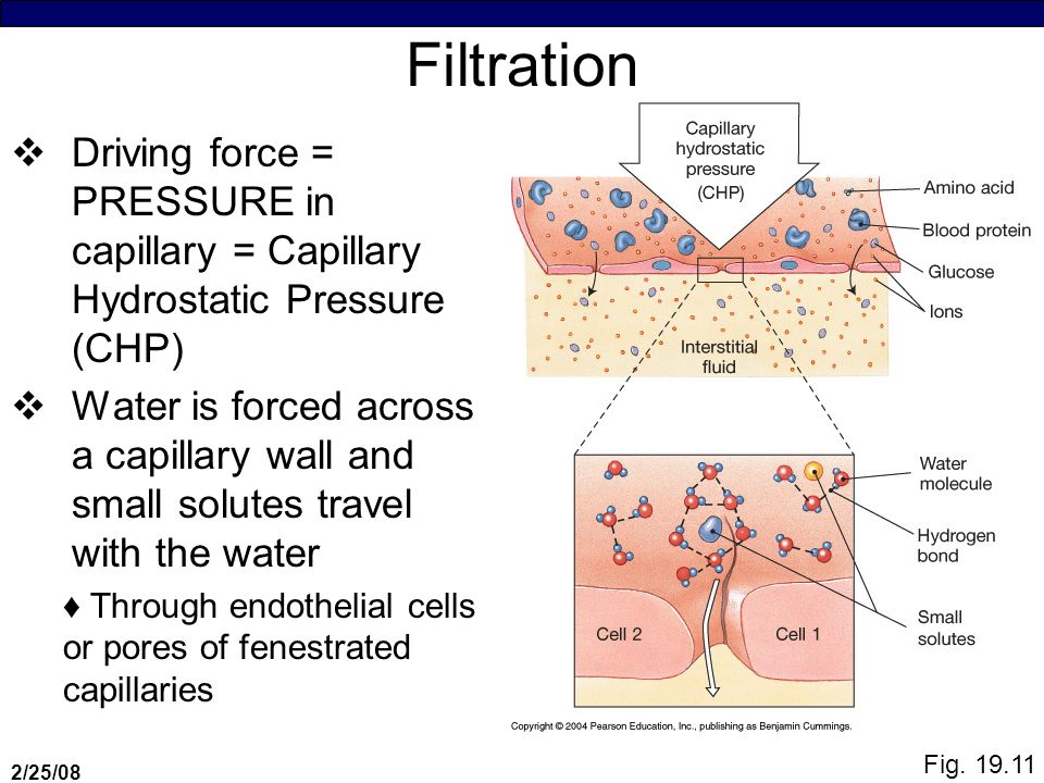
2/25/08 Filtration Driving force = PRESSURE in capillary = Capillary Hydrostatic Pressure (CHP) Water is forced across a capillary wall and small solutes travel with the water ♦Through endothelial cells or pores of fenestrated capillaries Fig. 19.11
18
2/25/08 Reabsorption Occurs as a result of osmosis ♦Diffusion of water across selectively permeable membrane Remember, water molecules move toward soln with higher solute concentration Process by which dissolved solutes is moved Osmotic pressure = amount of pressure that must be applied to prevent osmotic movement across a membrane ♦BCOP = blood colloid osmotic pressure = osmotic pressure of blood Remember…hydrostatic pressure forces water OUT of solution, whereas osmotic pressure draws water INTO a solution

19
2/25/08 Pressures have to be balanced so that fluid in and out can be coordinated Fig. 19.11

20
2/25/08 Filtration & Reabsorption If BHP > BOP in the blood vessel, fluid is pushed out If BHP < BOP fluid enters in IFHP & IOP – low & stable because there are fewer proteins in interstitial fluid Net Filtration Pressure is the difference between the net hydrostatic pressure and the net osmotic pressure: ♦Net filtration = net hydrostatic – net colloid pressurepressure osmotic pressure If positive fluid moves OUT of capillary If negative fluid moves INTO capillary

21
2/25/08 Equilibrium Analogy to filtration experiment ♦Charcoal = cells & proteins ♦CuSO 4 = nutrients & O 2 Cells & Proteins remain in blood vessel H 2 O, hormones, other chemicals, nutrients, O2, glucose, ions = pushed out Equilibrium between arterial & venus ends maintains proper pressure differences Excess fluid expelled into tissues causes an increase in interstitial fluid – if in excess, causes EDEMA ♦Excess fluid buildup, swollen ankles, etc Read in book and follow handout

22
2/25/08 Equilibrium Equilibrium between arterial & venus ends maintains proper pressure differences Excess fluid expelled into tissues causes an increase in interstitial fluid – if in excess, causes EDEMA ♦Excess fluid buildup, swollen ankles, etc Read in book and follow handout Different demands for gas and nutrient exchange For proper delivery – the cardiovascular system depends on: 1. Cardiac Output 2. Peripheral Resistance 3. Blood Pressure

23
2/25/08 Equilibrium Different demands for gas and nutrient exchange For proper delivery – the cardiovascular system depends on: 1. Cardiac Output 2. Peripheral Resistance 3. Blood Pressure

24
2/25/08 Equilibrium Need constant control of these factors to maintain homeostasis = CARDIOVASCULAR REGULATION Controlled by ♦Autoregulation Mechanisms Local factors change pattern of blood flow w/in capillary beds Response to chemical changes in interstitial fluids ♦Neural Mechanisms Respond to changes in arterial pressure or blood gas levels @ a specific site stimulates cardiovascular centers of ANS ♦Endocrine Mechanisms Releases hormones that enhance short-term adjustments and direct long-term changes in cardiovascular performance

25
2/25/08 Equilibrium

26
2/25/08 Auto Regulation Local changes ♦Sphincters near capillaries are adjusted ♦Depend on local VASODILATION & VASOCONSTRICTION chemicals (nitrous oxide) – know examples from text (p 547) Vasodilator: factor(s) that promote the dilation of precapillary sphincters Local vasodilators act at the tissue level & accelerate blood flow through the tissue of origin
 – know examples from text (p 547) Vasodilator: factor(s) that promote the dilation of precapillary sphincters Local vasodilators act at the tissue level & accelerate blood flow through the tissue of origin")
27
2/25/08 Neural Mechanisms Neural Mechanisms ♦Sympathetic division controls… Smooth muscle tone (vasomotor tone) Increased sympathetic impulses ( vasoconstriction) Decreased sympathetic impulses ( vasodilation) ♦Receptors
 Increased sympathetic impulses ( vasoconstriction) Decreased sympathetic impulses ( vasodilation) ♦Receptors")
28
2/25/08 Capillaries Fig. 19.5

Similar presentations


. The Circulatory System is known as a CLOSED SYSTEM because the blood is contained within either the heart.>")


 Cardiovascular system (fast) (a) cardiac output increase c.o., increase pressure (b) peripheral.>")


