Download presentation
Presentation is loading. Please wait.

1
Dr: Dalia Galal Hamouda
lecture 8 blood bank Hemolytic Diseases: 1-Hemolytic disease of the Newborn and Fetus 2- Hemolytic anaemias Dr: Dalia Galal Hamouda

2
Haemolytic diseases of the fetus &newborn

3
Haemolytic diseases are those conditions in which the red cells of the individual do not survive normally in vivo. Haemolysis lead to red cell destruction and may be associated with anaemia. If red cell destruction occur inside the blood vessels it is called intravascular haemolysis. Intravascular haemolysis is mediated by complement activation and results in free hemoglobin (Hb) in circulation (not good for the kidney). Red cell destruction in the Reticulo-endothelial system (e.g. Liver and spleen) is known as extravascular haemolysis (e.g. aged red cells). Extravascular haemolysis is macrophage mediated red cell destruction. The liver is vital for the removal of toxic metabolites which are produced following red cell haemolysis.
 in circulation (not good for the kidney). Red cell destruction in the Reticulo-endothelial system (e.g. Liver and spleen) is known as extravascular haemolysis (e.g. aged red cells). Extravascular haemolysis is macrophage mediated red cell destruction. The liver is vital for the removal of toxic metabolites which are produced following red cell haemolysis.")
4
Role of the liver in RBC haemolysis
1) Intravascular haemolysis: Releases free hemoglobin which is toxic to the kidney. Hb is immediately bound by haptoglobin in the plasma. Hb-haptoglobin complex is removed by hepatic cells. 2) Extravascular haemolysis: Free Hb haem molecules bilirubin. Free unconjugated bilirubin is transported to the liver where it is conjugated to glucuronic acid and eventually removed ( in faeces).
 Intravascular haemolysis: Releases free hemoglobin which is toxic to the kidney. Hb is immediately bound by haptoglobin in the plasma. Hb-haptoglobin complex is removed by hepatic cells. 2) Extravascular haemolysis: Free Hb haem molecules bilirubin. Free unconjugated bilirubin is transported to the liver where it is conjugated to glucuronic acid and eventually removed ( in faeces).")
5
Fetal liver Foetus liver cannot metabolise bilirubin which is toxic (cannot be filtered by kidney and can damage the brain tissues). Bilirubin in fetal circulation is transferred to the mother circulation for its excretion. - Newborn baby liver enzymes are not functioning before day 10 post-delivery

6
Haemolytic disease of the fetus and newborn (HDFN)
• HDFN is triggered by maternal antibodies to paternally inherited antigens in the fetus. • The most frequently described antibody in HDFN is the RhD antibody. • If the mother is RhD negative and the father is RhD positive the baby may inherit the D antigen and express it on fetal RBC. • Fetal red cells (RhD +) enter the maternal circulation as a result of fetomaternal haemorrhage (FMH) at the time of delivery. • The mother immune system reacts and form anti-D antibodies for many years. • Second pregnancy: Maternal IgG anti-D crosses the placenta and sensitizes the D+ fetal cells.
 enter the maternal circulation as a result of fetomaternal haemorrhage (FMH) at the time of delivery. • The mother immune system reacts and form anti-D antibodies for many years. • Second pregnancy: Maternal IgG anti-D crosses the placenta and sensitizes the D+ fetal cells.")
7
Haemolytic disease of the fetus and newborn (HDFN)
Synthesised fetal RBCs are removed from fetal circulation and destroyed by macrophages in the Reticulo-endothelial system . • This cause severe anaemia and referred to as erythroblastosis fetalis. • During pregnancy, excess free bilirubin is transported to mother for metabolism and excretion. • Severe fetal anaemia causes edema of the fetal liver and spleen, which are the organs for both erythropoiesis and cell destruction. • In the most severe cases of HDFN, Hydrops fetalis (severe oedema) may result in intrauterine death and stillbirth. • At delevery the maternal system is no longer available to remove and excrete bilirubin & neonatal liver is not fully functioning free toxic bilirubin can damage the brain (kernicterus).
 may result in intrauterine death and stillbirth. • At delevery the maternal system is no longer available to remove and excrete bilirubin & neonatal liver is not fully functioning free toxic bilirubin can damage the brain (kernicterus).")
8
HDFN caused by ABO blood group incompatibility
• HDFN occurs very frequently due to ABO incompatibility between the mother and the fetus. • The ABO antibodies in the maternal blood are IgG anti- A,B in a group O mother carrying a group A or B fetus. • This type of HFDN does not cause problem to the fetus in the uterus but may cause jaundice and mild anaemia post-delivery. • The disease usually requires special treatment such as phototherapy and/ or the administration of immunoglobulin.

9
HDFN caused by Rh blood group incompatibility
• Maternal anti-D is the most frequent cause of Rh HDFN and causes the most severe cases of Rh HDFN, followed by anti-c. • The other antibodies within the Rh system may also be implicated, in addition to combinations of antibodies, e.g. anti-C plus anti-D, anti-C plus anti-e, or anti-c plus anti-E.

10
Antenatal testing • Pregnant woman blood should be sent to the lab for antenatal screening tests: 1- ABO grouping. 2-Rh typing. 3- antibody screening. 4- antibody identification. • Parental red cell typing • Fetal amniocentesis & Doppler technique

11
Postnatal tests for HDFN

12
Haemolytic anaemias

13
Alloimmune hemolysis • Hemolytic disease of the newborn (also known as HDN or erythroblastosis fetalis) – Rh D hemolytic disease of the newborn (also known as Rh disease) – ABO hemolytic disease of the newborn (the indirect Coombs test may only be weakly positive) – Anti-Kell hemolytic disease of the newborn – Rh c hemolytic disease of the newborn – Rh E hemolytic disease of the newborn – Other blood group incompatibility (RhC, Rhe, Kidd, Duffy, MN, P and others) • Alloimmune hemolytic transfusion reactions
 – Rh D hemolytic disease of the newborn (also known as Rh disease) – ABO hemolytic disease of the newborn (the indirect Coombs test may only be weakly positive) – Anti-Kell hemolytic disease of the newborn – Rh c hemolytic disease of the newborn – Rh E hemolytic disease of the newborn – Other blood group incompatibility (RhC, Rhe, Kidd, Duffy, MN, P and others) • Alloimmune hemolytic transfusion reactions")
14
Examples of autoimmune hemolysis
• Warm antibody autoimmune hemolytic anemia – Idiopathic – Systemic lupus erythematosus – Evans' syndrome (antiplatelet antibodies and hemolytic antibodies) • Cold antibody autoimmune hemolytic anemia – Idiopathic cold hemagglutinin syndrome – Infectious mononucleosis – Paroxysmal cold hemoglobinuria
 • Cold antibody autoimmune hemolytic anemia – Idiopathic cold hemagglutinin syndrome – Infectious mononucleosis – Paroxysmal cold hemoglobinuria")
15
Drug-induced immune-mediated hemolysis
• Methyldopa (IgG mediated type II hypersensitivity) • Penicillin (high dose) • Quinidine (IgM mediated activation of classical complement pathway and Membrane attack complex, MAC)
 • Penicillin (high dose) • Quinidine (IgM mediated activation of classical complement pathway and Membrane attack complex, MAC)")
16
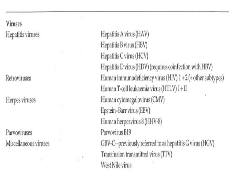
Infections agents reported to have been transmitted by blood transfusion (2 species only)
")
Similar presentations


i.>")
>")
 & COOMBS TEST>")