Download presentation
Presentation is loading. Please wait.

1
Chapter 15.4: Electrocardiogram

2
Electrocardiogram (ECG/EKG)
A recording of electrical changes in a heartbeat Action potentials generate measurable electrical currents Three waves (changes in electrical current): P Wave QRS Complex T Wave
: P Wave. QRS Complex. T Wave.")
3
EKG Waves P wave QRS Complex T Wave Atrial depoloarization
Seconds Millivolts (mV) EKG Waves P wave Atrial depoloarization Action potential moving throughout atria Initiates atrial contraction QRS Complex Ventricular depolarization Action potential spreading through ventricles Initiates ventricular contraction T Wave Ventricular repolarization Occurs just before ventricle relaxation
 EKG Waves. P wave. Atrial depoloarization. Action potential moving throughout atria. Initiates atrial contraction. QRS Complex. Ventricular depolarization. Action potential spreading through ventricles. Initiates ventricular contraction. T Wave. Ventricular repolarization. Occurs just before ventricle relaxation.")
5
Chapter 15.5: The Cardiac Cycle

6
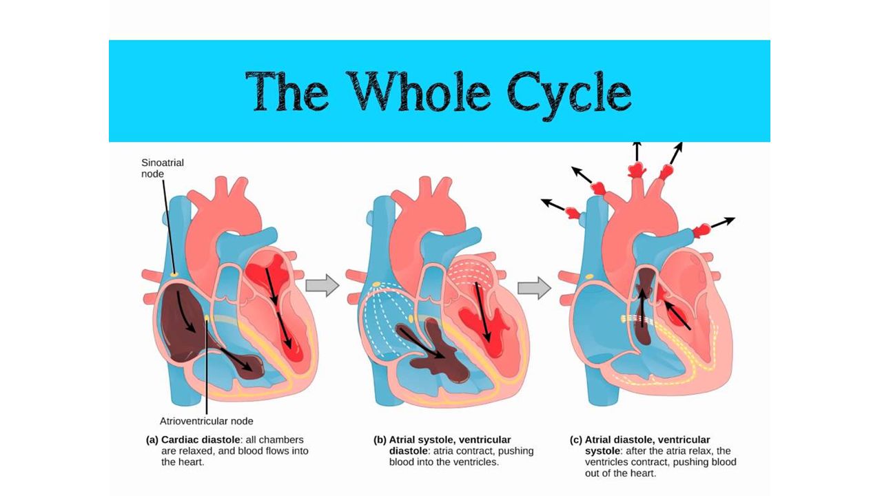
Cardiac Cycle All of the events associated with one heart beat
Three main events: Relaxation (diastole) period All 4 chambers relaxed Ventricles repolarizing (T-Wave) AV valves open and 75% of blood empties into ventricles 1 Relaxation period 3 Ventricular systole 2 Atrial systole
 period. All 4 chambers relaxed. Ventricles repolarizing (T-Wave) AV valves open and 75% of blood empties into ventricles. 1. Relaxation. period. 3. Ventricular. systole. 2. Atrial. systole.")
7
Cardiac Cycle Three main events: 2) Atrial systole (contraction)
- Both atria contract - Occurs right after P-Wave - Forces last 25% of blood into ventricles 3) Ventricular systole (contraction) - Both ventricles contract - Occurs right after QRS Complex - Pushes blood out of the ventricles 1 Relaxation period 3 Ventricular systole 2 Atrial systole
 Ventricular systole (contraction) - Both ventricles contract. - Occurs right after QRS Complex. - Pushes blood out of the ventricles. 1. Relaxation. period. 3. Ventricular. systole. 2. Atrial. systole.")
9
Heart Sounds Caused by closing of valves
Lubb: a long, booming sound from AV valves closing Occurs right as ventricular systole begins Dupp: the second, short and sharp sound from SL valves closing Occurs at the end of ventricular systole

Similar presentations


 b.Atrioventricular node (AV node) c.Atrioventricular bundle (AV bundle) d.Right and left atrioventricular bundle.>")


 Transport O 2, nutrients, hormones, cell wastes, etc…>")


