Download presentation
Presentation is loading. Please wait.

1
Neurophysiology Neurons Gross Anatomy The Central Auditory Nervous System Frequency and Intensity encoding Central Auditory Processing Binaural Processing Central Control (Descending Systems) Cortical Processing
 Cortical Processing")
2
The Neuron Dendrites receive synaptic stimulation (neurotrans.) Action Potential generated in soma near axon AP conducted along axon from Node to Node (saltatory conduction) AP produces release of neurotransmitter at terminal boutons
 Action Potential generated in soma near axon AP conducted along axon from Node to Node (saltatory conduction) AP produces release of neurotransmitter at terminal boutons")
3
An Action Potential (or Spike)
")
4
Two Descriptors for Neurons Afferent (sensory)-- carrying signals toward the brain Efferent (motor) -- carrying signals from brain to periphery
-- carrying signals toward the brain Efferent (motor) -- carrying signals from brain to periphery")
5
Afferent Afferent & Efferent Neurons

6
4 Types of Cochlear Neurons INNER HAIR CELLS >Multiple (10 to 20) Afferent synapses >(Efferents synapse on afferent dendrites) OUTER HAIR CELLS: >Large Efferent synapses engulf base of cell >Small (& not very active) Afferent synapses
 Afferent synapses >(Efferents synapse on afferent dendrites) OUTER HAIR CELLS: >Large Efferent synapses engulf base of cell >Small (& not very active) Afferent synapses")
7
IHC Innervation Pattern

8
OHC Innervation Pattern

9
Inner hair cells Synapse at the base with up to 20 afferent neurons “Divergence” Efferents synapse on afferent dendrites under IHCs

10
IHC activation alters firing rate

11
Afferent neurons have their cell bodies in the Spiral Ganglion (4)
")
12
Central Nervous System Structures Nucleus = a group of nerve cell bodies Fiber Tract = a group of axons

13
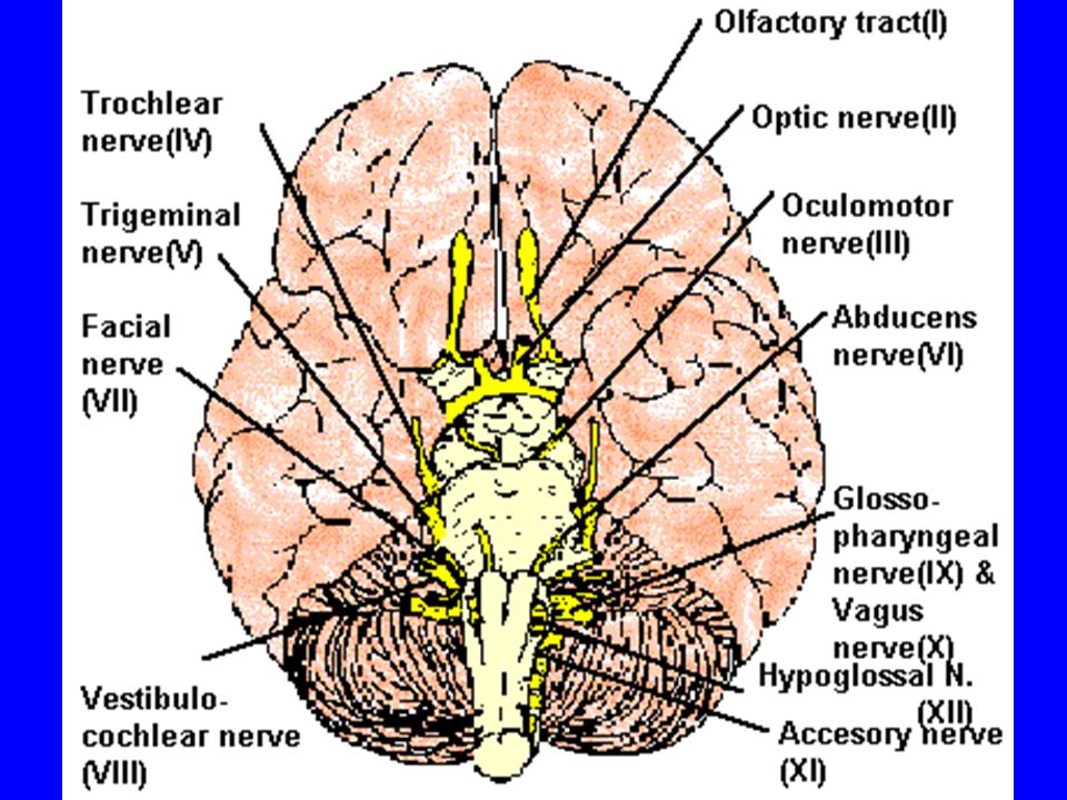
Major Components of the Central Auditory Nervous System (CANS) VIIIth cranial nerve Cochlear Nucleus Superior Olivary Complex Lateral Lemniscus Inferior Colliculus Medial Geniculate Body Primary Auditory Cortex Brainstem Thalamus Mid-brain Temporal Lobe
 VIIIth cranial nerve Cochlear Nucleus Superior Olivary Complex Lateral Lemniscus Inferior Colliculus Medial Geniculate Body Primary Auditory Cortex Brainstem Thalamus Mid-brain Temporal Lobe")
14
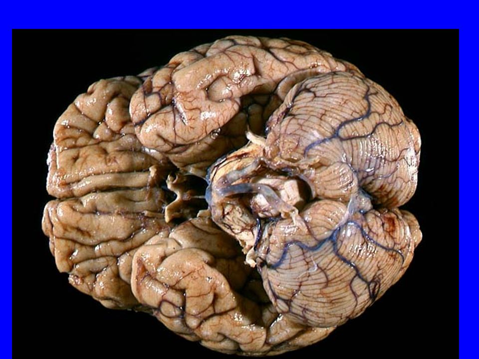
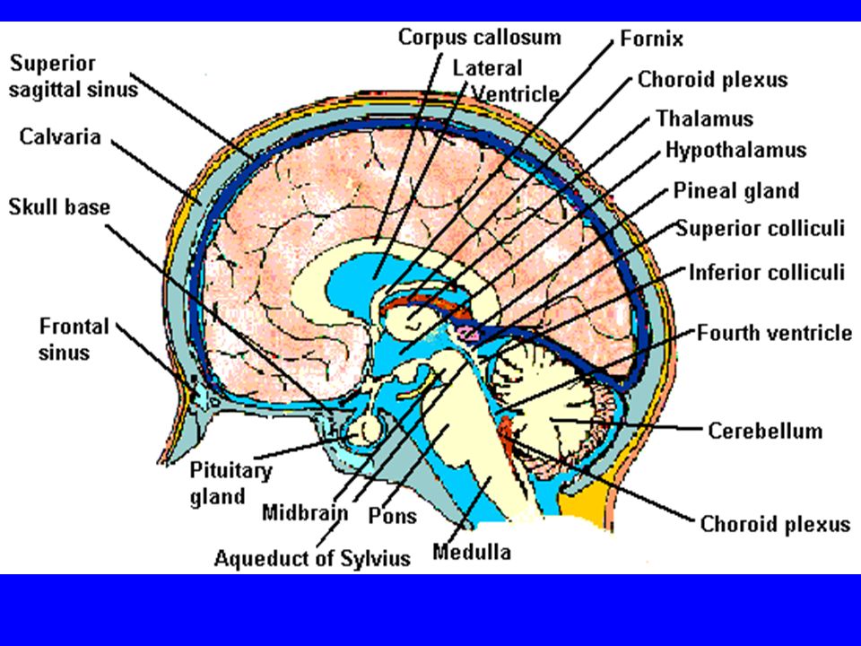
Mid-Saggital View of Brain Pons Cerebellum 4th Ventricle Thalamus Corpus Callosum

15
MedGen Body Inf Coll Lat Lemn SOC Coch Nuc VIIIth CN

16
Neural Web-Site http://rprcsgi.rprc.washington.edu/ neuronames/hierarchy.html

20
Section Thru Brainstem Shows Cochlear Nucleus

21
The Superior Olivary Complex

22
Connections To the Superior Olivary Complex

23
Superior Olivary Processing Supports Localization Lateral SO-- Interaural Intensity Differences Medial SO-- Interaural Time Differences (These are the two primary acoustic cues for localizing sounds)
")
24
Dorsal (back) Side of Brainstem Thalamus (medial geniculate) Inferior Colliculus 4th Ventricle Area of Pons
 Side of Brainstem Thalamus (medial geniculate) Inferior Colliculus 4th Ventricle Area of Pons")
25
Inferior Colliculus

26
Thalamus in Purple

27
Auditory Radiations Connect Medial Geniculate Body (in purple) to Primary Auditory Cortex (in blue)
 to Primary Auditory Cortex (in blue)")
28
Lateral-Superior view of brain

29
Primary Auditory Cortex (AI): superior surface of the temporal lobe
: superior surface of the temporal lobe")
30
Brain Photos Web-Sites http://rpiwww.mdacc.tmc.edu:80/se/anatomy /brain/ http://www.ets.uidaho.edu/med532/start.htm

31
Neurophysiological Measures Gross Evoked Potentials-- Voltage changes in response to auditory stimulation recorded from the scalp Single-Unit Measures-- Voltage (or other) changes recorded within a neuron
 changes recorded within a neuron")
32
Auditory Evoked Potentials Recorded in different time intervals (“epochs”) following a sound Earlier epochs come from lower in the system Later epochs come from higher in the system
 following a sound Earlier epochs come from lower in the system Later epochs come from higher in the system")
33
Examples of AEP Epochs Electrocochleography-- within 5 milliseconds Auditory Brainstem Response-- thru 10 ms Middle Latency Response-- thru 75 ms Auditory Late Response-- thru 200 ms

34
Auditory Brainstem Response Time (ms) Amplitude ( V) Wave V Latency I II III IV V Amp V 0 10
 Amplitude ( V) Wave V Latency I II III IV V Amp V 0 10")
35
GENERATORS of ABR WAVES I II III IV V =Distal VIIIth nerve =Medial VIIIth nerve =Cochlear Nucleus =Superior Olivary Complex =Lateral Lemniscus & Inferior Colliculus

36
Single-Unit Measures Post-Stimulus Time Histogram-- Shows firing rate changes over time Period or Interval Histograms-- Show phase-locking of neural firing

37
Tuning Curves Iso-Rate Function -- Shape similar to what we’ve already described (Fig 6.12 b) Iso-level Function -- Shows spike rate as a function of frequency-- peak at a single frequency (Fig 6.12a)
 Iso-level Function -- Shows spike rate as a function of frequency-- peak at a single frequency (Fig 6.12a)")
38
Two-Tone Suppression The response to one tone can be reduced or eliminated by introducing a second tone near the neuron’s CF. (Fig 6.16) Second tone can be either one which normally would excite the neuron or not
 Second tone can be either one which normally would excite the neuron or not.")
39
Two-tone Suppression Regular Tuning Curve

40
Frequency Coding The Place Code-- each neuron has a characteristic frequency Periodicity Pitch-- neurons phase-lock to stimuli

41
Intensity Coding Firing rate increases in single neurons Spread of activation to a wider range of neurons-- “Density of Discharges” Latency of Firing (shorter delay at higher levels)
")
42
Efferent (Descending) Control Cochlear Efferents come from Superior Olivary Complex --The Olivo-Cochlear Bundle (OCB) Uncrossed OCB-- synapses on dendrites under inner hair cells Crossed OCB-- synapses on outer hair cells Both use inhibitory neurotransmitters
 Control Cochlear Efferents come from Superior Olivary Complex --The Olivo-Cochlear Bundle (OCB) Uncrossed OCB-- synapses on dendrites under inner hair cells Crossed OCB-- synapses on outer hair cells Both use inhibitory neurotransmitters")
43
Uncrossed OCB-- synapses on dendrites under inner hair cells

44
Crossed OCB-- synapses on outer hair cells

45
Efferent Control (cont’d) The Acoustic Reflex Auditory Cortex and Thalamus also send descending fibers to auditory brainstem locations
 The Acoustic Reflex Auditory Cortex and Thalamus also send descending fibers to auditory brainstem locations")
46
The Acoustic Reflex Afferent: VIIIth nerve Cochlear Nucleus Superior Olivary Complex Efferent: VIIth nerve nucleus VIIth nerve Stapedius muscle

47
Primary Auditory Cortex (AI): superior surface of the temporal lobe
: superior surface of the temporal lobe")
48
6 Cortical Layers Thalamic inputs >IV project to pyramidal cells in layer III Divergence from III –within AI –other cortical areas –contra AI V and VI >>thalamus &IC

49
Cortical Neurons Tonotopically and Spatiotopically organized Highly Adaptable Sensitive to CHANGES in Frequency and Intensity –Coding virtual pitch –demodulating complex signals (e.g. speech)
.")
50
Cortical Processing Pattern Recognition Duration Discrimination Localization of Sounds Selective Attention

51
Cerebral Dominance/Laterality Language Processing in the left hemisphere. (Remember the right ear has the strongest connections to the left hemisphere) Most people show a right-ear advantage in processing linguistic stimuli
 Most people show a right-ear advantage in processing linguistic stimuli.")
Similar presentations


>")


>")


 contains three components: fiber bundles of the corticospinal tracts, pontine nuclei.>")


